검색 전체 메뉴
PDF
맨 위로
OA 학술지
New records of genus Scenedesmus (Chlorophyceae) found in Korea
  • 비영리 CC BY-NC
  • 비영리 CC BY-NC
ABSTRACT
New records of genus Scenedesmus (Chlorophyceae) found in Korea
KEYWORD
Chlorophyceae , newly recorded species , Scendesmus , South Korea
  • INTRODUCTION

    The family Scenedesmaceae is coccal green algae, which is arranged into microscopic, flat, spherical, and circular coenobia. This family is classified into 6 subfamilies and 28 genera (Komárek and Fott 1983). The subfamily Scenedesmoideae is composed of 8 genera. The genus Scenedesmus, which belongs to this subfamily, is the largest and taxonomically most difficult genus of coccal green algae. About 447 taxa of Scenedesmus has been recorded worldwide by Guiry and Guiry (2014) and 94 taxa have been recorded in Korea. Most of them were identified by traditional key characters such as cell shape and size, modes of cell arrangement in the coenobia, presence of spines and ridges on the cell wall and position of spines on the coenobia. But the morphological variability of its coenobia, cells, and cell wall sculptures has resulted in the description of many taxa and the present system is rather complicated (Comas and Komárek 1984).

    The morphological variabilities in Scenedesmus have been reported by some researchers after the 1960s (Trainor and Rowland 1968, Hegewald 1982, Comas and Komárek 1984, Hegewald and Silva 1988). The many structures of cell walls of Scenedesmus that are not visible under light microscopy are investigated using electron microscope. Hegewald and Silva (1988) reported that these electronic microscopical criteria have been used taxonomically by Komárek and Ludvik (1971), and Hindák (1990).

    The genus Scenedesmus species have been identified and classified to the ultrastructure by an electron microscope, but this study is based on morphological characteristics of Chlorococcales (Scenedesmaceae) as seen under the light microscope. The samples for this study were collected at lakes, ponds, swamps and rivers throughout Korea and briefly discussed with regard to their taxonomy, distribution, and ecology in South Korea (Fig. 1 and Table 1).

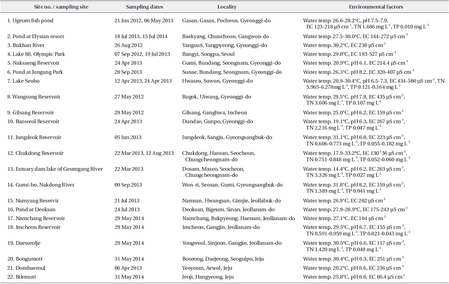
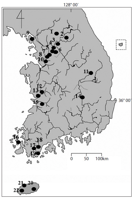
    [Table 1.] Information of sampling sites of the genus Scenedesmus in South Korea

    label

    Information of sampling sites of the genus Scenedesmus in South Korea

    MATERIALS AND METHODS

    The samples of the genus Scenedesmus (Chlorophyceae, Chlorococcales) were collected at 112 sites including ponds, swamp, reservoirs, lakes and rivers from 2012 to 2014. Sampling sites were located throughout the country. All samples were collected using 10 μm or 20 μm mesh-sized plankton nets with vertical and/or horizontal towing, or submerged benthic or soil algae with spoid or brush. The samples were immediately fixed with Lugol’s iodine solution (0.5%) to immobilize the cells and facilitate microscopic examination. To examine the fine structures and cellular shapes, and to identify and classify the chlorococcal species, temporary slides were made using the following steps: 1) the phytoplankton samples (chloroccocal algae) were mixed with glycerin in micro tubes, and 2) the mixed samples were placed by drop on slide glass, and fixed in position with cover slides. Permanent slides were made using the following steps: 1) the phytoplankton samples (chloroccocal algae) were mixed with liquid glycerol gelatin to mount on histochemical slides (Sigma-®); 2) the mixed samples were placed drop-wise on slide glass and fixed in position with a cover slide; and 3) samples were affixed at the margin of a cover glass with manicure (Thecashop-®). The temporary and permanent slides were observed at x200-x1,000 magnification using light microscope (LM) (Axioskop 20 and Axio Imager A2; Carl Zeiss, Oberkochen, Germany) while an attached digital camera (Zeiss Axiocam HRc; Carl Zeiss, Jena, Germany) was used to capture images. The scale bar in illustrations represents 10 μm.

    At each site, physical and chemical factors of water were recorded during the sampling period (Table 1). Water temp. (Water temperature, °C) and EC (electric conductivity) was measured in situ using a portable thermometer and EC meter (Orion 5-star; Thermo Fisher Scientific, Waltham, MA, USA), and pH was measured in situ using a pH meter (Ultrabasic-5; Denver Instrument, Bohemia, NY, USA) respectively. The data for total nitrogen (TN) and total phosphate (TP) concentrations at each sampling site was referred from the water information system of the Ministry of Environment (NIER 2014).

    The genus Scenedesmus (Chlorophyceae, Chlorococcales) was identified mainly based on Prescott (1962), Uherkovich (1966), Hirose et al. (1977), Hegewald and Schnepf (1979) Komárek and Fott (1983), Yamagishi and Akiyama (1984-1997), Hegewald and Silva (1988), and John and Tsarenko (2002).

    RESULTS AND DISCUSSION

      >  Order Chlorococcales A. Pascher Family Scenedesmaceae Oltmanns Genus Scenedesmus Meyen Subgenus Scenedesmus Hegewald

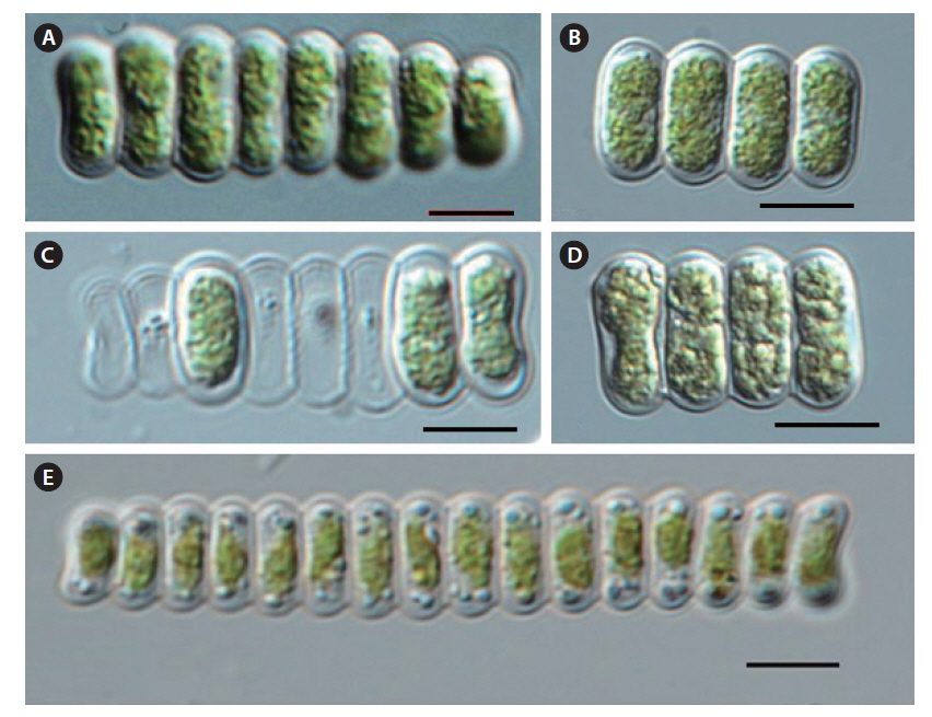
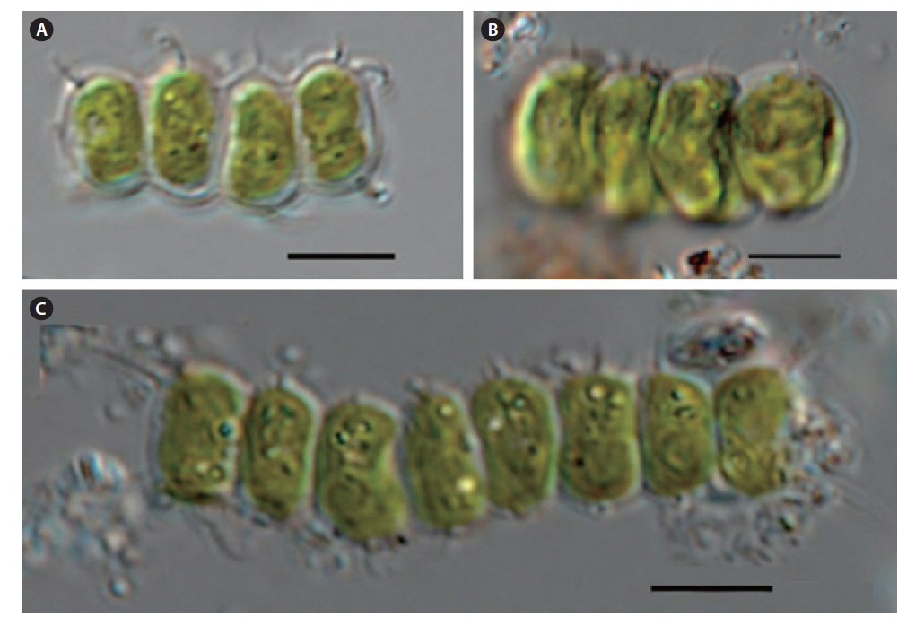
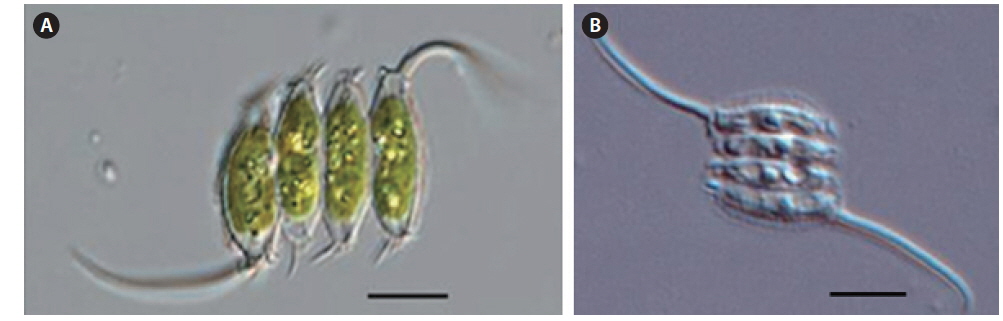
    Scenedesmus linearis Komarek (Figs. 2A-2E)

    References: Komárek and Fott 1983, p 832, taf. 226, fig. 6; Hegewald and Silva 1988. p 298, fig. 487.

    Description: Coenobia are 4-8-16 cells, linearly arranged cells, with 3-quarters of the cell length conjoined. cells are cylindrical with broadly rounded cell poles and slightly thick cell walls. The outsides of the marginal cells are slightly concave. Cell walls are smooth without teeth, spines, or granulates. Cells have a single chloroplast and a pyrenoid. Cells are 14-20 μm long and 5-8 μm wide.

    Occurrence sites: 3, 14, 18, 20, 21, 22 (Table 1 and Fig. 1)

      >  Subgenus Acutodesmus Hegewald

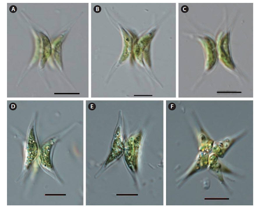
    Scenedesmus acuminatus var. elongatus G.M. Smith (Figs. 3A-3D)

    References: Uherkovich 1966, p 43, taf. III, Abb. 80; Komárek and Fott 1983, p 844, taf. 229, fig. 2; Hegewald and Silva 1988, p 52, fig. 65.

    Description: Coenobia are composed of 4-8 cells, commonly 4 cells, always forming a curved plate, with irregular twists. Cells are longer than S. acuminatus and proportionally narrower, and cells narrow and become fusiform, tapered, or pointed at the pole, while the outside is concave. The cell wall is smooth, without teeth, spines, or granulates. Cells have a single chloroplast and a pyrenoid. Cells are 50-60 μm long and 3-6 μm wide.

    Occurrence site: 4 (Table 1 and Fig. 1)

    Remarks: Komárek and Fott (1983) and Hegewald and Silva (1988) included this taxon into S. acuminatus. However, Komárek and Fott (1983) suggested that the figures related to S. acuminatus var. elongates are longer and more slender than S. acuminatus, and has slightly different morphological characteristics from S. acuminatus. Some figures of S. acuminatus var. tetradesmoides are similar to S. acuminatus var. elongatus, but it is shorter, wider, and less pointed than S. acuminatus var. elongatus. Some figures showed similarities to S. acuminatus var. tetradesmoides of Smith (1916). Hegewald (1979) collected the samples at the Haber River, Torfvenns and Dülmen. The cellular arrangements and morphological characteristics of the samples showed a striking difference from S. acuminatus. However, he included this taxon into S. acuminatus as a synonym. Many workers got confused about the morphological characteristics of cells and coenobia. This taxon is not merged into S. acuminatus, but separated into 4 taxa (S. acuminatus, S. acuminatus f. tortuosus, S. acuminatus var. elongates, S. acuminatus var. tetradesmoides) in this study, but further study is required to determine if it can be merged as a synonym or needs to be separated as an independent taxon.

    Scenedesmus acuminatus var. tetradesmoides G.M. Smith (Figs. 4A-4F)

    References: Komárek and Fott 1983, p 842, taf. 228, fig. 5; Hegewald and Silva 1988, p 55, fig. 70.

    Description: Coenobia are composed of 4-8 cells, commonly 4 cells, always forming a curved plate. Cells are arcuate or long crescent shaped, and adjoined side by side along a parallel axis, and radially arranged in apical view. Cells narrow and become fusiform, tapered, or pointed at the poles, and the outside is concave. The cell wall is smooth, without teeth, spines, or granulates. Cells have a single chloroplast, with a pyrenoid. Cells are 25-30 μm long and 5-8 μm wide.

    Occurrence sites: 4, 14 (Table 1 and Fig. 1).

    Remarks: Korshikov (1953) included this taxon into the basionym of S. acuminatus f. tetradesmoides. However, Komárek and Fott (1983) included S. acuminatus f. tetradesmoides and S. tetradesmiformis into this taxon as a synonym. The figure of this taxon in Komárek and Fott (1983) is slightly longer than the original figure in Smith (1916), and is narrower and longer than S. acuminatus. This taxon is identified and classified according to Komárek and Fott (1983).

      >  Subgenus Desmodesmus Chodat

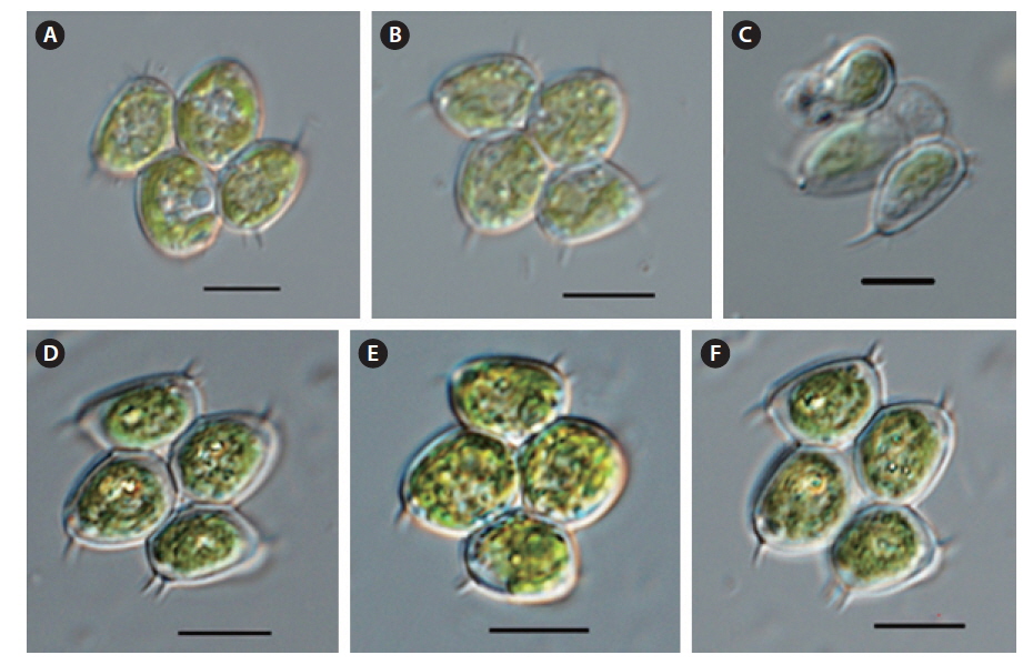
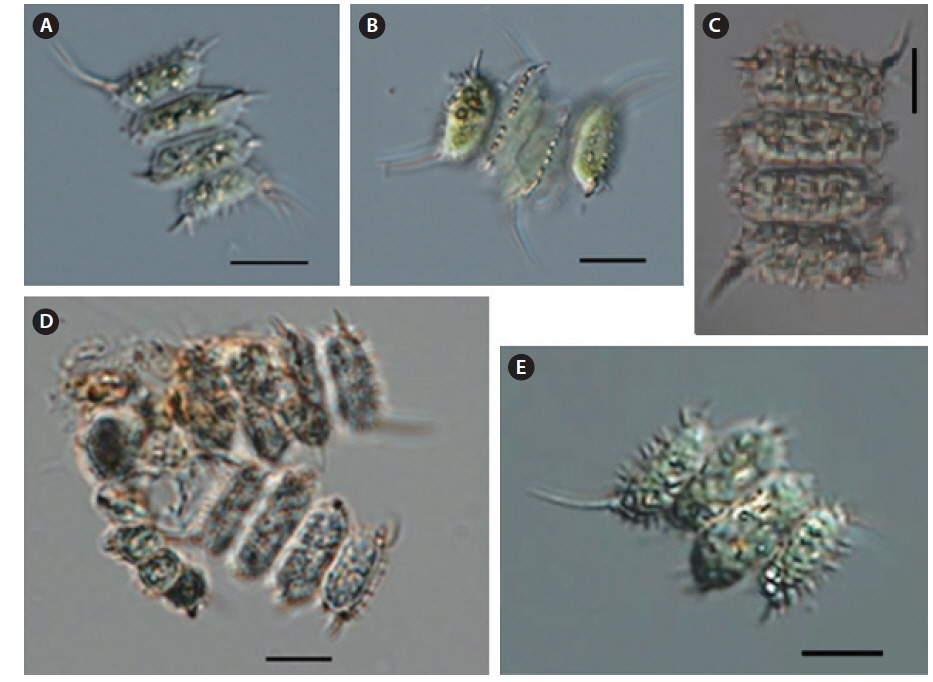
    Scenedesmus denticulatus var. disciformis Hortobagyi (Figs. 5A-5F)

    References: Hegewald and Silva 1988, p 200, fig. 315; John and Tsarenko 2002, p 392, pl. 95, fig. I.

    Description: Coenobia are composed of 4-8 cells, mostly arranged linearly. or with alternating cells in 1 or 2 rows, tightly joined without any space between the cells. Cells are broadly ovoid with 2-3 short teeth, sometimes outwardly directed and confined to the outer pole at inner cells and to both poles at the marginal cells, with or without fine granulates on the cell wall. Cells have a single chloroplast and a pyrenoid. Cells are 10-15 μm long and 7-10 μm wide.

    Occurrence sites: 4, 9, 12, 16, 19 (Table 1 and Fig. 1).

    Remarks: Komárek and Fott (1983) included the species having characteristics of S. denticulatus var. disciformis and S. denticulatus var. fenestratus into S. denticulatus as a synonym. But John and Tsarenko (2002) identified it as the mutant of S. denticulatus. The cells in this research were of wider widths, more egg-shaped, and had shorter protuberance at the end than Komárek and Fott (1983).

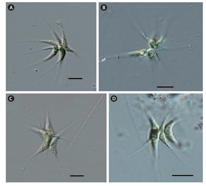
    Scenedesmus polydenticulatus Hortobagyi (Figs. 6A-6D)

    References: Komárek and Fott 1983, p 868, taf. 234, fig. 10; Hegewald and Silva 1988, p 398, fig. 643; Yamagishi and Akiyama 1995, p 15, pl. 73, figs. 1-6.

    Description: Coenobia are composed of 2-4-8 cells, mostly arranged linearly or in 1 or 2 rows of alternating cells that are rarely solitary, and tightly joined with or without a space between cells. Cells are broadly ovoid with 1-3-4 outwardly directed short teeth, and are confined to the outer pole at inner cells and to both poles at the marginal cells, with fine spines on the cell wall. Cells have a single chloroplast, with a pyrenoid. Cells are 15-25 μm long and 10-15 μm wide.

    Occurrence sites: 7, 2, 19, 21 (Table 1 and Fig. 1).

    Remarks: Komárek and Fott (1983) included S. denticulatus var. polyspinosus into S. smithii as a synonym. Taf. 234, fig. 4 in Komárek and Fott (1983) is referred to as the original figure of Biswas (1934), and taf. 234, fig. 10 is referred to as the original figure of S. polydenticulatus. These figures show different characteristics of S. spinulatus. Hegewald and Silva (1988) agreed with Komárek and Fott (1983), who included them as a synonym. And Hegewald and Silva (1988) merged S. polydenticulatus, S. denticulatus var. polyspinosus, S. smithii var. indicus, S. smithii var. linaeris and S. smithii var. polyspinosus into S. polydenticulatus as a synonym. S. smithii var. indicus and S. smithii var. linaeris do not have short spines or granulates on the cell wall, but S. smithii var. polyspinosus and S. denticulatus var. polyspinosus have short spines or granulates distributed on the cell wall and they are similar to S. spinulatus. This taxon is identified and classified as S. polydenticulatus on the basis of the distributed short spines on the cell wall in this study.

    Scenedesmus lefevrei Deflandre (Figs. 7A-7E)

    References: Komárek and Fott 1983, p 874, taf. 236, fig. 4; Hegewald and Silva 1988, p 295, fig. 481.

    Description: Coenobia are composed of 2-(4)-8 cells, linearly arranged in 1 row, with contact up to three-quarters of the cell length. Cells vary in shape from ellipsoidal to cylindrical, and are sometimes slightly asymmetrical, rounded, or tapered at the poles. The cells have short spines at the poles of the inner cells, while the marginal cells have slightly curved spines at the pole of the outer cells with a single longitudinal row of warty teeth or riblike structure on the side of each cell. Cells have a single chloroplast, with a pyrenoid. Cells are 15-25 μm long and 7-12 μm wide.

    Occurrence sites: 1, 7 (Table 1 and Fig. 1).

    Remarks: Deducenko-Scegoleva (1949) included S. lefevrei into the basionym of S. quadricauda var. lefevrei. Komárek and Fott (1983) included S. maculosus, S. quadricauda var. spinosus, S. decorus var. bicaudatus, S. lefevrei var. maguinii and S. lefevrei var. semiserratus f. bicaudatus into S. lefevrei as a synonym. However, Coenobia of S. maculosus and S. quadricauda var. spinosus are composed of 4 cells, with 2 long spines at the poles of their marginal cells. It is distributed along a longitudinal ridge structure. Considering the differences in characteristics of S. maculosus and S. quadricauda var. spinosus, these taxa are not merged into the synonym of S. lefevrei in this study.

    Kiriakov (1977) reported S. lefevrei var. semiserratus f. bicaudatus, the basionym of which was S. lefevrei var. maguinii. S. lefevrei var. semiserratus f. bicaudatus needs to be included into S. lefevrei as synonym, because the characteristics of this taxon are similar to S. lefevrei. Taf. X, abb. 424-427 in Uherkovich (1966) and the original figures of Deflandre (1924) showed different characteristics of long spines at the poles of the marginal cells. The original figure showed a long spine at one end and a short spine at the other end, with longitudinally distributed small spines on the cell walls at both sides. Uherkovich (1966) described the cells of this taxon as having the same length of long spines at the pole of outside cells, with longitudinal small spines on the inner cell wall, while there were no small spines on the sides of cell wall. It is possible to confuse the identification of this taxon on the basis of the two figures.

    Scenedesmus pannonicus Hortobagyi (Figs. 8A-8C)

    References: Komárek and Fott 1983, p 882, taf. 238, fig. 6; Hegewald and Silva 1988, p 369, fig. 598; John and Tsarenko 2002, p 397, pl. 95, fig. O.

    Description: Coenobia are composed of 4-8 cells, rarely 2 cells, arranged linearly or in slightly alternating cells. Cells are ellipsoid, cylindrical, or ovoid, with sides joined in linear series. The marginal cells have a short stout spine at the poles, while the inner cells have a stout spine at one pole or both poles. Cell walls are smooth. Cells have a single chloroplast, with a pyrenoid. Cells are 10-25 μm long and 6-12 μm wide.

    Occurrence sites: 1, 4, 5 (Table 1 and Fig. 1).

    Remarks: Komárek and Fott (1983) included S. naegelii, S. pannonicus f. heterocaudatus, S. longus, and S. intermedius var. balatonicus f. halophilus into S. pannonicu as synonyms. Among these taxa, S. pannonicus f. heterocaudatus is found to be similar to S. pannonicus, but other taxa differed from the original figures (Hegewald and Silva 1988). Therefore, one needs to double check before merging these taxa into a synonym. Hegewald et al. (1988) collected the samples from Madurai, India, and found some differences from the original sample. Coenobia are composed of 4-8 cells, with distributed longitudinal granulates or ridge structures on the cell wall. It encloses coenobia with gelatinous material. However, the identified taxon from this study has a similar cell structure to Hegewald et al. (1988), but differed from the ridge structure on the cell wall.

    Scenedesmus helveticus f. bicaudatus Korsikov (Figs. 9A-9B)

    References: Hegewald and Silva 1988, p 260, fig. 418.

    Description: Coenobia are composed of 2-4-8 cells, mostly arranged in a linear series or a slightly alternating manner. Cells are spindle, or elliptical shaped, and are tapered to conically rounded poles. The inner cells are with or without small teeth, and the marginal cells have elongated ends, with a straight or slightly curved long spine at one pole. The outer wall of marginal cells is straight or slightly convex, with wing-like structures on the side of the marginal cell. The cell wall is smooth, with continuous or interrupted longitudinal ribs. Cells have a single chloroplast, with a pyrenoid. Cells are 15-25 μm long and 5-8 μm wide.

    Occurrence sites: 11, 15, 17 (Table 1 and Fig. 1).

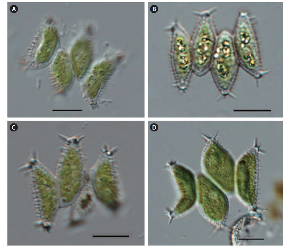
    Scenedesmus carinatus (Lemmermann) Chodat (Figs. 10A-10D)

    References: Komárek and Fott 1983, p 913, taf. 246, fig. 3; Hegewald and Silva 1988, p 141, fig. 569.

    Description: Coenobia are composed of (2)-4-8 cells, arranged in a linear series or slightly alternating row along the sides. Cells are long-fusiform, ellipsoidal, or naviculoid, with a conical or beaked pole. Cells have forked teeth spines on the poles and distinct longitudinal ribs. The marginal cells have long curved spines at the poles, and the outer side wall of marginal cells are straight or slightly convex. Cells have a single chloroplast, with a pyrenoid. Cells are 20-30 μm long and 4-8 μm wide.

    Occurrence sites: 2, 4, 6, 7, 9, 17 (Table 1 and Fig. 1).

    Remarks: Lemmermann (1899) reported the new species S. opoliensis var.carinatus. Chodat (1913) reported S. carinatus on the basis of a basionym S. opoliensis var.carinatus. Komárek and Fott (1983) described S. opoliensis var. carinatus again, and included S. carinatus var. hanoiensis, S. armatus (var. bolgariensis) f. crassicaudatus, and S. armatus (var. bolgariensis) f. bicaudato-granulatus into this taxon as synonyms. The former two taxa are similar to S. carinatus, but the latter taxon is different from S. carinatus.

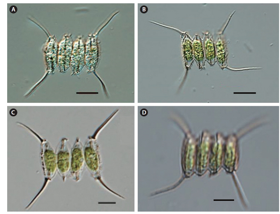
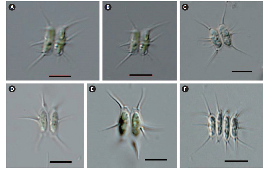
    Scenedesmus tenuispina Chodat (Figs. 11A-11F)

    References: Komárek and Fott 1983, p 916, taf. 247, fig. 1; Hegewald and Silva 1988, p 526, fig. 852.

    Description: Coenobia are composed of 2-4(-8) cells, arranged in a linear series or slightly alternating row along the sides. Cells are long-ovoid and ellipsoidal with pointed poles. One to two spines are on the poles of the inner cells. The marginal cells have 1-3 spines of equal cell length at the poles. The outside wall of marginal cells is straight or slightly convex, with 2-5 spines on the opposite side. Cells have a single chloroplast, with a pyrenoid. Cells are 10-15 μm long and 3-7 μm wide.

    Occurrence sites: 1, 4, 6, 8, 12, 13 (Table 1 and Fig. 1).

    Remarks: Uherkovich (1966) reported new species S. spinosus var. tenuispina on the basis of the basionym S. tenuispina. However, this new variety was invalid because there was no sufficient reference to basionym (Hegewald and Silva 1988). It was classified with S. tenuispina, not S. spinosus var. tenuispina according to Uherkovich (1966). This taxon has not been reported in Korea, because Uherkovich (1966) included S. tenuispina into S. spinosus, and Korean researchers have identified S. spinosus on the basis of Uherkovich (1966). S. tenuispina was identifed in this study according to Komárek and Fott (1983).

    Scenedesmus gutwinskii var. heterospina Bodrogkozy (Figs. 12A-12D)

    References: Uherkovich 1966, p 112, pl. XIX, fig. 783; Komárek and Fott 1983, p 918, taf. 247, fig. 4; Hegewald and Silva 1988, p 258, fig. 414.

    Description: Coenobia are composed of 2-4-8 cells, arranged in a linear series or slightly alternating row along the sides. Cells are long, ovoid and ellipsoidal, with conical or rounded poles. One to two teeth spines are found on the poles of the inner cells. The marginal cells have slightly curved or straight spines at one pole and short spines on other pole, with 3-8 straight or slightly convex, short spines on the outermost side. Cells have a single chloroplast, with a pyrenoid. Cells are 8-15 μm long and 3-7 μm wide.

    Occurrence sites: 1, 6, 7, 8 (Table 1 and Fig. 1).

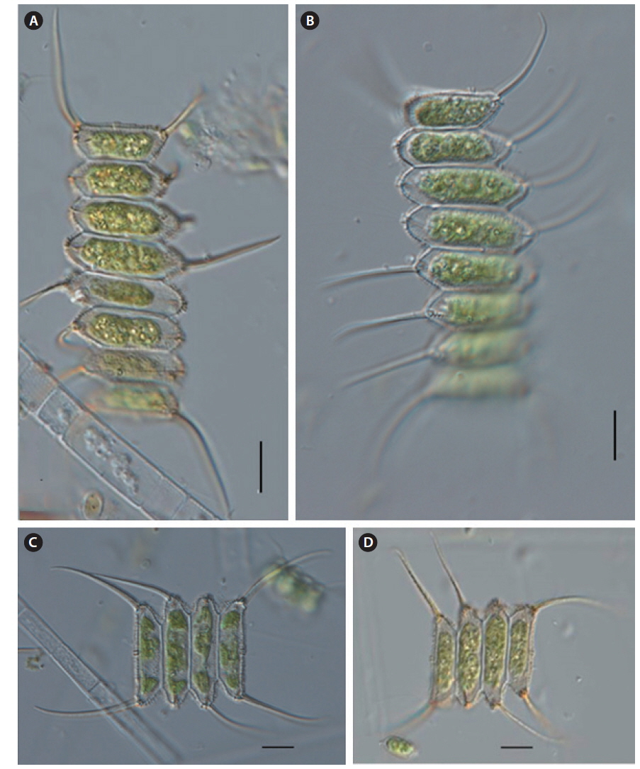
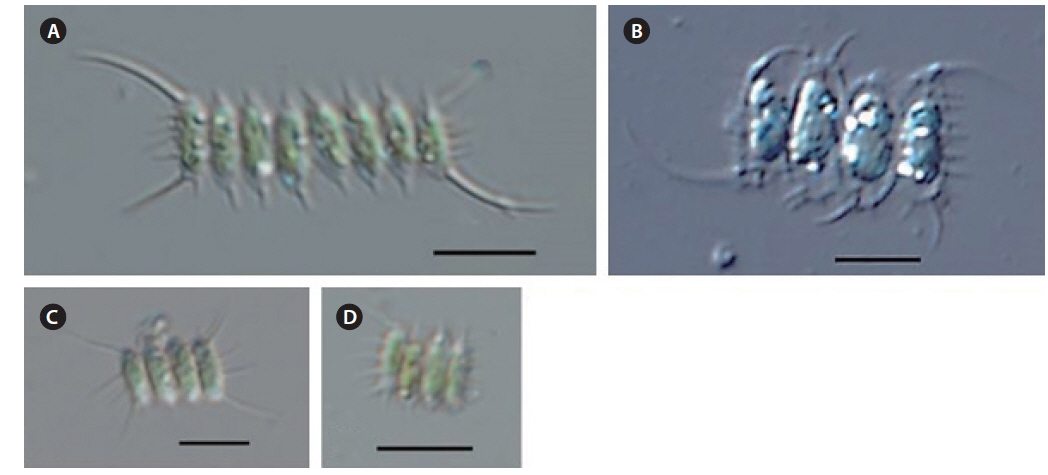
    Scenedesmus oahuensis var. clathratus f. longiclathratus G. Tell (Figs. 13A-12D)

    (Synonym: S. oahuensis var. longiclathratus)

    References: Komárek and Fott 1983, p 936, taf. 251, fig. 5; Hegewald and Silva 1988, p 329, fig. 539.

    Description: Coenobia are composed of 4-8 cells, arranged in a linear series. Cells are long cylindrical, or ellipsoidal-cylindrical, with conical or rounded poles. The inner cells may have long spines with continuous or interrupted longitudinal ribs at the poles. The marginal cells have a straight or slightly curved long spine at the poles, and the outer side wall of marginal cells are straight or slightly convex. There are distributed granulates on the side and wall of inner and marginal cells. Cells have a single chloroplast, with a pyrenoid. Cells are 25-35 μm long and 8-12 μm wide.

    Occurrence sites: 1, 4, 6, 7, 8, 13, 18 (Table 1 and Fig. 1).

    Remarks: Lemmermann (1905) reported new variety S. quadricauda var. oahuensis from the samples which were collected at Oahu, Hawaii. The new variety had the morphological characteristics of cells with rounded poles with distributed granulates on the cell wall and enclosed granulates on sides of the cell. Smith (1916) reported an independent species that had larger cells than S. quadricauda with distributed granulates on the cell wall. Hortobagyi (1973) separated this taxon from S. maximus on the basis of the spines at the poles with distributed granulates on the sides of the cells. Sieminska (1965) separated S. oahuensis var. montanensis from S. oahuensis because of the two short spines present at the poles of the inner cells, which were absent in typical ones. These spines were curved downwards, and the cell wall was a continuous longitudinal ridge. Hegewald (1979) separated S. maximus from S. quadricauda and included S. oahuensis var. granulatus, S. oahuensis var. montanensis, S. oahuensis var. symmetricus into S. maximus as synonyms. Komárek and Fott (1983) separated S. oahuensis and S. oahuensis var. montanensis from synonym taxa of Hegewald (1979). Jeon and Hegewald (2006) reported Desmodesmus tropicus var. longiclathratus on the basis of the basionym of S. oahuensis var. clathratus f. longiclathratus. It is classified with S. oahuensis var. clathratus f. longiclathratus according to Tell (1979) in this study, but further study is needed to merge as a synonym or separate as an independent taxon.

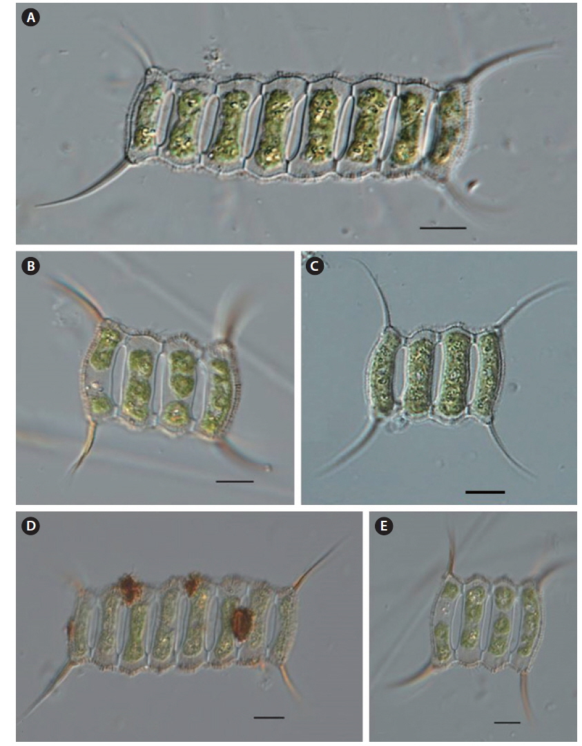
    Scenedesmus oahuensis var. clathratus Manguin (Figs. 14A-14F)

    References: Komárek and Fott 1983, p 940, taf. 252, figs. 6-7; Hegewald and Silva 1988, p 329, fig. 539.

    Description: Coenobia are composed of 4-8 cells, arranged in a linear series with intercellular perforation between cells along the polar region. Cells are long cylindrical or ellipsoidal-cylindrical, spool-shaped with rounded poles. The marginal cells have a straight or slightly curved long spine at the poles, while the outsides of marginal cells are straight or slightly convex. There are distributed granulates on the side and inner wall of marginal cells. Cells have a single chloroplast with a pyrenoid. Cells are 25-35 μm long and 8-12 μm wide.

    Occurrence sites: 1, 4, 6, 7, 8 (Table 1 and Fig. 1).

    Remarks: Manguin (1936) reported this taxon on the basis of the figure and description of Scenedesmus sp. and S. ornatus (Hegewald and Silva 1988). Komárek and Fott (1983) included S. papillatus, S. perforatus var. circumcinctus and S. perforates var. oahuensioides into a synonym of this taxon. The former two taxa are similar to S. oahuensis var. clathratus, but the latter taxon is similar to S. oahuensis. It is separated to S. perforatus var. oahuensioides as the synonym of this taxon in this study. S. perforatus has a similar shape to the coenobia of this taxon, but different cell wall with granulates on the sides of the cells. Therefore, this work separated it as an independent species from this taxon and S. perforates.

참고문헌
  • 1. Biswas K 1934 Some foreign weeds and their distribution India and Burma [Indian For] Vol.60 P.861-865 google
  • 2. Chodat R. 1913 Monographies d’algues en culture pure google
  • 3. Comas A, Komarek J 1984 Taxonomy and nomenclature of several species of Scenedesmus (Chlorellales). Arch Hydrobiol/Suppl 67 [Algol Stud] Vol.35 P.135-157 google
  • 4. Deducenko-Scegoleva NT 1949 Novye vidy I raznovidnosti roda Scenedesmus Meyen. Species atque varietates novae generis Scenedesmus Meyen. Bot Mat [Otd Sporovyk Rast Bot Gosudarstv Univ] Vol.67 P.117-133 google
  • 5. Deflandre G 1924 Additions a la flore algologique des environs de Paris [Bull Soc Bot France] Vol.71 P.1115-1130 google cross ref
  • 6. Guiry MD, Guiry GM 2014 AlgaeBase google
  • 7. Hegewald E 1979 Vergleichende Beobachtungen an Herbarmaterial und Freilandmaterial von Scenedesmus [Algol Stud] Vol.24 P.264-286 google
  • 8. Hegewald E 1982 Taxonomisch-morphologische Untersuchung von Scenedesmus-Isolaten aus Stammsammlungen [Algol Stud, Suppl] Vol.29 P.375-406 google
  • 9. Hegewald E, Engelberg K, Paschma R 1988 Beitrag zur taxonomie der gattung Scenedesmus, subgenus Scenedesmus (Chlorophyceae) [Nova Hedwigia] Vol.47 P.497-533 google
  • 10. Hegewald E, Hindak F, Schnepf E 1990 Studies on the genus Scenedesmus Meyen (Chlorophyceae, Chlorococcales) from South India, with special reference to the cell wall ultrastructure google
  • 11. Hegewald E, Schnepf E 1979 Geschichte und Stand der Systematik der Grunalgengattung Scenedesmus [Schweiz Zeit Hydrol] Vol.40 P.320-343 google
  • 12. Hegewald E, Silva PC 1988 Annotated Catalogue of Scenedesmus and Nomenclaturally Related Genera, Including Original Descriptions and Figures google
  • 13. Hindak F 1990 Studies on the Chlorococcal Algae (Chlorophyceae) google
  • 14. Hirose H, Akiyama M, Yamagishi T 1977 Illustrations of the Japanese Freshwater Algae google
  • 15. Hortobagyi T 1973 Der Formenkreis von Scenedesmus sooi Hortob [Acta Bot Acad Sci Hung] Vol.18 P.413-420 google
  • 16. Jeon SL, Hegewald E 2006 A revision of the species Desmodesmus perforatus and D. tropicus (Scenedesmaceae, Chlorophyceae, Chlorophyta) [Phycologia] Vol.45 P.567-584 google cross ref
  • 17. John DM, Tsarenko PM, John DM, Whitton BA, Brook AJ 2002 Order Chlorococcales P.327-409 google
  • 18. Kiriakov IK 1977 Rod Scenedesmus Meyen (Chlorophyta, Chlorococcales). Taksonomija, ecologija i razprostranenie v NR Bulgarija. Autoreferat google
  • 19. Komarek J, Fott B 1983 Das Phytoplankton des Sußwassers. Systematik und Biologie - 7 Teil, 1. Chlorophyceae (Grunalgen) google
  • 20. Komarek J, Ludvik J 1971 Die Zellwandultrastruktur als taxonomisches Merkmal in der Gattung Scenedesmus. 1. Die Ultrastrukturelementre [Algol Stud] Vol.5 P.301-333 google
  • 21. Korshikov OA 1953 The Freshwater Algae of the Ukranian SSR. V. Subclass Protococcineae. Vacuolales and Protococcales google
  • 22. Lemmermann E 1899 Das Phytoplankton sachsischer Teiche [Forsch-Bungsber Biol Stat Plon] Vol.7 P.96-135 google
  • 23. Lemmermann E 1905 Die Algenflora der Sandwich-Inseln. Ergebnisse einer Reise nach dem Pacific. H. Schauinsland 1896/97 [Bot Jahrb Syst] Vol.34 P.607-663 google
  • 24. Manguin E 1936 La flore algale de l’etang des Rablais (Sarthe); contribution a la connaissance de la microflore d’un etang calcaire de l’Ouest de la France [Bull Soc d’Agricult Sci Arts Sarthe] Vol.55 P.314-351 google
  • 25. 2014 Water Information System of the Ministry of Environment google
  • 26. Prescott GW 1962 Algae of the Western Great Lakes Area google
  • 27. Sieminska J 1965 Algae from Mission Wells Pond, Montana [Trans Amer Microsc Soc] Vol.84 P.98-126 google
  • 28. Smith GM 1916 A monograph of the algal genus Scenedesmus based upon pure culture studies [Trans Wisconsin Acad Sci Arts Letters] Vol.18 P.422-530 google
  • 29. Tell G 1979 Chlorophyceae d’eau douce rares et nouvelles de la Republique Argentine [Rev Algol NS] Vol.14 P.39-48 google
  • 30. Trainor FR, Rowland HL 1968 Control of colony and unicell formation in a synchronized Scenedesmus [J Phycol] Vol.4 P.310-317 google cross ref
  • 31. Uherkovich G. 1966 Die Scenedesmus-Arten Ungarns google
  • 32. Yamagishi T, Akiyama M 1997 Photographs of the Fresh-water Algae google
이미지 / 테이블
  • [ Fig. 1. ]  Showing the sampling sites of the genus Scenedesmus in South Korea.
    Showing the sampling sites of the genus Scenedesmus in South Korea.
  • [ Table 1. ]  Information of sampling sites of the genus Scenedesmus in South Korea
    Information of sampling sites of the genus Scenedesmus in South Korea
  • [ Fig. 2. ]  Scenedesmus linearis. Scale bars, 10 μm.
    Scenedesmus linearis. Scale bars, 10 μm.
  • [ Fig. 3. ]  Scenedesmus acuminatus f. elongatus. Scale bars, 10 μm.
    Scenedesmus acuminatus f. elongatus. Scale bars, 10 μm.
  • [ Fig. 4. ]  Scenedesmus acuminatus f. tetradesmoides. Scale bars, 10 μm.
    Scenedesmus acuminatus f. tetradesmoides. Scale bars, 10 μm.
  • [ Fig. 5. ]  Scenedesmus denticulatus var. disciformis. Scale bars, 10 μm.
    Scenedesmus denticulatus var. disciformis. Scale bars, 10 μm.
  • [ Fig. 6. ]  Scenedesmus polydenticulatus. Scale bars, 10 μm.
    Scenedesmus polydenticulatus. Scale bars, 10 μm.
  • [ Fig. 7. ]  Scenedesmus lefeveri. Scale bars, 10 μm.
    Scenedesmus lefeveri. Scale bars, 10 μm.
  • [ Fig. 8. ]  Scenedesmus pannonicus. Scale bars, 10 μm.
    Scenedesmus pannonicus. Scale bars, 10 μm.
  • [ Fig. 9. ]  Scenedesmus helveticus f. bicaudatus. Scale bars, 10 μm.
    Scenedesmus helveticus f. bicaudatus. Scale bars, 10 μm.
  • [ Fig. 10. ]  Scenedesmus carinatus. Scale bars, 10 μm.
    Scenedesmus carinatus. Scale bars, 10 μm.
  • [ Fig. 11. ]  Scenedesmus tenuispina. Scale bars, 10 μm.
    Scenedesmus tenuispina. Scale bars, 10 μm.
  • [ Fig. 12. ]  Scenedesmus gutwinskii var. heterospina. Scale bars, 10 μm.
    Scenedesmus gutwinskii var. heterospina. Scale bars, 10 μm.
  • [ Fig. 13. ]  Scenedesmus oahuensis var. clathratus f. longiclathratus. Scale bars, 10 μm.
    Scenedesmus oahuensis var. clathratus f. longiclathratus. Scale bars, 10 μm.
  • [ Fig. 14. ]  Scenedesmus oahuensis var. clathratus. Scale bars, 10 μm.
    Scenedesmus oahuensis var. clathratus. Scale bars, 10 μm.
(우)06579 서울시 서초구 반포대로 201(반포동)
Tel. 02-537-6389 | Fax. 02-590-0571 | 문의 : oak2014@korea.kr
Copyright(c) National Library of Korea. All rights reserved.