검색 전체 메뉴
PDF
맨 위로
OA 학술지
Screening and identification of bioorganic light-emitting substances from marine macrophytes
  • 비영리 CC BY-NC
  • 비영리 CC BY-NC
ABSTRACT
Screening and identification of bioorganic light-emitting substances from marine macrophytes
KEYWORD
bioorganic light-emitting material , gas chromatography-mass spectrometry , marine macrophytes , photo-luminescence spectrum
  • INTRODUCTION

    The history of displays begins with early cathode-ray tubes, moving on to the first and second generation of liquid crystal displays (LCDs), and finally reaching the current generation of organic light-emitting diode (OLED) devices (Wong and Ho 2009). The OLED is a self-emitting form, and improvements have resulted in an improved viewing angle and outstanding performance with thin layers. The device is a simple one, and power consumption has been greatly improved over time. However, shortages of materials and problems relating to device life expectancy have yet to be solved satisfactorily. South Korea is a current leader, alongside the United States and Japan, in terms of the number of patent registrations relating to OLEDs. However, with regard to the materials used to produce OLEDs (a core technology), South Korea has only 14% of all such patents, and so room for expansion exists with respect to OLED display technology (Ministry of Science and Technology 2006).

    The limitations of present OLED technology lie in the emission of light. The light-emitting materials used may affect the lighting field as well as the display field. For example, the green-colored light emitted by Alq3 is the most commonly used light-emitting material (Hall and Schlegel 2001). However, although blue light-emitting materials with chemical stability, low color purity, and high flexibility exist, these tend to show whitening. Red light-emitting materials can reach a wavelength of 650 nm, but their emitting capabilities are less than ideal for industrial use (Seo and Moon 2014). Previous studies have investigated the optical and semiconducting properties of OLED materials (Karakawa et al. 2007, Sasabe et al. 2008) and were undertaken to find a way to avoid the dependence on the current range of OLED materials. In the present study, we used substances derived from living organisms to solve this problem. We required the light-emitting substance to be solid at room temperature, contain a benzene ring within its structure, have a range of molecular weights from 100 to 500 and a range of melting points above 100°C, and show little or no reaction with moisture (Karakawa et al. 2007, Sasabe et al. 2008). To examine the efficiency of LEDs, photoluminescence (PL) spectra were used to measure the bandgap from the full valence band to the empty conduction band, and to examine the basic characteristics and optical properties of the derivatives (Karakawa et al. 2007).

    Few studies have reported on OLED materials derived from natural products / living organisms, and most OLED materials available at present are synthetic or a combination of organic and inorganic materials for increased functionality (Tajima et al. 2006). As “natural” OLED materials, chlorophyll a, chitin, cytochrome c, other proteins, and DNA have been studied (Steckl 2007, Singh et al. 2010), and researchers have continued to search for novel light-emitting materials, including biological products (Sanderson et al. 1987, Gomez et al. 2014). Bio-light-emitting materials derived from natural sources have been reported, including chitosan (d-glucosamine [2-amino-2-deoxy-d-glucose]), myoglobin, bovine serum albumin, deoxyribonucleic acid, cytochrome c, and chlorophyll (Hagen et al. 2006, Tajima et al. 2006, Steckl 2007, Singh et al. 2010, Rau et al. 2012). Adenine is a purine derivative that shows blue light emission in the 380-560 nm range, while cytosine can show high luminance at 380-560 nm. Emissions increase with purity at a reported maximum brightness of 2.16 cd m-2 at a wavelength of 386 nm (Lewis 2006, Lee et al. 2007, Kim et al. 2010, Shin 2013). PL studies on the photosynthetic pigment chlorophyll a have also been reported (Choi et al. 2001), with the maximum peak wavelengths reported as 373 and 668 nm, respectively, and with respective high emitting ranges at 380-400 and 660-800 nm with a luminance of 1.16 cd m-2 (Shin 2013).

    Among marine organisms, algae are a well-known source of novel substances, such as proteins, fatty acids, vitamins, and pigments (Volk 2008). They also produce a wide variety of secondary metabolites with different bioactivities (e.g., antifungal, antiviral, antibiotic). Algae also have potential for pharmaceutical research. In addition, those marine organisms that produce novel organic compounds are of particular interest.

    This study was undertaken to identify bio-light-emitting materials and to estimate the efficiency of the light emission using the PL spectra. We derived the diverse functional organic light-emitting substances from marine macrophytes.

    MATERIALS AND METHODS

      >  Samples

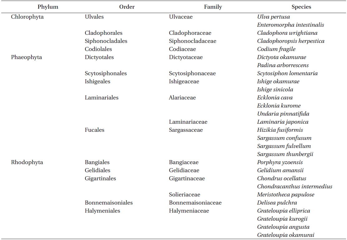
    Marine macrophytes were collected around the coastal areas of Ganjelgot in Ulsan City, Nameri in Gangneung, and Aewol in Jeju, South Korea. Materials were identified morphologically based on the “Marine Algae of Jeju” by Lee (2008) and Silva et al. (1996), Yoshida (1998) and Schneider and Wynne (2007) were used for further identification. The species of marine macrophytes examined comprised 5 Chlorophyta, 13 Phaeophyta, and 10 Rhodophyta (Table 1).

    [Table 1.] List of marine macrophytes collected in South Korean coastal areas

    label

    List of marine macrophytes collected in South Korean coastal areas

      >  Extraction and separation

    The collected marine macrophytes were rinsed, dried at room temperature, and then powdered. Each of the 100-g samples was powdered and extracted using 1,000 mL of 100% methanol (MeOH) over 25 h at room temperature. After filtration, the crude extract was concentrated to give a dark-green tar-like sticky residue (10 g) under reduced pressure in an evaporator, and then the MeOH extraction was purified by column chromatography using a Pyrex glass column (diameter : length = 24 : 40 cm) charged with a silica gel 60 (70 g). Elution was performed with a toluene and ethyl acetate in the following ratio: 7 : 3 in sequence. Fractionations were monitored by thin layer chromatography. Fractions of candidate organic compound materials were used to examine the samples in terms of their PL spectra, and promising candidates were selected based on their light-emitting properties.

      >  Identification

    Gas chromatography–mass spectrometry (GC-MS) analysis was performed using a Shimadzu GC MS-QP 2010-Plus (Shimadzu, Kyoto, Japan) and a 30 m × 0.25 mm × 0.25-μm Rtx-5MS capillary column (Shimadzu). The helium carrier gas was supplied at a head pressure of 35 psi to provide an initial flow rate of 1 mL min-1. The temperature of the split injector was 260°C and the split ratio was 1 : 50. The injection volume for the fraction samples was 1 mL. The GC oven was maintained at 60°C for 3 min and then programmed at 100°C (2 min hold), followed by 200°C (5 min hold) and finally 235°C (10-min hold). Fullscan mass spectra were collected from m/z 85 to 550 at a data acquisition rate of 10 spectra s-1. The MS transfer line was held at 250°C and the ion source temperature was 200°C. The electron energy was 70 eV. Isolated peaks were identified by comparison with the National Institute of Standards and Technology Library (Gaithersburg, MD, USA).

      >  PL spectra

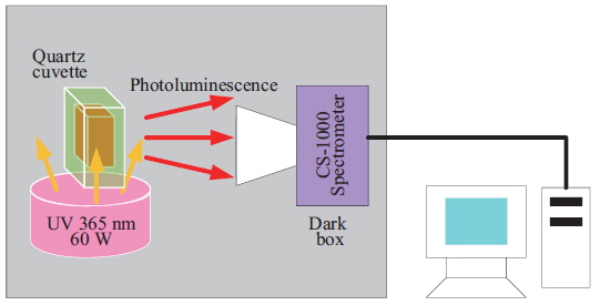
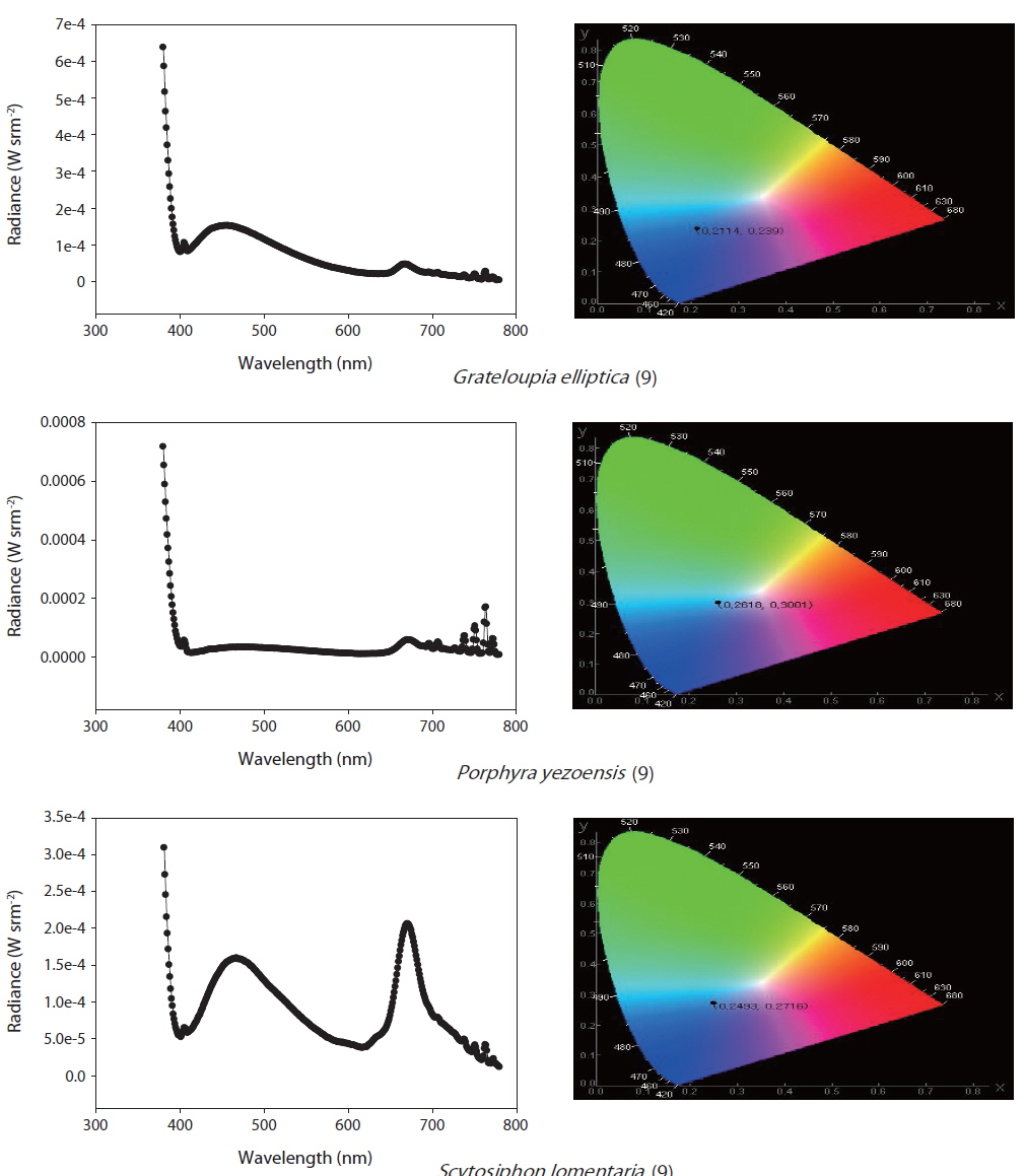
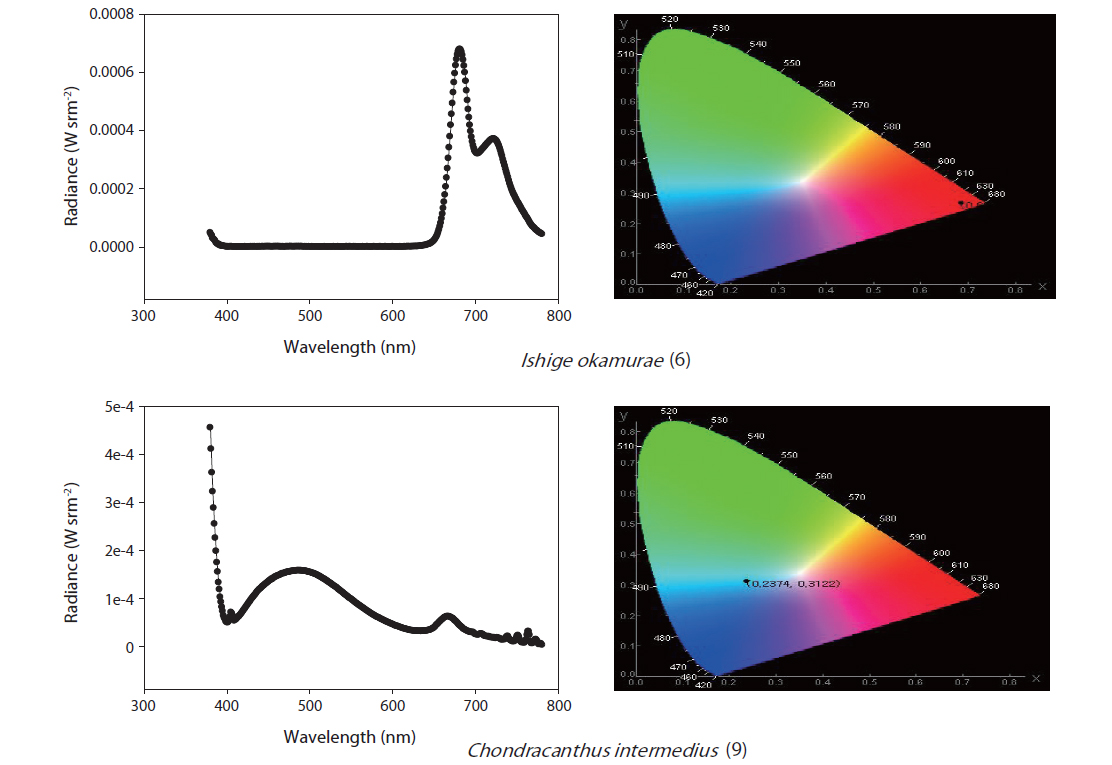
    The selected purified substances were examined in terms of their PL spectra using the apparatus shown in Fig. 1. The substances were exposed to UV radiation at 365 nm in a 1-mL crystal cuvette, and PL reactions were initiated. The CS-1000 spectra were used to measure the chromaticity, brightness, and light emission of the samples (Fig. 1).

    RESULTS

      >  Purification

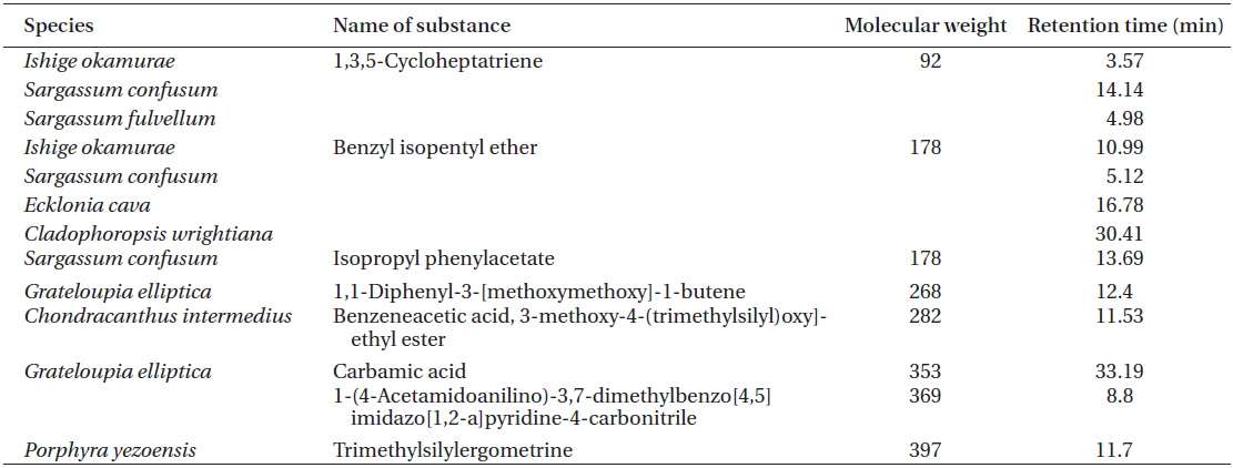
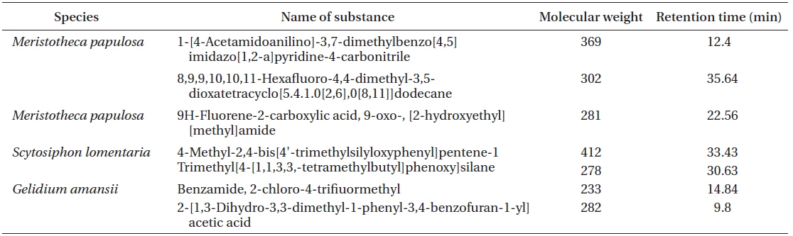
    In total, 1,221 solvent fractions were isolated from 28 marine macrophyte species. Sixteen solvent fractions with no pigments were selected and verified by PL reactions. The structures and material properties of the candidate substances were analyzed by GC-MS. In total, 14 bioorganic light-emitting materials identified from the 16 solvent fractions were selected for potential use as OLED materials (Tables 2 & 3).

    [Table 2.] List of OLED candidates identified by GC-MS

    label

    List of OLED candidates identified by GC-MS

    [Table 3.] List of OLED candidates identified by GC-MS

    label

    List of OLED candidates identified by GC-MS

      >  Identification

    1,3,5-Cycloheptatriene and benzyl isopentyl ether were found in Ishige okamurae and Sargassum confusum; S. confusum also contained isopropyl phenylacetate. 1,1-Diphenyl-3-[methoxymethoxy]-1-butene was identified from Grateloupia elliptica. Benzene acetic acid, 3-methoxy-4-[(trimethylsilyl)oxy]-ethyl ester, trimethylsilyl ergometrine, 9H-fluorene-2-carboxylic acid, 9-oxo-[2-hydroxyethyl] [methyl]amide, and benzamide, 2-chloro-4-trifluoromethyl were found in Chondracanthus intermedius, Porphyra yezoensis, Meristotheca papulosa, and Gelidium amansii. Carbamic acid and 1-(4-acetamidoanilino)-3,7-dimethylbenzo[4,5] imidazo[1,2-a]pyridine-4-carbonitrile were identified from G. elliptica. 1-[4-Acetamidoanilino]-3,7-dimethyl-benzo[4,5]imidazo[1,2-a]pyridine-4-carbonitrile and 8,9,9,10,10,11-hexafluoro-4,4-dimethyl-3,5-dioxatetracyclo [5.4.1.0[2,6],0[8,11]]dodecane were found in M. papulosa. 4-Methyl-2,4-bis[4′-trimethylsilyloxyphenyl] pentene-1 and trimethyl[4-[1,1,3,3,-tetramethylbutyl] phenoxy]silane were identified from Scytosiphon lomentaria, and benzamide, 2-chloro-4-trifluoromethyl, and 2-[1,3-dihydro-3,3-dimethyl-1-phenyl-3,4-benzofuran-1-yl]acetic acid were found in G. amansii.

      >  Photoluminescence spectra

    Two solvent fractions of M. papulosa showed purple and sea-green PL reactions (Fig. 2). A solvent fraction of G. amansii exhibited maximum light emission peaks at 380-400 nm and 600 nm, similar to the chlorophyll a PL reaction, but appeared dark blue (Fig. 2). Maximum light emission peaks were exhibited for G. elliptica at 380-400 and 430-500 nm (Fig. 3), and P. yezoensis at 390-400 and 680-800 nm with a blue color (Fig. 3), while S. lomentaria showed maximum light emission peaks at 380-400 and 680-700 nm (Fig. 4). I. okamurae (Rhodophyta) and C. intermedius showed maximum light emission peaks at 440-520 nm, with a color between blue and green, based on chromaticity coordinates.

    DISCUSSION

    Choi et al. (2001) reported on an OLED device consisting of a single layer of chlorophyll a and aluminum, with a coating of chlorophyll a using the Langmuir-Blodgett method. The OLED efficiency and thickness of the chlorophyll coating both increased. However, chlorophyll a is not used in commercial OLEDs because its chemical bonds break down with heat, and also the high costs associated with its commercial use. Chlorophyll a has a blue irradiance of electroluminescence from 300 to 400 nm, with low red band wavelengths and a tetrapyrrole ring with magnesium atoms in the porphyrin at its center. A great deal of research has investigated its PL properties. Chlorophyll a emits mainly at 380-400 and 660-800 nm, and its maximum emission wavelengths have been found to show a brightness of 1.16 cd m-2 at 373 and 668 nm.

    Generally, higher plants and animals, fungi, and yeast have cytochrome c as part of the electron transport system within their mitochondria. Reductive sulfate protein absorbance bands appear at 550, 520, and 415 nm, while an oxidative protein has an absorbance band at 407 nm. Tajima et al. (2006) reported on the use of cytochrome c in an OLED coating plate.

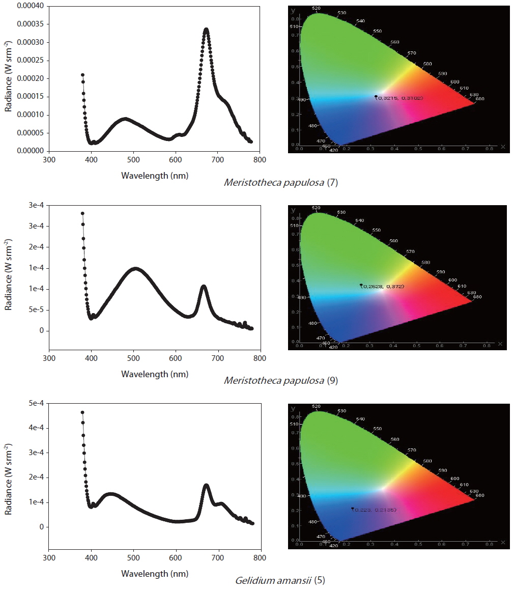
    To determine the methods for OLED candidate PL measurements, Alq3 is a commercially available light-emitting material with a known spectrum and chromaticity coordinates. Its PL spectrum is in the range of 480-700 nm, its luminance is 18.66 cd m-2 , and the maximum emission wavelength was found to be 531 nm in primary examination (Shin 2013). Furthermore, commercial N,N'-bis-(1-naphthalenyl)-N,N'-bis-phenyl-(1,1'-biphenyl)-4,4'-diamine (NPB) PL spectrum and color analysis chromaticity coordinates are available and can be used as a representative material with benzene rings and two amines. NPB shows a 400-700 nm PL spectrum, broad emissions with green and blue chromaticity coordinates, and color purity. The luminance is 10.15 cd m-2 and the maximum emission wavelength is 488 nm. Chitosan PL spectra and chromaticity coordinates (reference material) are also available. Chitosan is derived from crustacean shells and has been assessed as an OLED material, but requires a high purity. Unlike other substances, it emits from 380 to 400 nm (blue), showing high color purity up to 0.59 cd m-2 and a maximum light emission wavelength of 380 nm.

    In this study, we described the identification and physical properties of functional bio-light-emitting materials from marine macrophytes. The compounds were first identified as functional bio-light-emitting materials. In total, 16 solvent fractions were selected and confirmed by the PL reactions, and structures and material properties were analyzed for candidate substances by GC-MS. Fourteen bio-light-emitting materials identified from the 16 solvent fractions potentially contained OLED materials from 28 strains of marine macrophytes. Many benzene derivatives were identified, including benzylisopentyl ether, isopropyl phenylacetate, 1,1-diphenyl-3-[methoxymethoxy]-1-butene, benzene acetic acid, benzamide, 4-methyl-2,4-bis[4'-trimethylsilyloxy-phenyl]pentene-1, and trimethyl[4-[1,1,3,3,-tetramethylbutyl] phenoxy]silane benzamide. In addition, two fluorine compounds, 9H-fluorene-2-carboxylic acid and 8,9,9,10,10,11-hexafluoro-4,4-dimethyl-3,5-dioxatetracyclo[5.4.1.0[2,6],0[8,11]]dodecane, were recognized. One amine, carbamic acid, was discovered in G. elliptica. Two solvent fractions of M. papulosa showed purple and seagreen PL reactions. The solvent fraction of G. amansii exhibited maximum light emissions at 380-400 and 600 nm, similar to chlorophyll a but with a dark-blue color. The maximum light emissions shown by G. elliptica were at 380-400 and 430-500 nm, while those of P. yezoensis were at 390-400 and 680-800 nm with a blue color. S. lomentaria exhibited maximum light emissions at 380-400 and 680-700 nm. I. okamurae (Rhodophyta) and C. intermedius showed maximum light emissions at 440-520 nm, the color of which was between blue and green based on chromaticity coordinates.

    In conclusion, marine algae were screened to discover new OLED material candidates. As an example, we extracted the red alga G. amansii with an organic solvent system and confirmed the active fraction using PL methods and characterized the bio-light-emitting materials by GC-MS analysis. The OLED candidates identified in this study should be studied further with a view to improving their emission efficiencies.

참고문헌
  • 1. Choi H. G., Oh B. K., Lee W. H., Choi J. W. 2001 Depostion behavior and photoelectrochemical characteristics of chlorophyll a Langmuir-Blodgett films [Biotechnol. Bioprocess Eng] Vol.6 P.183-188 google cross ref
  • 2. Gomez E. F., Venkatraman V., Grote J. G., Steckl A. J. 2014 DNA bases thymine and adenine in bio-organic light emitting diodes [Sci. Rep.] Vol.4 P.7105 google cross ref
  • 3. Hagen J. A., Li W., Steckl A. J., Grote J. G. 2006 Enhanced emission efficiency in organic light-emitting diodes using deoxyribonucleic acid complex as an electron blocking layer [Appl. Phys. Lett.] Vol.88 P.171109 google cross ref
  • 4. Hall M. D., Schlegel H. B. 2001 Molecular orbital study of the first excited state of the OLED material tris (8-hydroxyquinoline) aluminum (III) [Chem. Mater.] Vol.13 P.2632-2640 google cross ref
  • 5. Karakawa M., Chikamatsu M., Nakamoto C., Maeda V., Kubota S., Yase K. 2007 Organic light-emitting diode application of fluorescent cellulose as a natural polymer [Macromol. Chem. Phys.] Vol.208 P.2000-2006 google cross ref
  • 6. Kim S. O., Lee K. H., Kim G. Y., Seo J. H., Kim Y. K., Yoon S. S. 2010 A high efficient deep blue fluorescent OLED based on diphenylaminofluorenylstyrene-containing emitting materials [Synth. Met.] Vol.160 P.1259-1265 google cross ref
  • 7. Lee Y. -H., Kim W. -J., Kim T. -Y., Hong J. -W., Kim T. -W. 2007 Electrical and optical characteristics of organic light-emitting diodes for various thickness of the PTFE layer [J. Korean Phys. Soc.] Vol.51 P.1016-1018 google cross ref
  • 8. Lee Y. P. 2008 Marine algae of Jeju P.2-418 google
  • 9. Lewis J. 2006 Material challenge for flexible organic devices [Mater. Today] Vol.9 P.38-45 google cross ref
  • 10. 2006 Technology trends of organic light emitting diodes P.59 google
  • 11. Rau I., Grote J. G., Kajzar F., Pawlicka A. 2012 DNA-novel nanomaterial for applications in photonics and in electronics [C. R. Phys.] Vol.13 P.853-864 google cross ref
  • 12. Sanderson K. J., Jameson P. E., Zabkiewicz J. A. 1987 Auxin in a seaweed extract: identification and quantitation of indole-3-acetic acid by gas chromatography-mass spectrometry [J. Plant Physiol.] Vol.129 P.363-367 google cross ref
  • 13. Sasabe H., Chiba T., Su S. -J., Pu Y. -J., Nakayama K., Kido J. 2008 2-Phenylpyrimidine skeleton-based electron-transport materials for extremely efficient green organic light-emitting devices [Chem. Commun.] Vol.44 P.5821-5823 google
  • 14. Schneider C. W., Wynne M. J. 2007 A synoptic review of the classification of red algal genera a half century after Kylin’s “Die Gattungen der Rhodophyceen” [Bot. Mar.] Vol.50 P.197-249 google
  • 15. Seo Y. -S., Moon D. -G. 2014 Effects of BCP electron transport layer thickness on the efficiency and emission characteristics of white organic light-emitting diodes [J. Korean Inst. Electr. Electron. Mater. Eng.] Vol.27 P.45-49 google
  • 16. Shin H. W. 2013 Development of new OLED materials using bioluminescence from marine organisms. Annual report P.60 google
  • 17. Silva P. C., Basson P. W., Moe R. L. 1996 Catalogue of the benthic marine algae of the Indian Ocean [Univ. Calif. Publ. Bot.] Vol.79 P.1-1259 google
  • 18. Singh T. B., Sariciftci N. S., Grote J. G. 2010 Bio-organic optoelectronic devices using DNA [Adv. Polym. Sci.] Vol.223 P.189-212 google
  • 19. Steckl A. J. 2007 DNA: a new material for photonics? [Nat. Photonics] Vol.1 P.3-5 google cross ref
  • 20. Tajima H., Matsuda M., Ikeda S. 2006 A biomolecular light-emitting diode [SPIE Newsroom] google cross ref
  • 21. Volk R. -B. 2008 Screening of microalgae for species excreting norharmane, a manifold biologically active indole alkaloid [Macrobiol. Res.] Vol.163 P.307-313 google cross ref
  • 22. Wong W. -Y., Ho C. -L. 2009 Functional metallophosphors for effective charge carrier injection/transport: new robust OLED materials with emerging applications [J. Mater. Chem.] Vol.19 P.4457-4482 google cross ref
  • 23. Yoshida T. 1998 Marine algae of Japan P.1222 google
이미지 / 테이블
  • [ Table 1. ]  List of marine macrophytes collected in South Korean coastal areas
    List of marine macrophytes collected in South Korean coastal areas
  • [ Fig. 1. ]  Schematic diagram of photoluminescence analysis.
    Schematic diagram of photoluminescence analysis.
  • [ Table 2. ]  List of OLED candidates identified by GC-MS
    List of OLED candidates identified by GC-MS
  • [ Table 3. ]  List of OLED candidates identified by GC-MS
    List of OLED candidates identified by GC-MS
  • [ Fig. 2. ]  Photoluminescence spectra of candidate light-emitting materials (order of extracted fraction).
    Photoluminescence spectra of candidate light-emitting materials (order of extracted fraction).
  • [ Fig. 3. ]  Photoluminescence spectra of candidate light-emitting materials (order of extracted fraction).
    Photoluminescence spectra of candidate light-emitting materials (order of extracted fraction).
  • [ Fig. 4. ]  Photoluminescence spectra of candidate light-emitting materials (order of extracted fraction).
    Photoluminescence spectra of candidate light-emitting materials (order of extracted fraction).
(우)06579 서울시 서초구 반포대로 201(반포동)
Tel. 02-537-6389 | Fax. 02-590-0571 | 문의 : oak2014@korea.kr
Copyright(c) National Library of Korea. All rights reserved.