검색 전체 메뉴
PDF
맨 위로
OA 학술지
Pythium aphanidermatum에 의한 Chewing Fescue에 잎마름병 발생 Occurrence of Pythium Blight Caused by Pythium aphanidermatum on Chewing Fescue
ABSTRACT
Pythium aphanidermatum에 의한 Chewing Fescue에 잎마름병 발생

Pythium blight occurred by Pythium aphanidermatum on chewing fescue cv. “Jamestowm II” from early June, 2010 and 2011 at the test field in Daegu University in Gyeongbuk Province, Korea. Disease symptoms on the turfgrass were leaf blights dying from the leaf tip and root rot, which appeared patches of brown to dark brown color or gray brown color in the field. The pathogens (40-1 isolate) of Pythium blight was isolated from the diseased leaf and crown tissue and cultured on potato-dextrose agar (PDA) for identification. Lobulate sporangia were inflated, complex structures, and filamentous sporangia were usually indistinguishable from vegetative hyphae. Sequences of ribosomal RNA gene of the fungus were homologous with similarity of 100% to those of P. aphanidermatum isolates in GenBank database. Pathogenicity was also confirmed on the chewing fescue, creeping betgrass and Kentucky bluegrass by Koch's postulates. This is the first report of Pythium blight on chewing fescue caused by P. aphanidermatum in Korea.

KEYWORD
실 같은 포자 , 균사 , 잎 같은 포자 , 잎마름병
  • 병 발생과 증상

    Pythium spp.에 의한 잎마름과 뿌리썩음병으로 보이는 병이 대구대학교 잔디 시험포장에 chewing fescue 3품종에서 2010과 2011년 6월 초순에 발생하였고(Fig. 1A), 2012년과 2013년 7월 하순경에는 경상북도 상주시에 소재한 골프장에 러프 잔디로 심은 Kentucky bluegrass에서 전형적인 잎마름병이 심하게 발생하였다(Fig. 1B). 발병 초기 증상은 잎의 선단부가 갈색으로 변하고 발병된 잔디의 잎은 말라서 부정형과 타원형의 작은 patch를 형성하였다(Fig. 1A, B). Chewing fescue종에 나타난 잎마름병 증상이 나타난 patch의 크기는 주로 3-5 cm로 나타나지만, 점차 여러 병반 patch들이 주위 병반과 합쳐지면서 큰 모양의 patch가 나타나기도 하였다(Fig. 1A). 7월에 하순에 골프장 러프 잔디로 심어진Kentucky bluegrass종에서 나타난 patch 증상은 초기에는 chewing fescue 품종에서와 비슷한 증상을 보이다가 점차 심해지면서 잔디가 움푹 패여 뿌리까지 죽는 patch형태로 나타났으며, 병든 잔디 잎은 짙은 갈색과 회갈색으로 말라 죽었고, patch의 크기는 직경이 3-7 cm에 달했다(Fig. 1B).

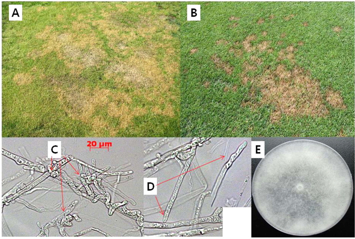
    병원균은 토양 속에 있거나 토양과 닫는 부분에 생존하 면서 잎, 뿌리 및 지하경에서 병을 일으킨다(Dick, 1990; Matthews, 1931). 초기증상은 노란색과 괴사된 patch를 형성하고 점차 넓은 지역으로 확대된다. 감염된 식물은 뿌리, 근부 및 번식체에 짙은 갈색으로 퇴화되고 잔디는 죽는다(Couch, 1995). 발병 증상은 마름증상과 뿌리썩음으로 인하여 patch로 나타나는데, 온도가 높고 습도가 높은 봄철부터 가을철까지 잔디가 생육하는 기간에는 흔하게 나타난다(Feng and Dernoeden, 1999; Hodges and Coleman, 1985). Patch의 크기는 보통 2-5 cm이나 가끔은 15 cm까지 크게 나타나지만. 잎마름병 증상은 30-35℃ 조건에서는 갑자기 나타나고, 특히 밤 온도가 20도 이상이고, 비 오는 날이나 습도가 90%이상으로 14시간 유지가 되면 잘 나타난다. 이른 아침에는 잔디 잎은 수침상이 나타나며, patch는 어두운 색으로 보이고, 그 모양이나 크기는 불규칙적이고 일정하지 않다(Abad et al., 1994; Couch, 1995; Dick, 1990; Matthews, 1931).

    Pythium에 의한 병은 생육기에 잔디를 주로 짧게 예초를 할 경우에 잘 나타나는데, 이때 나타나는 증상은 작게 나타나서 온도와 습도가 알맞으면 빠르게 크게 확대되며, 봄철과 가을철은 습한 조건에서 증상은 작고, 점차 갈색의 patchs로 확대가 되며, 보통 직경의 크기는 4-7 cm가 된다(Couch, 1995; Saladini et al., 1983).

    P. aphanidermatum에 의한 병은 주로 묘의 잎마름과 관련이 있는 중요한 종으로 알려져 왔으나(Montheith, 1933; Muse et al., 1974), 일부 발표에 의하면 면부병(솜털 같은 균사에 의한 마름병: cottony blight)의 유발하는 우점 종으로 보고되었고(Hendrix et al., 1970; Saladini et al., 1983), 뿌리썩음병(root rot)의 주요 병원균으로도 보고하였다(Abad et al., 1994).

    병원균의 분리와 동정

    병원균은 2010년과 2011년에 경상북도 대구대학교 시험 포장과 상주시 골프장 러프 잔디에서 6월 상순과 7월 하순에 전형적인 피시움잎마름 증상을 보이는 chewing fescue “Jamestowm II” 품종과 Kentucky bluegrass의 3종 혼합 품종에서 이병식물의 잎과 줄기가 포함된 것을 채집하였다. 병원균 분리를 위하여 이병 잎과 줄기의 감염 조직을 잘라 1% sodium hypochlorite에서 1분간 표면 살균 후, 70% ethanol에서 다시 30초간 살균하였다. 표면 살균한 이병 조직은 멸균수로 세척하여, 멸균된 거름종이(Whatman No. 1)에 올려놓아 수분을 제거하고, water agar(WA)에 항생제 용액(살균증류수 100 ml에 pimaricin 100 mg과 ampicillin 1000 mg 함유)을 1%을 첨가하여 만든 배지에 치상 하여 25℃ 항온기에서 24일간 배양하였다. 치상한 이병 조직에서 자라나온 균사는 선단 부분의 균사를 떼어서 PDA(potato dextrose agar) 배지로 옮겨 25℃ 항온기에서 배양한 20개 균주 중 생리적 반응이 동일한 균주인 40-1을 대표 균주로 보관하면서 본 실험을 수행하였다. 공시 균주의 보관은 25℃ 항온기에서 계대 배양을 하면서 시험하거나, 24시간 배양한 균총의 선단부에서 직경 5 mm의 균사 조각을 떼어내어 cap tube(직경: 1 cm; 높이: 10 cm)에 5조각씩 넣고, 보관용액 5 ml(살균수: 5 ml; streptomycin (3,000 μg): 0.5 ml)를 넣어 25℃ 항온기에 보관하며 실험에 사용하였다.

    형태적인 관찰을 위하여 공시 균주를 CPA(carrot potato agar)배지에 접종하여 25℃ 항온기에 7일간 배양한 후 균사를 조사한 결과 lobulate sporangia(Fig. 1C)를 관찰할 수 있었고, 영양생장 균사로부터 가는 실 같은 포자낭(filamentous sporangia)을 관찰할 수 있었다(Fig. 1D). 그리고 온도 별(20℃, 25℃, 28℃, 30℃)로 배양한 결과, 균사생장에 차이를 보였다. 균사는 격막(septum) 없었고, 균총은 형태적으로 차이를 보이지 않았지만, 솜털처럼 왕성하게 생장하는 것을 볼 수 있었다(Fig. 1E).

    모든 Pythium spp.은 잔디에서 병원성을 가지며, 균사는 흰색으로 솜털처럼 자라며, 동체성균사(homothallic)로 난포자를 생산하고, 인공배지에서는 유주자낭을 형성한다(Van der Plaats-Niterink, 1981). 조직에서 관찰되는 난포자의 직경은 15-20 um이며, 형태적인 동정은 웅성생식기관인 장정기(antheridia), 장난기 및 포자낭의 특성에 주로 근거를 둔다(Dick, 1990; Matthews, 1931).

    rDNA의 ITS 염기서열 분석

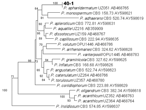
    분자생물학적 동정을 위하여 25℃ 항온기에서 2일간 배양한 후 DNA를 추출하여 rDNA의 ITS1-ITS4 프라이머을 쌍으로 증폭하였다(White et al., 1990). DNA의 증폭을 위하여 universal primer인 ITS5(5'-GGAAGTAAAAGTCG TAACAAGG-3)와 ITS4(5'-TCCTCCGCTTATTGA TAT GCG-3')를 사용하였다(White et al., 1990). PCR산물은 2% agarose gel을 만들어 50V에서 120분 동안 전기영동 하여 밴드를 확인하고, PCR 산물은 PCR purification kit를 사용하여 순화하여, Eugentech(대전)에 sequencing을 의뢰하였다. 리보소옴 ITS 지역의 DNA 염기서열을 분석하기 위하여 ITS1(5'-TTCGTAGGTGAACCTGCGG-3')와 ITS4(5'-AACATGCGTGAGATTGTAAGT-3') primers 사용하여 분석하였다(White et al., 1990).

    공시 균주인 40-1을 ITS 염기서열을 분석한 결과, 증폭한 rDNA-ITS지역의 크기는 800-960bp이었다. Phylogenetic tree는 MEGA version 4를 사용하여 P. aphanidermatum UZ051 AB468765 염기서열과 GenBank의 염기서열을 참고로 하여 작성하였다(Uzuhashi et al. 2010). phylogenetic tree는 Tamura-Nei parameter model을 이용하여 Neighbor-Joining(NJ) 분석을 통하여 구축하였다.

    공시 균주인 40-1을 rDNA의 ITS 지역을 분석을 한 결과 P. aphanidermatum UZ051 AB468765 균주와 100% 일치하는 결과를 보임으로써 공시균주인 40-1은 P. aphanidermatum로 동정되었다(Fig. 2).

    Pythium spp.의 분류를 위해 7종에 대한 ITS-RFLP 분석을 한 결과, 종간에 차이를 볼 수 있었고, 그 길이는 820 bp-1020 bp이었으나, P. aphanidermatum, P. arrhenomanesP. perplocum 이 820 bp 이었다고 하였다(Kageyama et al., 1998).

    배양적인 특성

    분리 동정한 병원균을 PDA배지에 접종 후 25℃ 항온기에서 24시간 자란 균사조각을 떼어내어 광학현미경하에서 격막 등을 관찰하였고, 생리적인 특성은 생육 온도와 배지종류에 대한 반응을 조사하였다.

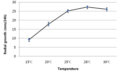
    공시 균주 인 40-1은 생육 온도에 대한 반응을 조사하기 위하여 PDA배지에서 24간 배양한 균총의 가장자리 부분을 직경 5 mm 크기의 균사 조각을 떼어내어 PDA배지에 접종 후 15℃, 20℃, 25℃, 28℃ 및 30℃에서 24시간 배양하여 균총 직경을 조사하였다.

    공시 균주 인 40-1은 생육은 온도에 따라서 큰 차이를 보였다(Fig. 3). 28℃에서 가장 왕성한 생장을 보였다. 본 시험에서는 온도의 차이를 28℃와 30℃로 세분화한 것은 구체적으로 생육 적온을 조사하기 위하여 생육이 왕성한 25℃와 30℃ 사이인 28℃에서 균사 생장량을 조사한 결과, 24시간 동안 균사가 28 mm 생장하는 것을 볼 수 있었다.

    Kageyama et al. (1998)에 의하면 7종의 Pythium을 CPA배지에서 15℃부터 5도 차이로 35℃까지 배양한 결과, 온도 차이에 따른 생육의 차이는 없었다고 하였으며, 모든 균은 30℃에서 최대 생장을 보였고, 하루의 최대 생장은 32-34 mm이었다. 그러나 다른 연구에서는 25℃-30℃에서 생장이 왕성하다고 하였다(Couch, 1995; Montheith, 1933).

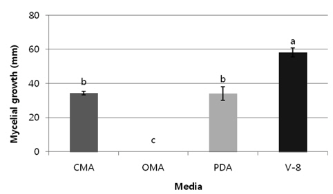
    배지의 종류에 대한 균사생장 반응을 조사하기 위하여 생육온도와 같은 방법으로 공시 균주를 CMA (corn meal agar), OMA (oat meal agar), PDA 및 V-8 (V-8 juice agar) 배지에 접종한 후 25℃에서 24시간 배양 후 균사 생장량을 측정하였다(Fig. 4). V-8배지에서 가장 왕성한 성장을 보였다. 균사의colony 형태관찰이나, 형태적인 동정을 위한 난포자나 포자낭생산을 위해서는 CMA배지, CPA 및 V-8배지에서 배양하는 것이 좋다고 한다(Abad et al., 1994).

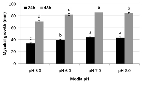
    배지의 pH에 따른 균사의 생육을 조사하기 위해서는 PDA배지에 KOH와 HCl을 이용하여 pH를 5, 6, 7 및 8로 조절하고 공시 균을 접종한 후 25℃에서 24시간과 48시간 배양 후 균사 생장량을 측정하였다(Fig. 5). pH7에서 균사 생육이 자장 좋은 것으로 조사되었다.

    병원성 검정

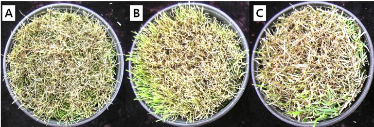
    병원성을 검정하기 위하여 포장에서 병이 발생한 chewing fescue 품종인 “Jamestowm II”, Kentucky bluegrass 혼용 품종(Midnight 33%, Moonlight 33%, prosperity 33%) 및 creeping bentgrass 품종인 “Penn Cross” 에서 병원성 시험을 실시하였다. 잔디는 관행 관리에 의해 1년 이상 자란 시험포장 잔디를 홀 컷터로 잘라내어 화분에 옮긴 후 온실에서 20일간 관리한 잔디에 병원균을 접종하였다(Chang, 2011; Chang and Lee, 2013). PDA 배지에서 자란 활력이 양호한 균총의 가장자리 균사를 지름 5 mm의 크기로 자른 균총 1조각을 PDB (Potato Dextrose Broth) 배지에 접종 한 뒤 25±1℃ 암 상태에서 5일간 배양하였다. 배양된 균사는 수확하여 멸균된 거름종이에 올려놓아 수분을 제거하고 동일한 량의 균사를 다시 멸균수에 넣고 30초간 균일하게 균사를 마쇄하였다. 접종원은 균사 생체 중 0.1 g· ml-1가 함유된 1 ml를 포트당 접종을 하고, 미리 준비한 플라스틱 상자 바닥에 1 cm 높이로 물을 채운 접종한 포트를 넣고 뚜껑을 닫고 밀봉하여 3일간 25±1℃ 암 상태의 생장상(growth chamber)에서 배양한 후 온실로 옮겨 발병 정도를 조사하였다.

    인공 접종한 병원균은 3종류의 잔디에서 병원성을 나타내었다(Fig. 6). 감염 초기에 잎의 선단부가 갈변하고 이병 부위가 수침상으로 변하면서 점차 갈색이 퇴화하고 시간이 경과됨에 따라 patch 형태를 유지하면서 잎과 줄기는 고사하여 최종 회갈색이 되었다(Fig. 6A, B ,C). 인공접종하여 발병된 병반부에서 다시 병원균을 재 분리하여 PDA배지에서 배양한 후 병원균의 특성과 광학현미경에서 균사의 격막과 균사모양을 관찰한 결과 동일한 균으로 확인되었다(Fig. 1D).

    따라서 본 병은 한지형 잔디인 chewing fescue 종에서 P. aphanidermatum에 의해 잎마름증상과 뿌리썩음증상을 동반한 잎마름병으로 국내에서 처음 보고하며, 병명은 기존 병명과 동일한 잎마름병으로 보고하고자 한다.

참고문헌
  • 1. Abad Z.G., Shew H.D., Lucas L.T. 1994 Characterization and pathogenicity of Pythium, species isolated from turfgrass with symp-toms of root and crown rot in North Carolina [Pytopathology] Vol.84 P.913-921 google cross ref
  • 2. Andrews E.A. 1943 Seedling blight and root rot of grasses in Minnesota [Phytopathology] Vol.33 P.234-239 google
  • 3. Chen W., Hoy J.W. 1993 Molecular and morphological comparison of Pythium arrhenomanes and P. graminicola [Mycol. Res.] Vol.97 P.371-1378 google
  • 4. Couch H.B. 1995 Disease of Turfgrass google
  • 5. Chang T. 2011 Difference of susceptibility on bentgrass cultivars to pink snow mold caused by Microdochium nivale [Asian J. Turfgrass Sci.] Vol.25 P.177-183 google
  • 6. Chang T., Lee Y.S. 2013 Occurrence of brown patch on Kentucky bluegrass caused by Rhizoctonia solani AG-1 IB [Weed Turf. Sci.] Vol.2 P.88-94 google cross ref
  • 7. Dick M.W. 1990 Keys to Pythum google
  • 8. Feng Y., Dernoeden P.H. 1999 Pythum species associated with root dysfunction of creeping bentgrass in Maryland [Plant Dis.] Vol.83 P.516-520 google cross ref
  • 9. Freeman T.E., Larsen P.O., Joyner B.G. 1980 Seedling disease of turf-grasses incited by Pythium P.41-44 google
  • 10. Hendrix Jr. F.F., Campbell W.A., Mon-crief J.B. 1970 Pythium species associated with golf turfgrass in the South and Southeast [Plant Dis. Rep.] Vol.54 P.419-421 google
  • 11. Hodges C.F., Coleman L.W. 1985 Pythium-induced root dysfunction of secondary roots of Agrostis palustris [Plant Dis.] Vol.69 P.336-340 google
  • 12. Hsiang T., Wu C., Yang L., Liu L. 1995 Pythium root rot associated with cool-season dieback of turfgrass in Ontario and Quebec [Can. Plant Dis. Surv.] Vol.75 P.191-195 google
  • 13. Kageyama K., Uchino H., Hyakumachi M. 1998 Characterization of hyphal swelling group of Pythium: DNA polymorphisms and cultural and morphological characteristics [Plant Dis.] Vol.82 P.218-222 google cross ref
  • 14. Kim J.W., Park E.W. 1997 Pythium spp. isolated from turfgrass at golf courses in Korea [Kor. J. Mycol.] Vol.25 P.276-290 google
  • 15. Matthews V.D. 1931 Studies on the genus Pythium google
  • 16. Matumoto C., Kageyama K., Suga H., Hyakumachi M. 2000 Intraspecific DNA polymorphisms of Pythium irregular [Mycol. Res.] Vol.104 P.1333-1341 google cross ref
  • 17. Montheith J. 1933 A Pythium disease of turf [Phytopathology] Vol.23 P.23-24 google
  • 18. Muse R.R., Schmitthenner A.F., Parthka R.E. 1974 Pythium spp. Associated with foliar blighting of creeping bentgrass [Phytopathology] Vol.64 P.252-253 google cross ref
  • 19. Saladini J.L., Schmitthenner A.F., Larsen P.O. 1983 Prevalence of Pythium species as-sociated with cottonyblighted and healthy turfgrass in Ohio [Plant Dis.] Vol.67 P.517-519 google cross ref
  • 20. Uzuhashi S., Tojo M., Kakishima M. 2010 Phylogeny of the genus Pythium and description of new genera [Mycoscience] Vol.51 P.337-365 google cross ref
  • 21. Van der Plaats-Niterink A.J. 1981 Monograph of the genus Pythium. Studies in Mycology No. 21 P.242 google
  • 22. White T.J., Bruns T., Lee S., Taylor J., Innis M.A., Gelfand D.H., Sninsky J.J., White T.J. 1990 Amplification and direct sequencing of fungal ribosomal RNA genes for phylogenetics P.315-322 google
이미지 / 테이블
  • [ Fig. 1. ]  Disease symptoms on chewing fescue (A) and Kentucky bluegrass (B) caused by Pythium aphanidermatum. A: Symptoms on early June were brownish colored and leaf dried patches; B: Patch symptoms by leaf dries (blighted) and damping off on late July; C: Lobulate sporangia are inflated, complex structures; D: Filamentous sporangia are usually indistinguishable from vegetative hyphae. Bars=20 μm with X400. E: Mycelial growth on PDA medium for 72 hour after inoculation.
    Disease symptoms on chewing fescue (A) and Kentucky bluegrass (B) caused by Pythium aphanidermatum. A: Symptoms on early June were brownish colored and leaf dried patches; B: Patch symptoms by leaf dries (blighted) and damping off on late July; C: Lobulate sporangia are inflated, complex structures; D: Filamentous sporangia are usually indistinguishable from vegetative hyphae. Bars=20 μm with X400. E: Mycelial growth on PDA medium for 72 hour after inoculation.
  • [ Fig. 2. ]  Taxonomic position of 40-1 strain based on internal transcribed spacer (ITS) region of ribosomal RNA. Pythium strains were compared with strains of Uzuhashi et al. (2010). ITS region sequences were first analyzed using the Tamura-Nei parameter distance calculation model with gamma-distributed substitution rates, which were then used to construct the Neighbor-Joining tree with MEGA version 4.0.2. Numbers below or above nodes are bootstrap values.
    Taxonomic position of 40-1 strain based on internal transcribed spacer (ITS) region of ribosomal RNA. Pythium strains were compared with strains of Uzuhashi et al. (2010). ITS region sequences were first analyzed using the Tamura-Nei parameter distance calculation model with gamma-distributed substitution rates, which were then used to construct the Neighbor-Joining tree with MEGA version 4.0.2. Numbers below or above nodes are bootstrap values.
  • [ Fig. 3. ]  Mycelial growth on potato dextrose agar. Mycelial growth was measured sizes of colony diameter with digmatic caliper on 24 hour after inoculation using a 5 mm cork borer. Error bars indicate standard deviation and showed significant difference at P =0.05 level according to Tukey’s HSD test.
    Mycelial growth on potato dextrose agar. Mycelial growth was measured sizes of colony diameter with digmatic caliper on 24 hour after inoculation using a 5 mm cork borer. Error bars indicate standard deviation and showed significant difference at P =0.05 level according to Tukey’s HSD test.
  • [ Fig. 4. ]  Mycelial growth on several media. Mycelial growth was measured sizes of colony diameter with digmatic caliper on 24 hour after inoculation using a 5 mm cork borer. CMA: Corn Meal Agar; OMA: Oat Meal Agar; PDA: Potato Dextrose Agar; V-8: V-8 Juice Agar. Error bars indicate standard deviation and mean in column followed by the different letter that indicates significant difference at P =0.05 level according to Tukey’s HSD test.
    Mycelial growth on several media. Mycelial growth was measured sizes of colony diameter with digmatic caliper on 24 hour after inoculation using a 5 mm cork borer. CMA: Corn Meal Agar; OMA: Oat Meal Agar; PDA: Potato Dextrose Agar; V-8: V-8 Juice Agar. Error bars indicate standard deviation and mean in column followed by the different letter that indicates significant difference at P =0.05 level according to Tukey’s HSD test.
  • [ Fig. 5. ]  Response of mycelial growth on different pH media. Mycelial growth was measured sizes of colony diameter with digmatic caliper on 24hr. and 48hr. after inoculation using a 5 mm cork borer. Error bars indicate standard deviation and mean in column followed by the different letter that indicates significant difference at P =0.05 level according to Tukey’s HSD test.
    Response of mycelial growth on different pH media. Mycelial growth was measured sizes of colony diameter with digmatic caliper on 24hr. and 48hr. after inoculation using a 5 mm cork borer. Error bars indicate standard deviation and mean in column followed by the different letter that indicates significant difference at P =0.05 level according to Tukey’s HSD test.
  • [ Fig. 6. ]  Pathogenicity test on chewing fescue cultivar “Jamestone II” (A), creeping bentgrass cultivar “Penn cross” (B) and Kentucky bluegrass mixed cultivars (Midnight 33%, Moonlight 33%, and prosperity 33%) (C). Inoculation was applied with mycelium suspension of 1ml by injection in the pots. Disease symptom showed on 5 days after inoculation.
    Pathogenicity test on chewing fescue cultivar “Jamestone II” (A), creeping bentgrass cultivar “Penn cross” (B) and Kentucky bluegrass mixed cultivars (Midnight 33%, Moonlight 33%, and prosperity 33%) (C). Inoculation was applied with mycelium suspension of 1ml by injection in the pots. Disease symptom showed on 5 days after inoculation.
(우)06579 서울시 서초구 반포대로 201(반포동)
Tel. 02-537-6389 | Fax. 02-590-0571 | 문의 : oak2014@korea.kr
Copyright(c) National Library of Korea. All rights reserved.