검색 전체 메뉴
PDF
맨 위로
OA 학술지
Docosahexaenoic acid (DHA)를 다량 함유하는 해양미생물의 분리 및 동정 Isolation and Identification of DHA-Rich Marine Microorganism
  • 비영리 CC BY-NC
  • 비영리 CC BY-NC
ABSTRACT
Docosahexaenoic acid (DHA)를 다량 함유하는 해양미생물의 분리 및 동정

Docosahexaenoic acid (DHA, 22:6n-3) and ecosapentaenoic acid (20:5n-3) have attracted increasing attention since the first epidemiological report on the importance of n-3 essential fatty acids. It is thought that DHA has important functions in brain and retinal tissues. Thraustochytrids, a group of marine protists, are capable of heterotrophic growth, and are potential omega-3 producers for industrial use, especially the members of the Schizochytrium and Thraustochytrium genera. The aims of this work were to isolate, identify and screen thraustochytrids from 17 different locations. Twenty-three isolates were screened for biomass, total fatty acid (TFA) and DHA content. Analysis of the fatty acid methyl esters revealed four distinct clusters biomass ranged from 8.68 - 9.36 g L−1, and lipid and DHA contents ranged from 3.11 - 4.10 g L−1 and 1.05 - 1.93 g L−1 biomass, respectively. B-12 isolates were screened for biomass (9.36 g L−1), TFA (4.10 g L−1) and DHA (47.01%, w/w) content. C-6 isolates were also screened for biomass (8.92 g L−1), TFA (3.30 g L−1) and DHA (49.41%, w/w) content. The 18S rRNA gene sequencing results identified Schizochytrium mangrovei as B-12 and Crypthecodium cohnii as C-6.

KEYWORD
Schizochytrium mangrovei , Crypthecodium cohnii , docosahexaenoic acid (DHA) , Marine Microorganism
  • 서 론

    Docosahexaenoic acid (DHA)는 eicosapentaenoic acid (EPA), α-linolenic acid와 함께 오메가-3계 지방산에 속하며, serum triacylglycerides 및 LDL-cholesterol의 함량을 낮추고 HDL-cholesterol 수치를 높임으로써 동맥경화 및 혈전증 등과 같은 심혈관계 질환의 예방에 효과가 있는 것으로 보고되고 있다 (Haglund et al., 1991; Horrocks and Yeo, 1999; Lee and Kim, 2001; Ohr, 2003). 또한 두뇌, 신경계 및 안구조직의 필수지방산으로 특히, 유아의 시력 및 운동능력 개발 등에 중요한 요소이며, 뇌의 구조적 지질에 가장 풍부한 구성요소 중의 하나이다(Kanazawa et al., 1982; Drats and Deose, 1986; Lee et al., 2003).

    오메가-3계 고도불포화지방산의 주요 공급원은 어유(Fish oil)이며, 어유에는 7-17%의 DHA가 포함되어있다(Kremer et al., 1987; Stenson et al., 1991). 그러나 식품첨가제로 사용되는 어유는 특유의 비린내와 맛 그리고 산화에 의한 안전성 문제등으로 그 사용이 제한적이다(Tanaka et al.,1992; Carlson, 1996; Gill and Valiverty, 1997). 고도불포화지방산은 해조류나 곰팡이 또는 박테리아가 생합성하여 체내에 에너지원으로 저장하거나 몸체의 구성요소로 사용한다. 해산어류는 오메가-3 고도불포화지방산을 직접 생합성할 수 없기 때문에 체내에서 필요한 고도불포화지방산은 미생물의 섭취를 통하여 충족시키고 있다(Castell et al., 1972; Bremer, 1995; Bremer and Talbot, 1995; Tanaka et al.,1999 ). 따라서 어유를 구성하고 있는 지방산의 양과 조성은 해양미생물 즉, 먹이사슬의 일차생물 오메가-3 고도불포화지방산 생합성 형태에 따라서 결정된다고 할 수 있다(Miller and Jones, 1983; Braden and Carroll, 1986).

    오메가-3계 고도불포화지방산은 고등어, 꽁치 등과 같은 등푸른 생선으로부터 추출하여 산업적으로 이용하고 있으나, 어유 특유의 비린내 및 특정 지방산의 비율조절의 어려움 등과 같은 단점을 가지고 있다(Park et al., 1994; Lee et al., 1997; Cho et al., 2004). DHA 같은 고도불포화지방산은 인위적인 화학합성 과정을 통한 생산이 매우 어렵기 때문에 천연물질로부터 획득해야 하는데, 현재 공업적으로는 이들 지방산을 어유에서 추출하여 분리 정제 후 사용하고 있다. 생산되는 어유의 대부분은 수소첨가반응에 의해 마가린과 소프트닝으로 제조되는데, 이 과정에서 이중결합의 위치가 바뀌고 cis형에서 trans형으로 입체 이성화가 일어나는 등 대부분의 오메가-3계 지방산은 파괴 또는 변형된다(Singh and Ward, 1997). 이러한 천연물로부터 추출공정을 통한 고도불포화지방산 생산시 발생하는 여러 한계점을 극복하고자 고도불포화지방산 생산능력이 보고된 미생물을 통하여 경제적으로 유용한 고도불포화지방산을 생산하려는 생물학적 연구가 많이 진행 중이다(Yazawa et al., 1998; Tonon et al., 2002; Owen and Ajay, 2005). 특히, 해양식물성 플랑크톤과 Entromophthorrales속, Thraustochytrid속, Mucoreles속 그리고 Ascomycetes속 등이 DHA를 생산하는 미생물로 알려져 있다(Moss, 1986; Sathe et al., 1993; Sharma et al., 1994; Yipp et al., 1995). 이들 중에서 Thraustochytrid과에 속하는 미생물은 DHA 함량이 매우 높으며, DHA와 유사한 다른 오메가-3 고도불포화지방산의 함량이 낮아 순수한 DHA 분리하는데 상대적으로 유리하다고 보고되고 있다(Bajpai et al., 1991a,b; Watanabe et al., 1997; Yokochi et al., 1998).

    DHA의 주요 공급원으로서 사용되는 어유는 바다로부터 공급되는 양은 한정되어 있고, 수요는 급격히 증가되고 있는 추세이다(Stenson et al., 1991). 이러한 점을 착안하여 이번 연구에서는 DHA를 다량 함유하는 해양미생물(Thraustochytrid 科)을 분리 동정하여 DHA의 새로운 공급원으로서 이용하고자 한다.

    재료 및 방법

      >  해양미생물의 분리 및 배양조건

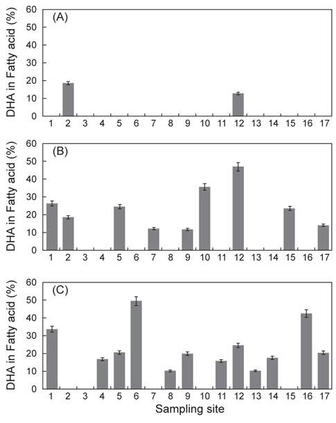
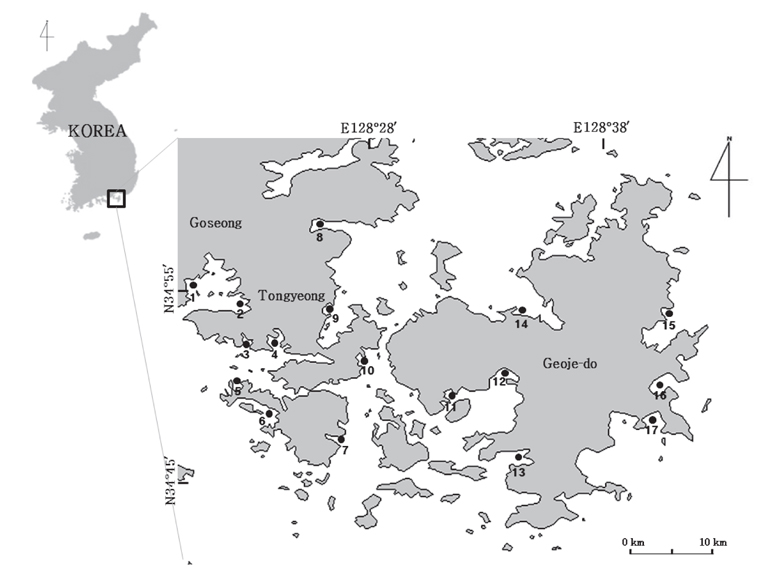
    DHA를 다량 함유하는 해양미생물의 분리를 위한 시료는 2006년 4월부터 10월까지 경상남도 통영, 고성 및 거제 일대 해안으로부터 3가지 방법으로 시료를 채취하였다(Fig. 1). 첫째, 부유생물 시료는 3 μm 플랑크톤네트를 이용하여 해수를 여과시킨 여과잔여물을 이용하였고, 둘째, 저질층 시료는 유기퇴적물이 비교적 풍부한 곳을 선정하여 직접 채니하였다. 셋째, 조류(algae)는 해안가 연안에 부유성 조류를 시료로 채집하였다. 채취한 각각의 시료는 10℃ 로 유지되는 Ice Box에 넣어서 실험실로 운반하여 사용하였다.

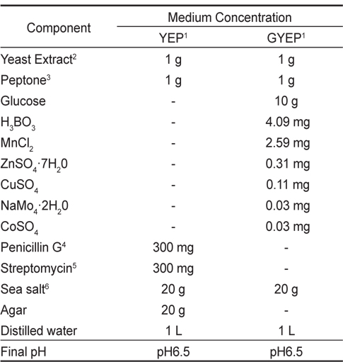
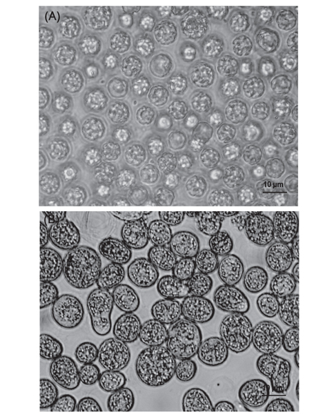
    유용미생물의 분리는 Table 1에 나타낸 바와 같이 yeast extract (Becton, Dickinson and Company sperks, MD, USA) 1.0 g/L, peptone (Acumedia Manufacturers, Inc. Lansing, MI, USA) 1.0 g/L를 주요 성분으로 하는YEP 배지를 사용하였으며(Wong et al., 2005), 이 때 세균의 증식억제를 위해 penicillin-G (P3032, Sigma, USA)와 streptomycin sulfate (S-9137, Sigma, USA)를 각각 300 mg씩 별도로 멸균 처리된 증류수에 용해시켜 0.22 μm 여과지(Millipore)로 여과하여 분리용 평판배지에 첨가하였다. 분리용 YEP 배지를 이용하여 채취된 각 시료들을 도말하여 25℃ 세포배양기(BOD Incubator DS-11B, Dasol Scientific Co., Ltd.)에서 72시간 배양하였다. 유용미생물은 평판배지에 형성된 집락(colony)을 현미경을 이용하여 Thraustochytrid 과(科)와 유사형태를 가진 해양미생물을 선발하였다. 선발된 균체의 배양은 glucose 10 g/L, yeast extract 1.0 g/L 그리고 peptone 1.0 g/L 를 주요 성분으로 하는 GYEP 배지를 부분적으로 보완하여(Table 1) pH 6.5로 조정된 평판배지에서 25℃에서 72시간 배양한 후, 이들 균주를 동일한 액상배지로 25℃, 120 rpm의 진탕배양기(KSI-200L, KOENCON)에서 72시간 배양하였다. 균체의 지방산 분석 시료는 균체 배양액을 원심분리기(HA-1000-3, Hanil science Industrial Co., Ltd.)로 5,000 rpm에서 10분간 원심분리한 후, 배양액은 버리고 침전된 세포를 회수하여 동결 건조하였다.

    [Table 1.] Composition of YEP1 and GYEP1 medium

    label

    Composition of YEP1 and GYEP1 medium

      >  Total lipid 추출

    총 지질 추출은 Bligh and Dyer (1959) 방법에 준하였다. 균체 중의 지질 추출 시료를 비커에 5 g 취하여 세포분쇄기(Homogenizer AM-12, Nihonseiki Kaisha Ltd. Tokyo, Japan)에서 15,000 rpm으로 5분간 분쇄한 후, chloroform : methanol (2:1, v/v) 혼합 용매를 시료의 2 배량 넣어 하루 동안 방치하였다. 다음날 chloroform 층만을 분리하기 위하여 둥근 플라스크 위에 깔때기를 놓고, 여과지를 깔아 그 위에 Na2SO4를 넣어 서서히 chloroform층만 흘러내리게 하였다. 분리된 chloroform 층은 진공회전농축기(Rotavapor R-114, BUCHI)를 사용하여 40℃ 이하에서 용매를 완전히 증발시킨 후, 추출된 총 지질의 무게를 측정하였다. 모든 작업은 질소 기류 하에서 행하였다.

      >  지방산 분석

    추출된 지질 50 mg 을 cap tube에 취하고, 0.5 N NaOH-methanol 용액 1.5 mL 가하여 질소를 충진한 다음, 100℃에서 8분간 가열하여 검화하였다. 방냉 후 12% BF3-methanol 2 mL 가한 후, tube 뚜껑을 닫고, 100℃에서 11분간 가열하여 methyl화 하였다. 약 30℃로 냉각한 후, iso-octane 1 mL 첨가하고 30초간 vortex mixer로 혼합하였다. 즉시 3 mL의 포화식염수를 가한 다음 흔들어 방치하여 iso-octane층이 분리되도록 하였다. iso-octane층을 시료 병에 옮긴 후, 다시 iso-octane 1 mL 첨가한 다음 흔들어 재추출하여 시료 병에 모으고 이를 지방산 methylester 시료로 하였다.

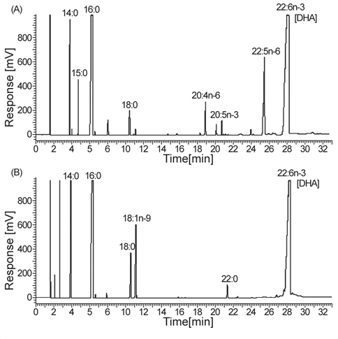
    지방산 분석에 사용하는 GLC는 OmegawaxTM-320 fusedsilica capillary column (30 m×0.32 mm×0.25 μm, i.d., Supelco Co., Bellefonte, PA, USA)를 장착한 Clarus 600 (Perkin Elmer Co. Ltd., USA)을 이용하였다. 분석은 column 조건의 경우 185℃에서 8분간 유지하고 3℃/min씩 230℃까지 상승시킨 후, 10분간 유지하였다. 이 때 주입기는 250℃, 검출기는 270℃ 그리고 carrier gas는 헬륨(He)을 사용하였다. 지방산의 분석은 동일조건에서 분석한 표준품의 ECL과 비교하여 동정하였고, 지방산 표준품은 14:0, 16:0, 18:1, 18:2, 18:3, 20:0, 22:1, 24:0 (Sigma Chemical Co., St. Louis, MO, USA)과 GC-MS로 동정된 menhaden oil을 사용하였다.

       18S rDNA sequencing

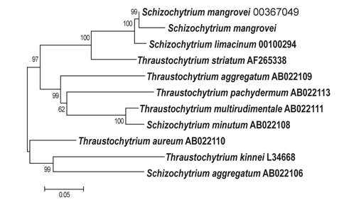
    분리된 해양미생물의 18S ribosomal DNA sequencing은 Wizard genomic DNA purification kit (Promega, USA)를 이용해 chromosomal DNA를 분리한 후, 18S rRNA sequencing에 사용하는 universal primer인 NS1 (5'-GTAGTCATATGCTTGTCTC-3')와 NS8 (5'-TCCGCAGGTTCACCTACGGA-3') primer를 사용하여 PCR 증폭하였다. 증폭된 PCR 산물은 전기영동장치(Wizard SV Gel)와 PCR clean-up system (Promega, USA)을 이용하여 정제하였다. 정제된 PCR 산물은 ABI PRISM 3700 DNA Analyzer (Applied Biosystem, Foster, CA, USA)를 이용하여 염기서열을 분석하였다. 그 결과는 NCBI의 BLASTN 프로그램을 이용하여 GENEBANK의 ribosomal DNA sequence와 비교하였으며, sequence의 상동성은 Clustal X와 Mega 2 program을 이용하여 비교 분석하였다(Thompson et al., 1994).

      >  통계처리

    모든 자료는 SPSS (12.0) 프로그램을 이용하여 분산분석 (one-way ANOVA)을 실시하여 Duncan's multiple range test (Duncan, 1955)로 평균간의 유의성(P<0.05)을 검정하였다.

    결과 및 고찰

      >  해양미생물의 분리

    Thraustochytrid 科로 추정되는 해양미생물은 Fig. 2에서 보는 바와 같이 여과해수 잔여물에서 2종, 유기퇴적물에서 9종 그리고 조류에서 12종으로 총 23종을 분리하였다. 여과해수 잔여물에서는 A-2, A-12 지점에서 2개의 균주를 분리하였으며 DHA는 각각 18.64% 와 12.75%로 나타났다. 유기퇴적물에서 채취한 시료는 B-1, B-2, B-5, B-7, B-9, B-10, B-12, B-15 그리고 B-17지점에서 9개의 균주를 분리하였으며 DHA는 각각 26.40, 15.75, 24.59, 12.26, 11.85, 35.65, 47.01, 23.53 그리고 14.14% 로 B-12지점에서 분리한 균주의 DHA가 47.01%로 가장 높게 나타났으며(P<0.05), B-10지점에서 분리한 균주도 35.65%로 높게 나타났다. 해안가 부유하는 조류에서 채취한 시료는 C-1, C-4, C-5, C-6, C-8, C-9, C-11, C-12, C-13, C-14, C-16 그리고 C-17 지점에서 12개의 균주를 분리하였으며 DHA는 각각 33.60, 16.77, 20.55, 49.41, 10.23, 19.88, 15.75, 24. 59, 10.26, 17.60, 42.54 그리고 20.33%로 C-6 지점에서 분리한 균주가 49.41%로 가장 높게 나타났으며(P<0.05), C-1 과 C-16지점에서 분리한 균주도 33.60%와 42.54%로 나타났다. 분리된 균주 23종 중에서 DHA 함량이 30% 이상인 B-10, B-12, C-1, C-6 그리고 C-16지점에서 분리한 균주 5종을 1차 선발하였다(Table 2).

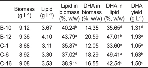
    [Table 2.] Biomass, lipid and DHA production of isolated species culture

    label

    Biomass, lipid and DHA production of isolated species culture

      >  Total lipid 및 지방산 조성

    분리 선발된 균주 5종의 총 지질 함량에 관한 결과는 Table 2에 나타낸 바와 같이 B-10, B-12, C-1, C-6 그리고 C-16지점에서 분리된 균주에 따른 biomass는 각각 9.12, 9.36, 8.68, 8.92 그리고 9.08 g/L로 B-12지점에서 분리된 균주가 9.36 g/L로 가장 높게 나타났으며(P<0.05), total lipid는 각각 40.24, 43.79, 35.87, 37.02 그리고 38.91%로 B-12지점에서 분리된 균주가 43.79 %로 가장 높게 나타났다(P<0.05). DHA생산량은 B-10, B-12, C-1, C-6 그리고 C-16지점에서 분리된 균주에서 각각 1.31, 1.93, 1.05, 1.63 그리고 1.50 g/L로 B-12지점에서 분리한 균주가 1.93 g/L로서 DHA 생산량이 가장 많이 하는 것으로 나타났으며(P<0.05), 다음으로 C-6지점에서 분리한 균주가 1.63 g/L로 많은 것으로 나타났다(P<0.05). 선발된 균주 5종의 지방산 조성 분석 결과는 Table 3에 나타낸 바와 같이 B-10, B-12, C-1, C-6 그리고 C-16 지점에서 분리된 5종의 해양미생물의 DHA는 각각 35.65, 47.01, 33.60, 49.41 그리고 42.54%로 C-6 지점에서 분리한 균주가 49.41%로 가장 높게 나타났으며(P<0.05), 다음으로 B-12지점에서 분리된 균주가 47.01% 높게 나타났다(P<0.05). 오메가-3 계열은 B-10, B-12, C-1, C-6 그리고 C-16 지점에서 분리한 균주가 각각 40.03, 49.04, 34.27, 51.47 그리고 44.54%로 C-6지점에서 분리한 균주가 51.48%로 가장 높게 나타났으며(P<0.05), 다음으로 B-12지점에서 분리된 균주가 47.04%로 높게 나타났다(P<0.05). 오메가-6 계열은 B-10, B-12, C-1, C-6 그리고 C-16 지점에서 분리한 균주가 각각 6.61, 13.65, 0.07, 0.00 그리고 13.41%로 C-6지점에서 분리한 균주에서 오메가-6 계열 지방산이 전혀 나타나지 않아 DHA 만을 추출하기에는 C-6지점에서 분리한 균주가 가장 적합한 것으로 나타났다. 이로서 23여종의 분리한 균주 중에 B-12와 C-6 지점에서 분리한 균주 2종이 DHA 생산 균주로서 적합한 것으로 나타났다(Fig. 3).

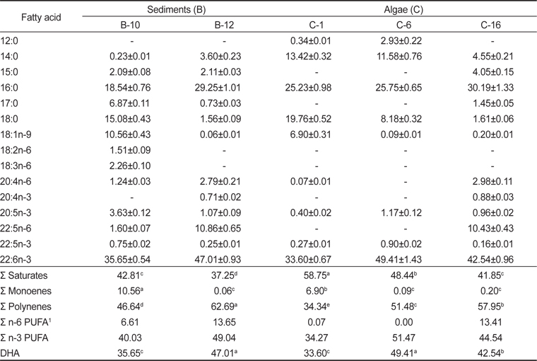
    [Table 3.] Fatty acid compositions of isolated species cultured for 3 days (% of total fatty acids)

    label

    Fatty acid compositions of isolated species cultured for 3 days (% of total fatty acids)

       18S rDNA sequencing

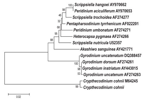
    B-12그리고 C-6 지점에서 분리 선발된 2종에 대하여 18S rDNA sequencing 분석을 실시한 결과는 Figs. 3, 4에 나타난 바와 같다. B-12지점에서 분리된 균주는 Schizochytrium mangrovei로 동정되었으며(Fig. 3), C-6지점에서 분리된 균주는 Crypthecodium cohnii 로 동정되었다(Fig. 4).

    오메가-3계 고도불포화지방산을 생산하는 미생물에 대한 관심은 EPA나 DHA의 생합성 유전자들이 분석되고 동정됨으로서 이루어졌다(Yazawa, 1996; Tanaka et al., 1999). 고도불포화지방산 생합성에 관여하는 유전자에 대한 연구는 EPA를 생산하는 Shewanella sp. strain SCRC-2738로부터 38 kb의 genomic fragment를 분석하고 cloning 함으로서 시작되었다. 최근 보고되어진 고도불포화지방산 유전자 산물과 13C-labelling 실험에서 보여주는 multiple enzyme domains을 바탕으로 할때, 고도불포화지방산 생산은 polyketide 합성반응과 일부 동일한 효소를 사용하고 있음이 증명되고 있으며 DHA를 생산하는 해양미생물인 Moritella marina strain MP-1과 Schizochytrium sp.로 부터 DHA 생합성 유전자들이 밝혀졌다(Metz et al., 2001).

      >  Thraustochytrids 과의 특성

    최근 미생물에 의한 지질 생산에 관하여 다시 관심을 갖게 되었는데 이는 실질적인 기술에 의하여 미생물에 의한 지질 생산이 가능한 것으로 보고하고 있다(Wu et al., 2005; Zhu et al., 2007). 인지질과 sterol 지방산 함량과 조성은 여러 가지 환경조건 즉, 온도, pH, 배지의 조성의 영향을 받는데 식물 조직세포에서 고도불포화지방산은 12℃에서 80%가 형성되나 20℃와 30℃에서는 각각 51%와 30%의 불포화지방산이 합성된다고 보고하고 있다(Browse and Slack, 1993). 온도가 낮으면 불포화지방산의 양이 많아지는 반면 배양 온도가 높으면 포화지방산의 양이 많아진다고 보고되고 있다(Knivett and Cullen, 1965). 이전에 보고된 Thraustochytrids의 종에 따른 적정 배양 온도는 다양하게 나타났는데 Thrausochytrium antarcticum, Thrausochytrium kerguelensis, Thrausochytrium rossii의 3종은 최적배양온도가 4-9℃로 나타났으며(Bahnweg, 1979), Thrausochytrium aureum과 Thrausochytrium roseum의 경우는 각각 20-25℃와 25-30℃로 보고되고 있다(Goldstein, 1963). 또한 Schizochytrium limacinum는 25℃였으며, Schizochytrium sp.의 최적배양온도는 28℃이였다는 연구결과를 발표하였다(Bajpai and Bajpai, 1993; Yokochi et al., 1998). 이후 Schizochytrium sp.와 Schizochytrium mangrove 2종 모두 25℃로 보고하였다(Fan et al., 2002). 이외의 여러 해양미생물의 경우에도 배양온도가 균체의 성장에 영향을 미치나 최적온도는 균주에 따라 서로 다르다는 연구결과가 보고되었다(Yongmanitchai and Owen, 1989). 또한 이전에 보고된 Thraustochytrids과의 종에 따른 최적 염분농도는 T. antarcticum, T. kerguelensis, T. rossii 3종의 경우는 각각 15-30, 20-30 그리고 15-20 psu로 나타났으며(Bahnweg, 1979), T. aureumT. roseum의 경우 각각 20-35 psu와 25-35 psu였다(Goldstein, 1963). 그리고 S. limacinum는 15-30 psu였으며(Honda et al., 1998), Schizochytrium sp.와 S. mangrove는 각각 7.5-22.5 psu와 22.5-30 psu로 보고하였다(Hammond et al., 2001; Fan et al., 2002).

    이러한 미생물에 의한 지질 생산의 장점은 태양에너지가 필요하지 않고, 날씨에 관계없이 연중 생산이 가능하며, 배양조건에 따라 생산되는 지질의 조성을 조절 할 수 있다는 것이다(Hulan et al., 1989; Ward, 1995; Abril et al., 2003). 유지(油脂)미생물의 지질축척은 배지의 영양소, 대개는 질소가 고갈되었을 때 시작되며, DHA 생산에 미치는 glucose 영향을 검토한 결과 glucose 농도가 낮으면 지방산과 DHA 생성이 촉진된다고 한다(Honda et al., 1998). 또한 지질의 농도는 대수증식기에는 감소되었지만, 배지성분이 제한되어 생육이 지연되면 지질의 생산은 증가되고, DHA 농도는 감소된다는 보고도 있다(Yazawa, 1996; Honda et al., 1998). 따라서 해양미생물의 종에 따른 고도불포화지방산 생성 능력이 우수한 종을 찾아내는 것도 중요하지만, 배양 환경에 따른 물리적인 조건과 영양성분에 따른 생리적인 조건을 적절하게 조절하는 것이 무엇보다 중요하며 이러한 점을 고려하여 더 많은 연구가 이루어진다면 더욱 우수한 양질의 지질과 보다 많은 양의 DHA를 생산할 수 있을 것으로 사료된다.

참고문헌
  • 1. Abril RJ, Garrett SG, Zeller WJ, Sander WJ, Mast RW 2003 Safety assessment of DHA-rich microalgae from Schizochytrium sp. V. Target animal safety/toxicity study in growing swine, Regue [Toxicol Pharmacol] Vol.37 P.73-82 google
  • 2. Bahnweg G 1979 Studies on the physiology of Thraustochytriales I. Growth requirements and nitrogen nutrition of Thraustochytrium spp., Schizochytrium sp., Japonochytrium sp., Ulkenia spp. and Labyrinthuloides spp [Veroff Inst Meeresforsch Bremerh] Vol.17 P.245-268 google
  • 3. Bajpai PK, Bajpai P, Ward OP 1991 Optimization of production of docosahexaenoic acid(DHA) by Thraustochytrium aureum ATCC 34304 [JAOCS] Vol.68 P.509-514 google
  • 4. Bajpai PK, Bajpai P, Ward OP 1991 Production of docosahexaenoic acid by Thraustochytrium aureum [Appl. Microbiol Biotechnol] Vol.35 P.706-710 google
  • 5. Bajpai P, Bajpai PK 1993 Eicosapentaenoic acid(EPA) production from microoranisms: a review [J Biotech] Vol.30 P.161-183 google
  • 6. Bligh EG, Dyer WJ 1959 A rapid method of total lipid extraction and purification [Can J Biochem Physiol] Vol.37 P.911-917 google
  • 7. Braden LM, Carroll KK 1986 Dietary ploysaturated fat in relation to mammarial carcinogenesis in rats [Lipids] Vol.21 P.285-288 google
  • 8. Bremer GB 1995 Lower marine fungi (Labyrinthulomycetes) and the decay of mangrove leaf litter [Hydrobiologia] Vol.295 P.89-95 google
  • 9. Bremer GB, Talbot G 1995 Celluloytic enzyme activity in the marine protist Schizochytrium aggregatum [Bot Mar] Vol.38 P.37-41 google
  • 10. Castell JD, Sinnhuber RO, Wales JH, Lee DJ 1972 Essential fatty acids in the diet of rainbow trout (Salmo gairdneri): growth, feed conversion and some gross deficiency symptoms [J Nutr] Vol.102 P.77-86 google
  • 11. Carlson SE 1996 Arachidonic acid status of human infants: influence of gestational age at birth and diets with very long chain n-3 and n-6 fatty acids [J Nutr] Vol.126 P.1092-1098 google
  • 12. Cho EJ, Lee JH, Lee KT 2004 Optimization of enzymatic synthesis condition of structured lipids by response surface methodology [Korean J Food Sci Technol] Vol.36 P.531-536 google
  • 13. Drats EA, Deose AJ 1986 Health Effects of Polyunsaturated Fatty Acids in Seafoods P.319 google
  • 14. Duncan DB 1955 Multiple range and multiple F test [Biometric] Vol.11 P.1-42 google
  • 15. Fan KW, Vrijmoed LLP, Jones EBG 2002 Physiological Studies of Subtropical Mangrove Thraustochytrids [Botanica Marina] Vol.45 P.50-57 google
  • 16. Gill I, Valiverty R 1997 Polyunsaturated fatty acids: occurrence, biological activities and application [Trends Biotechnol] Vol.15 P.401-409 google
  • 17. Goldstein S 1963 Development and nutrition of new species of Thraustochytrium [Amer. J Bot] Vol.50 P.271-179 google
  • 18. Haglund O, Luostarinen R, Wallin R, Wibell L, Saldeen T 1991 The effects of fish oil on triglycerides, cholesterol, fibrinogen and malondialdehyde in humans supplemented with vitamin E [J Nutr] Vol.121 P.165-169 google
  • 19. Hammond BG, Mayhew DA, Naylor MW, Ruecker FR, Mast RW, Sander WJ 2001 Safety assessment of DHA-rich microalgae from Schizochytrium sp. I. Subchronic rat feeding study [Regul Toxicol Pharmacol] Vol.33 P.192-204 google
  • 20. Honda D, Yokochi T, Nakahara T, Erata M, Higashihara T 1998 Schizochytrium limacinum sp. nov., a new thraustochytrid from a mangrove area in west Pacific Ocean [Mycol Res] Vol.102 P.439-448 google
  • 21. Horrocks LA, Yeo YK 1999 Health benefits of docosahexaenoic acid(DHA) [Phamacol Res] Vol.40 P.211-225 google
  • 22. Hulan HW, Ackman RG, Ratnayake WMN, Proudfoot FG 1989 Omega-3 fatty acid levels and general performance of commercial broilers fed practical levels of redfish meal [Poultry Sci] Vol.68 P.153-162 google
  • 23. Kanazawa A, Teshima SI, Sakamoto M 1982 Requirements of essential fatty acids for larval ayu [Bull Jap Soc Sci Fish] Vol.48 P.587-590 google
  • 24. Kremer JM, Jubiz W, Michalek A 1987 A fish-oil fatty acid supplementation in active rheumatoid arthritis [Ann Int Med] Vol.106 P.497-503 google
  • 25. Lee HA, Yoo IJ, Lee BH 1997 Research and development trends on ω-3 fatty acid fortified foodstuffs [J Korean Soc Food Sci Nutr] Vol.26 P.161-174 google
  • 26. Lee JH, Kim HS 2001 Effects of dietary docosahexaenoic acid levels on the brain phospholipids and serum and liver lipid compositions in rats [Korean J Nutr] Vol.34 P.132-140 google
  • 27. Lee H, Kizito SA, Weese SJ, Craig-schmidt MC, Lee Y, Wei CI, An H 2003 Analysis of headspace volatile and oxidized volatile compounds in DHA-enriched fish oil on accelerated oxidative storage [J Food Sci] Vol.68 P.2169-2177 google
  • 28. Miller JD, Jones E 1983 Observations on the association of thraustochytrid marine fungi with decaying seaweed [Bot Mar] Vol.26 P.345-351 google
  • 29. Moss ST, Moss ST 1986 Biology and phylogeny of the labyrinthulales and thraustochytriales P.105-129 google
  • 30. Ohr LM 2003 Fats for healthy living [Food Technol] Vol.57 P.91-96 google
  • 31. Owen PW, Ajay S 2005 Omega-3/6 fatty acids: Alternative sources of production [Process Biochemistry] Vol.40 P.3627-3652 google
  • 32. Park BS, Hwang BJ, Lee SJ, Lee YC 1994 Omega-fatty acid P.58-59 google
  • 33. Sathe PV, Raghukumar S, Raghukumar C, Sharma S 1993 Thraustochytrid and fungal component of marine detritus. I. Field studies on decomposition of brown alga Sargassum cinereum [J Ag Indian J Mar Sci] Vol.22 P.159-167 google
  • 34. Sharma S, Raghukumar C, Raghukumar S, Sathe-Pathak V, Chandramohan D 1994 Thraustochytrid and fungal component of marine detritus II. Laboratory studies on decomposition of the brown alga Sargassum cinereum [J Ag J Exp Mar Biol Ecol] Vol.175 P.227-242 google
  • 35. Singh A, Ward OP 1997 Microbial production of docosahexaenoic acid (DHA, C22:6) [Adv Appl Microbial] Vol.45 P.27-311 google
  • 36. Stenson WF, Cort D, Beeken W, Rodgers J, Burakoff R 1991 Trial of fish oil supplemented diet in ulcerative colitis [World Rev Nutr Diet] Vol.66 P.51-71 google
  • 37. Tanaka T, Hirano J, Funada T 1992 Concentration of docosahexaenoic acid in glyceride by hydrolysis of fish oil with candida cylindracea lipase [J Am Oil Chem Soc] Vol.69 P.1210-1214 google
  • 38. Tanaka M, Ueno A, Kawasaki K 1999 Isolation of clustered genes that are notably homologous to the eicosapentaenoic acid biosynthesis gene cluster from the decosahexaenoic acid producing bacterium vibrio marinus strain MP-1 [Biotchnol Lett] Vol.21 P.939-945 google
  • 39. Thompson JD, Higgins DG, Gibson TJ 1994 CLUSTAL W: improving the sensitivity of progressive multiple sequence alignment through sequence weighting, positionspecific gap penalies and weight matrix choice [Nucleic Acids Res] Vol.22 P.4673-4680 google
  • 40. Tonon T, Harvey D, Larson TR, Graham IA 2002 Long chain polyunsaturated fatty acid production and partitioning to triacylglycerols in four microalgae [Phytochemistry] Vol.61 P.15-24 google
  • 41. Ward OP 1995 Microbial production of long-chain PUFAs [INFORM] Vol.6 P.683-688 google
  • 42. Watanabe K, Ishikawa C, Ohtsuka I, Kamata M, Tomita M, Yazawa K, Muramatsu H 1997 Lipid and fatty acid composition of a novel docosahexaenoic acid producing marine bacterium [Lipids] Vol.32 P.975-978 google
  • 43. Wong MKM, Vrijmoed LLP, Au DWT 2005 Abundance of thraustochytrids on fallen decaying leaves of Kandelia candel and mangrove sediments in Futian National Nature Reserve, China [Botanica Marina] Vol.48 P.374-378 google
  • 44. Wu ST, Yu ST, Lin LP 2005 Effect of culture conditions on docosahexaenoic acid production by Schizochytrium sp. S31 [Process Biochemistry] Vol.40 P.3103-3108 google
  • 45. Yazawa K 1996 Production of eicosapentaenoic acid from marine bacteria [Lipids] Vol.31 P.297-300 google
  • 46. Yazawa K, Araki K, Okazaki N, Watanabe K, Ishikawa C, Inoue A, Numao N, Kondo K 1998 Production of Eicosapentaenoic acid by marine bacteria [J Biochem] Vol.103 P.5-7 google
  • 47. Yipp MW, Hau CH, Walthew G 1995 Conservation evaluation of nine Hong Kong mangals [Hydrobiologia] Vol.295 P.323-333 google
  • 48. Yongmanitchai W, Owen PW 1989 Omega-3 fatty acids: Alternative sources of production [Process Biochem] Vol.24 P.117-125 google
  • 49. Yokochi T, Honda D, Higashihara T, Nakahara T 1998 Optimization of docosahexaenoic acid production by Schizochytrium limacium SR21 [Appl Microbiol Biotechnol] Vol.49 P.72-76 google
  • 50. Zhu L, Zhang X, Ji L, Song X, Kuang C 2007 Changes of lipid content and fatty acid composition of Schizochytrium limacinum in response to different temperatures and salinities [Process Biochemistry] Vol.42 P.210-214 google
이미지 / 테이블
  • [ Fig. 1. ]  Sampling sites of experimental organism.
    Sampling sites of experimental organism.
  • [ Table 1. ]  Composition of YEP1 and GYEP1 medium
    Composition of YEP1 and GYEP1 medium
  • [ Fig. 2. ]  Mean percentage DHA of Thraustochytrid isolates collected from (A) sea water (B) sediments and (C) algae grown at 25℃ for 3 days at 120 rpm in GYEP medium.
    Mean percentage DHA of Thraustochytrid isolates collected from (A) sea water (B) sediments and (C) algae grown at 25℃ for 3 days at 120 rpm in GYEP medium.
  • [ Table 2. ]  Biomass, lipid and DHA production of isolated species culture
    Biomass, lipid and DHA production of isolated species culture
  • [ Table 3. ]  Fatty acid compositions of isolated species cultured for 3 days (% of total fatty acids)
    Fatty acid compositions of isolated species cultured for 3 days (% of total fatty acids)
  • [ Fig. 3. ]  Gas chromatogram of fatty acid methyl esters from (A) B-12: Schizochytrium mangrovei and (B) C-6: Crypthecodium cohnii
    Gas chromatogram of fatty acid methyl esters from (A) B-12: Schizochytrium mangrovei and (B) C-6: Crypthecodium cohnii
  • [ Fig. 4. ]  Phylogenetic tree of B-12: Schizochytrium mangrovei based on 18S rDNA sequencing.
    Phylogenetic tree of B-12: Schizochytrium mangrovei based on 18S rDNA sequencing.
  • [ Fig. 5. ]  Phylogenetic tree of C-6: Crypthecodium cohnii based on 18S rDNA sequencing.
    Phylogenetic tree of C-6: Crypthecodium cohnii based on 18S rDNA sequencing.
  • [ Fig. 6. ]  Transmission electron microgram of (A) B-12: Schizochytrium mangrovei and (B) C-6: Crypthecodium cohnii.
    Transmission electron microgram of (A) B-12: Schizochytrium mangrovei and (B) C-6: Crypthecodium cohnii.
(우)06579 서울시 서초구 반포대로 201(반포동)
Tel. 02-537-6389 | Fax. 02-590-0571 | 문의 : oak2014@korea.kr
Copyright(c) National Library of Korea. All rights reserved.