Growth and Osteoblastic Differentiation of Mesenchymal Stem Cells on Silk Scaffolds
- Author: Cho Hee-Yeon, Baik Young-Ae, Jeon Suyeon, Kwak Yoon-Hae, Kweon Hae Yong, Jo You Young, l Lee Kwang Gil, Park Young Hwan, Kang Dongchul
- Organization: Cho Hee-Yeon; Baik Young-Ae; Jeon Suyeon; Kwak Yoon-Hae; Kweon Hae Yong; Jo You Young; l Lee Kwang Gil; Park Young Hwan; Kang Dongchul
- Publish: International Journal of Industrial Entomology Volume 27, Issue2, p303~311, 31 Dec 2013
-
ABSTRACT
In this study, we compared the efficiency of osteoblast differentiation media (ODM) containing three distinct reagent combinations in osteoblastic differentiation of human bone marrow-derived mesenchymal stem cells (hBMSCs) in monolayer culture. In addition, we analyzed growth and differentiation of hBMSCs on silk scaffolds and examined the bone-forming activity of a nanofibrous silk scaffold in a tibia diaphysis defect model of a rat hind limb with intramedullary nailing. Although all three ODM increased alkaline phosphatase activity to a comparable extent, the ODM containing bone morphogenetic protein-2 (BMP-2) was found to be significantly less effective in promoting mineral deposition than the others. Growth of hBMSCs on sponge-form silk scaffolds was faster than on nanofibrous ones, while osteoblastic differentiation was apparent in the cells grown on either type of scaffold. By contrast, bone formation was observed only at the edge of the nanofibrous scaffold implanted in the tibia diaphysis defect, suggesting that use of the silk scaffold alone is not sufficient for the reconstitution of the long bone defect. Since silk scaffolds can support cell growth and differentiation in vitro, loading MSCs on scaffolds might be necessary to improve the bone-forming activity of the scaffold in the long bone defect model.
-
KEYWORD
Silk fibroin , Mesenchymal stem cell , Osteoblastic differentiation
-
Surgical interventions including iliac autograft and allograft are necessary to reconstitute large bone defects resulting from trauma, age-associated bone loss, and occasionally, primary and secondary cancers (Zimmermann and Moghaddam, 2011). However, the supply of bone graft is limited by donor site morbidity for autograft and a shortage of natural resources for allografts. Bone tissue engineering and synthetic bone substitutes have been developed to cope with the clinical demands (Amini
et al ., 2012). Bone tissue engineering provides osteoinductive and/or osteoconductive materials to promote natural healing of bone defects and to produce artificial bone substitutes (Schroeder and Mosheiff, 2011). Cells, biomaterials, and regulatory signals are considered to be the three key components in bone tissue engineering (Drosseet al ., 2008).Silk fibroin has been used as a suture material for a long time . Silk fibroin is a natural product with well-known biocompatibility and versatile processibility, which renders it widely applicable for tissue engineering (Vepari and Kaplan, 2007). In addition to its utility in textiles and surgical sutures, silk has been used as a biomaterial for tissue engineering of bone, tendon, eardrum, neural conduits, and blood vessels as three-dimensional (3-D) scaffolds, powders, films, and tubes (Kasoju and Bora, 2012). Three-dimensional silk scaffolds fabricated with silk fibroin alone or silk composites with diverse biomaterials and minerals have been employed in bone tissue engineering and are shown to be osteoinductive and osteoconductive in numerous cases including calvarial defect models (Park
et al ., 2010; Zhang et al ., 2010). Silk scaffolds loaded with various cells and growth factors have also recently been tested in bone tissue engineering (Kirker- Headet al ., 2007; Meinelet al ., 2005).Mesenchymal stem cells (MSCs) can be differentiated into osteoblasts, chondrocytes, adipocytes, myoblasts, and neuronal cells (Dimarino
et al ., 2013). MSCs have been widely used as a cell source for bone tissue engineering (Wanget al ., 2013). MSC-loaded scaffolds are implanted into bone defects to promote bone regeneration in vivo (Gamieet al ., 2012). Alternatively, differentiation of MSCs loaded on scaffolds for the production of a bioengineered transplantable bone tissue has been attempted (Hofmannet al ., 2013; Meinelet al ., 2005). An MSC-scaffold complex develops bone tissue more efficiently than the scaffold itself at a critical defect of the femoral condyle (Caoet al ., 2012). The bonereconstituting capacity of MSC-loaded silk or silk composite scaffolds has also been examined in various experimental settings, which again underscores that MSCs can be used for bone tissue engineering (Wanget al ., 2006).MSCs differentiate into osteoblasts in response to combinatorial treatments using dexamethasone (DEX), ascorbic acid (AA) and β-glycerophosphate (βG) with or without BMPs in vitro (Kim
et al ., 2008; Pittengeret al ., 1999; Wolfeet al ., 2008). However, diverse combinations of the reagents are being tested for osteoblastic differentiation of MSCs in vitro. In this study, the differentiation efficiency of three distinct combinations of the reagents in ODM was directly compared for growth and differentiation of hBMSCs in monolayer culture. In addition, we analyzed the growth and differentiation of hBMSCs on different silk 3-D scaffolds and examined the bone-forming activity of a 3-D silk scaffold in a tibia diaphysis defect model of a rat hind limb by an intramedullary nailing procedure that is widely used to fix tibia fractures in humans.> Preparation of silk scaffolds
A silkworm cocoon , harvested from Rural Development Administration (Suwon, Korea), was used for the experiments. The cocoon was sliced and degummed twice with 0.5% on the weight of cocoon (o.w.c.) Marseilles soap and 0.3% o.w.c. sodium carbonate solution at boiling temperature for 1 h and then washed with distilled water. The degummed cocoon was dissolved in CaCl2:H2O:ethanol (1:8:2). The silk fibroin (SF) solution was obtained after dialysis against distilled water for 4 d and then stored in a refrigerator until use (Kweon
et al ., 2011). All chemicals were used as received without any further purification.Fibroin solutions (9% w/w concentrations) were obtained by concentrating the above fibroin solution. These solutions together with porogen (300–500 μm NaCl particles) were put into glass disks and then frozen in a −70℃ freezer for 12 h to fabricate sponge-type scaffolds 85-1 and 88-1. The composites were lyophilized and then immersed in ethanol and water to induce crystallization and to remove porogen.
Nanofibrous 3-D scaffolds were fabricated by electrospinning of 12% w/w dope solution. The dope was prepared and electrospun into a metal bath filled with methanol as a collector for SF nanofiber as described in Ki
et al . (2008). Porous nanofibrous scaffolds were prepared by mixing the electrospun SF nanofiber dispersion with NaCl particles of 100–200 μm and 300–500 μm as porogens for scaffolds 1020 and 3050, respectively (Kiet al ., 2008; Parket al ., 2010). A glass vessel (φ 6 mm) was filled with the nanofiber dispersion containing NaCl particles and lyophilized. After cross-linking with glutaraldehyde vapor and subsequent neutralization of the glutaraldehyde with 0.1 M glycine in 0.2 M Na2CO3 buffer (pH 9.2), the nanofibrous foam was washed with PBS several times, lyophilized again, and cut into disks (φ 6 mm × 3 mm thick).hBMSCs (ScienCell Research Laboratories, Carlsbad, CA) were grown in αMEM (Invitrogen, Carlsbad, CA) supplemented with 16.5% fetal bovine serum (Lonza, Basel, Switzerland), 100 units/mL penicillin, and 100 μg/mL streptomycin (JBI, Korea) at 37℃ in a humidified 5% CO2 incubator. hBMSCs at ~80% confluency were subcultured by trypsinization and plating 5 × 10⁵ cells on a 10-cm culture dish every 3–4 d.
> Osteoblastic differentiation of hBMSCs
Osteoblastic differentiation of hBMSCs was carried out either in a 96-well plate or on the silk scaffolds. hBMSCs up to passage 10 were plated at 8,000 cells/well in a 96-well plate and treated with ODM on the next day. Alternatively, hBMSCs (5 × 10⁴) inoculated on a silk scaffold (φ 6 mm × 3 mm thick for 10/20 and 30/50 or a quarter of φ 9 mm diameter × 1.5 mm thick for 85-1 and 88-1) were grown for a week before treatment with ODM. Three different combinations of dexamethasone (DEX), ascorbic acid (AA), and β-glycerophosphate (βG) were used for osteoblastic differentiation of hBMSCs, namely, ODM 1–50 μM AA, 10 mM βG, 100 nM DEX (Pittenger
et al ., 1999); ODM 2–50 μg AA, 10 mM βG, 100 nM DEX, 100 ng/ mL BMP-2 (Kimet al ., 2008); and ODM 3–100 μM AA, 20 mM βG, 10 nM DEX (Wolfeet al ., 2008). ODM was refreshed every 3–4 d for up to 2 wk.The viability of hBMSCs seeded in a 96-well plate at 8,000 cells/well was measured by methylthiazolyldiphenyltetrazolium bromide (MTT; Sigma-Aldrich, St Louis, MO, USA) assay as described in Huynh
et al . (2011). The MTT assay was modified from the 96-well plate protocol to monitor hBMSC viability in a silk scaffold (inoculated at 5 × 10⁴ cells in the same dimension as above). Briefly, the cell-containing scaffold was incubated with 0.5 mL of 0.5 mg/mL MTT solution for 3 h at 37℃. An equal amount of formazan solubilizer (10% SDS in 0.01N HCl) was added and incubated at 37℃ overnight. An aliquot (200 μL) of solubilized formazan was transferred to a 96-well plate, and the absorbance at 570 nm, with the reference absorbance at 650 nm, was measured with a Multiskan GO Spectrophotometer (Thermo Scientific, Rockland, IL, USA).Alkaline phosphatase (ALP) activity was measured directly for each well in a 96-well plate (Kim
et al ., 2009). After the medium was aspirated out, a 150-μL ALP reaction mixture containing 140 μL of alkaline buffer (Sigma-Aldrich) and 10 μL substrate solution (0.225 Mp -nitrophenyl phosphate and 1.5 mM MgCl2) was added directly to each well. The reaction was stopped by transferring a 20-μL fraction of the reaction to 80 μL 0.2N NaOH pre-aliquoted in a 96-well plate. Absorbance at 405 nm was measured with a Multiskan GO Spectrophotometer. ALP activity was normalized with viable cell amount measured by the MTT assay.> Alizarin S and von Kossa staining
Cells in a 96-well plate and in a silk scaffold were fixed with PBS-buffered 10% formalin for 30 min at RT. After washing with distilled water twice, the plate and scaffold were incubated in 1% Alizarin S solution or 5% silver nitrate for 30 min at RT. Then, they were washed with distilled water and dried in air. Stained Alizarin S was eluted by incubation in 100 μL of 100 mM cetylpyridinium chloride for 1 h and quantified by measuring absorbance of the eluate at 570 nm with a Multiskan GO Spectrophotometer. Cells grown in a silk scaffold were visualized by staining with 1 μg of 4′,6′-diamidino- 2-phenylindole (DAPI)/mL PBS after permeabilization with 0.2% Triton X-100.
> In vivo bone formation assay
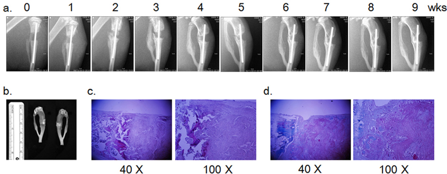
The in vivo-bone reconstitution capacity of a nanofibrous silk 3-D scaffold 3050 was assessed by implanting the scaffold into a rat tibia diaphysis defect. The tibia of the right hind limb of a rat (~350 gm male Sprague Dawley, RaonBio, Korea) anesthetized by inhalation of isoflurane (Hana Pham Co., Ltd, Korea) was exposed, and a ~3 mm longitudinal defect was generated by a sagittal saw (INT-200S, INTEC). An intramedullary nail (φ 0.9~1.2 mm, Solco Biomedical, Korea) loaded with the silk scaffold (3050) was installed into the tibia defect to fill the gap. Then, the site of surgery was closed with a black silk suture (Ailee Co., Ltd., Korea), and ceftriaxone (4 mg in 100 μL) was injected intradermally for prophylactic purpose. Bone formation was monitored weekly by radiography with EZX-60 portable dental X-ray (Genoray Co., Ltd., Korea). Animal care and operating procedures were approved by the Hallym University Medical Center Institutional Animal Care and Use Committee, Korea (HMC2012-0-1026-1).
> H & E and Masson trichome stain
Nine wks post operation, the lower limb was surgically extirpated, and soft tissue was removed with a scalpel. After removal of the intramedullary nail, the tibia was decalcified by incubation in Calci-clear Rapid (National Diagnostics, Atlanta, GA) at room temperature overnight. Paraffinembedded tissue was processed by the standard procedure of hematoxylin and eosin (H & E) staining. Masson trichrome staining was carried out with the Trichrome Stain Kit (Sigma- Aldrich) according to the manufacturer’s protocol.
> Osteoblastic differentiation of hBMSCs
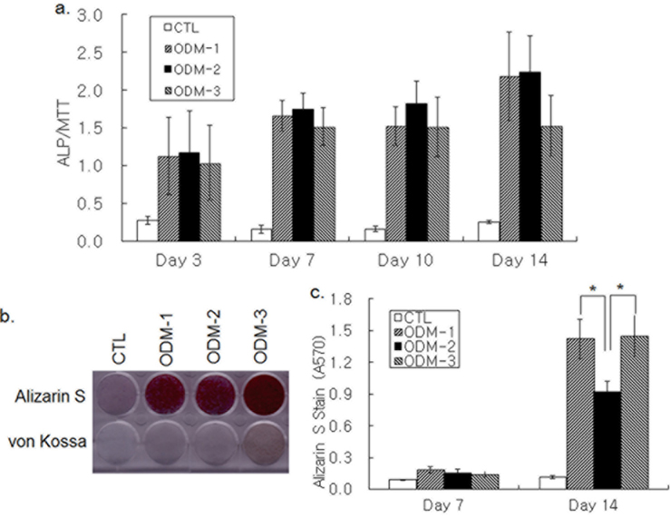
Osteoblastic differentiation was induced by treatment of hBMSCs grown in 96-well plates with three different combinations of DEX, AA, βG, and BMP-2. ALP activity was increased in 3 d of ODM treatment and kept increasing until d 14 (Fig. 1a). Although the ALP activity increase on d 14 by ODM-3 appeared lower than that by ODM-1 and ODM-2, the difference was not significant statistically. Deposition of bone mineral by differentiating hBMSCs was determined by Alizarin S and von Kossa staining. An increase in both Alizarin S and von Kossa staining was not apparent in 7 d, but all three differentiation media increased the staining intensity strongly on d 14 (Fig. 1b). Colorimetric measure of Alizarin S precipitate showed that Alizarin S staining was stronger in the ODM-1 and ODM-3-treated groups than in the ODM- 2-treated one, suggesting that ODM-2 was less efficient for calcium deposition than ODM-1 and ODM-3 (Fig. 1c). Moreover, the intensity of von Kossa staining was the strongest with ODM-3 treatment (Fig. 1b). In summary, although all three differentiation media tested were similarly effective in ALP activity increase, there was a significant difference in their potencies to induce mineral deposition.
> Growth of hBMSC and osteoblastic differentiation on silk scaffolds
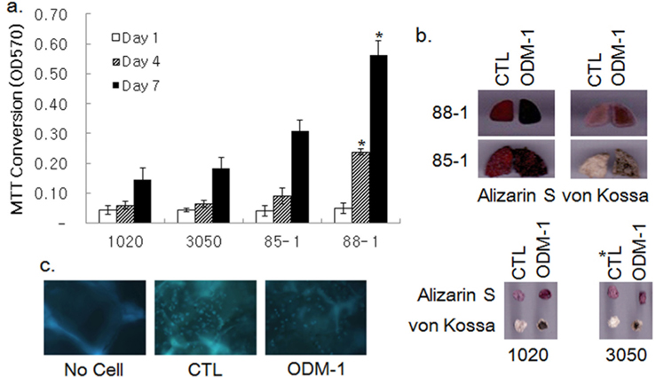
The growth of hBMSCs in various silk 3-D scaffolds was examined by MTT assay. Fibroin nanofibrous scaffolds with pores of φ 100–200 (1020) and 300–500 (3050) μm, and two sponge-type scaffolds prepared by the salt-leaching method with φ 300~500 μm pores (85-1 and 88-1) were tested for their capability to support cell growth for a wk. Cell growth in the scaffolds was attenuated until d 4 and then increased rapidly until d 7 post inoculation, except for scaffold 88-1, in which cell growth was relatively steady (Fig. 2a). Although cell growth in the 3050 scaffolds appeared to exceed that in the 1020 ones, the difference was not statistically significant. The growth of cells in the sponge-type scaffolds was ~1.5 to 3-fold higher than that in the 3050 nanofibrous scaffolds. Among the sponge-type scaffolds, the 88-1 scaffold supported cell growth better than the 85-1 scaffold did.
Osteoblastic differentiation of hBMSCs in the silk scaffolds was examined by treatment of ODM-1 followed by Alizarin S and von Kossa staining. Alizarin S and von Kossa staining demonstrated that treatment of ODM-1 induced differentiation of hBMSCs cultivated both on nanofibrous scaffolds (1020 and 3050) and sponge-type scaffolds (85-1 and 88-1; Fig. 2b). DAPI staining verified the presence of differentiated cells in scaffold 88-1 (Fig. 2c). Thus, all of the silk scaffolds tested could support growth and osteoblastic differentiation of hBMSCs in vitro.
> In vivo bone reconstitution capacity of silk 3-D scaffolds
Since a nanofibrous silk scaffold supports calvaria bone regrowth, bone forming activity of the 3050 nanofibrous scaffold was examined in a long bone defect model of rat tibia diaphysis (Park
et al ., 2010). Intramedullary nailing was employed to fix the silk scaffold in the tibia defect of rat hind limb. Although silk-filled gaps were shrunken by ~1 mm and dorsal contact with the tibia by the forward bending of broken bone displaced the silk scaffold ventrally 1 wk post operation, the position of the silk scaffold was maintained up to 9 wk after that point (Fig. 3a). Weekly monitoring of bone formation by X-ray imaging showed that opaqueness in the nanofibrous silk scaffolds did not increase with time. Lower limbs recovered from operated animals after 9 wk had silk scaffold protruding from the implanted site and reunion of the bone at dorsal region by the forward bending (Fig. 3b). H & E staining of the recovered tibia revealed new bone formation only at the boundary between implanted silk and undisturbed bone, although most of the silk scaffold remained intact (Fig. 3c and d). Formation of the bone tissue at the edge was also apparent from Masson trichrome staining (Fig. 3d). However, new bone formation within the silk scaffold was not observed even after 9 wk post operation. Remaining silk scaffolds were fragmented and infiltrated with histiocytes and multinucleate giant cells, signifying foreign body reaction (Fig. 3c). Masson trichrome stain visualized stream-like collagen within the silk scaffold deposited probably by percolated fibroblasts (Fig. 3d). Although new bone formation was evident at the edge of the silk scaffold, the implanted nanofibrous silk scaffold alone did not support bone ingrowth in the long bone defect model effectively.MSCs established from various sources, including bone marrow, dental pulp, adipose tissue, peripheral blood, and umbilical cord blood have the potential to differentiate into osteoblasts with appropriate treatments (Gamie
et al ., 2012). In this study, hBMSCs were induced to differentiate into osteoblasts by three different combinations of DEX, AA, and βG, with or without BMP-2. Although all three ODM increased ALP activity by a similar extent, Alizarin S and von Kossa staining showed that BMP-2-containing ODM-2 was significantly less efficient than ODM-1 and ODM-3 in mineral deposition. Inefficient mineral deposition in ODM-2-treated cells might be ascribed to BMP-2 because subtle difference in concentrations of DEX, AA, and βG in ODM-1 and ODM-3 did not make a significant difference in mineral deposition.Attachment, growth, and differentiation of MSCs in 3-D scaffolds are required to produce bone tissue in vitro and to reconstitute bone tissue in vivo (Murphy
et al ., 2013). Both nanofibrous and sponge-type silk 3-D scaffolds supported cell growth and differentiation. Differentiation of MSCs inoculated on the 3-D scaffolds was similar among the scaffolds. However, direct comparison of cell growth capacity in the scaffolds showed that cell growth was significantly faster in sponge-type scaffolds than in the nanofibrous ones, although both nanofibrous and sponge-type silk scaffolds are reported to be suitable to support cell ingrowth (Kimet al ., 2005; Parket al ., 2010). Cell growth would depend on surface area, surface property, material exchange rate, and yet unidentified properties of a scaffold (Murphyet al ., 2013). However, it is not clear which one(s) of them should be critical to make the difference in cell growth between the two types of scaffolds.Although the nanofibrous scaffold 3050 supported growth and osteoblastic differentiation of inoculated MSCs in vitro, bone tissue formation was limited at the edge of the implanted 3050 scaffolds in the in vivo tibia diaphysis defect model. Unlike the long bone defect model, the nanofibrous scaffold served as a guide of bone regrowth in the rat calvaria defect model (Park
et al ., 2010). The calvaria differs from the tibia in the mechanism of bone formation, mechanical properties, resorption, and composition (van den Boset al ., 2008). In addition, scaffolds implanted in the calvarial defect were thin, stable, and not weight bearing, which should provide favorable condition in regeneration of the bone defect. In contrast, scaffolds in the tibial defect were 3-mm-thick and were exposed to continuous pressure, which might explain the poor regenerative capacity of the long bone defect with the scaffold. Although reconstitution of the long bone defect with composite scaffold of bone mineral and synthetic biomaterials has been reported occasionally, incorporation of MSCs and/or growth factors in guiding scaffolds has been demonstrated to enhance bone formation in vivo significantly (Berneret al ., 2013; Kuzyket al ., 2010). It is possible that loading MSCs into the nanofibrous scaffold 3050 might be required to facilitate healing of the long bone defect.Healing a bone fracture requires firm stabilization of the fracture throughout the healing process (Marsell and Einhorn, 2011). Large animals, including sheep, rabbit, pig, and dog, have been employed in segmental bone defect model of the long bone partly because steady fixation of the fracture can be achieved with intramedullary nailing and plaster casting (Cancedda
et al ., 2007). The utility of small animals such as mouse and rat in study of the long bone defect model, especially in the tibia model, is limited because of the lack of suitable fixation measures (Histinget al ., 2011). Although intramedullary nailing is widely used to stabilize bone fractures in humans and large animals, its utility in the rat tibia model has recently been reevaluated as an alternative of external fixation (Sigurdsenet al ., 2009). Intramedullary nailing was applied to stabilize osteotomized tibia in this study and resulted in minor longitudinal shrinkage of defect and ventral curvature. However, direct application of straight intramedullary nail in the tibia model could maintain the defect location after the first wk of initial displacement. Shrinkage along the defect length might be ascribed to the repositioning of the nail toward the wide proximal region. Improved procedures such as locking the proximal region of the nail by a pin or a screw might be required to fix the nail in the desired positionIn summary, the current study demonstrates that BMP-2- containing ODM (ODM-2) is less effective for promoting differentiation-associated mineral deposition of human MSCs than BMP-2-deficient media. Although silk scaffoldsboth in the nanofibrous and sponge formsupport growth and differentiation of MSCs in vitro, a nanofibrous silk scaffold by itself is not sufficient to reconstitute tibial defect in the rat hind limb. Since the nanofibrous scaffold is osteoconductive in the rat calvaria model and could sustain MSC ingrowth and differentiation in vitro (Park
et al ., 2010), MSC loading on implanting scaffold should improve bone-forming activity of the scaffold in the tibia defect model.-
[Fig. 1.] Osteoblastic differentiation of hBMSCs by three ODM with distinct combinations of DEX, AA, βG, and BMP-2. a. ALP activity of hBMSCs on indicated days after treatment of ODM-1, 2, and 3. ALP activity was normalized against the MTT value to compensate cell number variation. Results shown are average ± standard error of five experiments. b. Alizarin S and von Kossa staining of differentiationinduced hBMSCs by treatment with the indicated ODM for 14 d. c. Intensity of Alizarin S staining. Absorbance at 570 nm of cetylpyridinium chloride-extracted Alizarin S was measured to determine intensity of Alizarin S deposited by differentiating hBMSCs. Results shown are average ± standard error of seven experiments. * depicts p < 0.05.
-
[Fig. 2.] Growth and differentiation of hBMSC on silk 3-D scaffolds. a. MTT assay results for cells grown on specified silk 3-D scaffolds for the indicated periods. Results shown are average ± standard error of three experiments. * depicts p < 0.05. b. Differentiation of hBMSC grown on silk 3-D scaffolds. Alizarin S and von Kossa staining results of silk 3-D scaffolds loaded with hBMSC. Osteoblastic differentiation of hBMSCs grown on the silk scaffolds was induced by ODM-1 treatment. c. DAPI staining of cells grown and differentiated on a silk scaffold. Silk scaffolds 88-1 with differentiated hBMSC were stained with DAPI after fixation and permeabilization, and were observed with a LSM 510 confocal microscope (Carl Zeiss, Oberkochen, Germany).
-
[Fig. 3.] Bone forming activity of a silk 3-D scaffold in the rat tibia diaphysis defect model. a. Weekly post-operation follow up by radiography. A nanofibrous silk scaffold (3050) was implanted into a 3-mm tibia diaphysis defect by intramedullary nailing (φ 0.9 mm). Callus formation and bone formation was monitored by X-ray imaging weekly from the day of operation (wk 0). b. Crus skeleton with implanted silk scaffold extirpated nine wk after surgery. c. H & E staining of a longitudinal section of the silk scaffold in the tibia diaphysis defect. d. Masson trichrome staining of a longitudinal section of the silk scaffold in the tibia diaphysis defect