검색 전체 메뉴
PDF
맨 위로
OA 학술지
Anti-inflammatory Effect of an Ethanolic Extract of Myagropsis yendoi in Lipopolysaccharide-Stimulated BV-2 Microglia Cells
  • 비영리 CC BY-NC
  • 비영리 CC BY-NC
ABSTRACT
Anti-inflammatory Effect of an Ethanolic Extract of Myagropsis yendoi in Lipopolysaccharide-Stimulated BV-2 Microglia Cells
KEYWORD
Anti-neuroinflammation , Myagropsis yendoi , NF-κB , BV-2 microglia
  • Introduction

    Inflammation is a complex response to an exterior challenge or cellular injury that leads to the release of a complex array of inflammatory moderators, eventually restoring tissue structure and function. However, prolonged inflammation can be harmful, contributing to the pathogenesis of many diseases such as cancer, diabetes, arthritis, atherosclerosis, and Alzheimer’s (Makarov, 2001; Packard and Libby, 2008; Cunningham, 2012). As the primary immune cells in the brain, microglia play an important role in modulating the innate immune response to support the integrity of the central nervous system. The developing brain is insulted by a variety of factors, including inflammation, excitotoxicity, and oxidative stress. Under these conditions, activated microglia secrete excess pro-inflammatory mediators such as nitric oxide (NO) and pro-inflammatory cytokines such as tumor necrosis factor-α (TNF-α), interleukin (IL)-1β, and IL-6 (Bozinovski et al., 2002; Silva et al., 2008). These mediators and cytokines exacerbate neuronal tissue injury and subsequent neurodegeneration. Therefore, modulation of activated microglia is a logical therapeutic strategy to inhibit neuroinflammation.

    NO is primarily produced by enzymatic action of inducible nitric oxide synthase (iNOS). Expression of this enzyme as well as pro-inflammatory cytokines is regulated by the activation of nuclear factor-kappa B (NF-κB) (Bozinovski et al., 2002); the iNOS gene promoter contains an NF-κB consensus sequence. In most cell types, NF-κB is located in the cytosol as an inactive complex bound to inhibitory kappa B (IκB) family, and is activated by diverse external stimuli including inflammatory cytokines, bacterial components, and viral infection (Pahl, 1999). Lipopolysaccharide (LPS) stimulates the phosphorylation, ubiquitination, and proteasomal degradation of IκB, resulting in the translocation of NF-κB into the nucleus by dissociation of the NF-κB–IκB-α complex (Janssen-Heininger et al., 2000). The activation of NF-κB is also controlled by cellular signaling kinases such as mitogen-activated protein kinases (MAPKs) and phosphoinositide 3-kinase/AKT (Cantley, 2002; Jang et al., 2005). The MAPK family, including extracellular signal-regulated kinase (ERK), p38 MAPK, and c-Jun NH2-terminal kinase (JNK), regulates transcription of inflammatory genes via NF-κB activation (Herlaar and Brown, 1999; Jang et al., 2005). Elucidating the detailed molecular mechanisms of this regulation is important for counteracting the harmful effects of pro-inflammatory mediators (Jana et al., 2007).

    Marine macroalgae contain an abundance of polysaccharides, minerals, and polyunsaturated fatty acids known to be beneficial to human health. Recently, diverse studies have revealed that compounds such as phlorotannins (e.g., bieckol), chromenes (sargachromenol, sargaquinoic acid, and sargahydroquinoic acid), and pigments (e.g., fucoxanthin), in extracts from Sargassum spp. and Myagropsis myagroides have strong antioxidant, anti-inflammatory, and antidiabetic activities in vivo and in vitro (Joung et al., 2012a, 2012b; Gwon et al., 2013; Kang et al., 2013; Kim et al., 2008, 2013b; Yang et al., 2013). Myagropsis yendoi, which like Sargassum spp. inhabits the subtidal zone of the coast of East Asia, belongs to the phaeophyta family Sargassaceae. Here, we illustrate the strong anti-inflammatory activity by ethanolic extracts of M. yendoi (MYE). To the best of our knowledge, no previous reports have described the anti-inflammatory activity of MYE. Hence, we further investigated the molecular mechanism of the anti-inflammatory activity of MYE using LPS-stimulated BV-2 microglia cells. Our results suggest that dietary supplements of MYE could help to prevent neuroinflammatory diseases.

    Materials and Methods

      >  Reagents

    LPS (Escherichia coli O55:B5), 2,2-diphenyl-l-picrylhydrazyl (DPPH), 4’,6-diamidino-2-phenylindole (DAPI), and dimethyl sulfoxide (DMSO) were purchased from Sigma Chemical Co. (St. Louis, MO, USA). Dulbecco’s modified Eagle’s medium (DMEM), penicillin–streptomycin mixture, 0.25% trypsin-EDTA, and fetal bovine serum (FBS) were obtained from Gibco-BRL Life Technologies (Grand Island, NJ, USA). A CellTiter 96 AQueous One Solution Cell Proliferation assay kit, dual luciferase assay kit, murine NF-κB promoter/luciferase DNA, pRL-TK DNA, and Moloney murine leukemia virus (M-MLV) reverse transcriptase were purchased from Promega (Madison, WI, USA). Primary and secondary antibodies were obtained from Cell Signaling Biotechnology (Danvers, MA, USA) and Santa Cruz Biotechnology (Santa 28Cruz, CA, USA), respectively. Enhanced chemiluminescence (ECL) detection kit was purchased from GE Healthcare (Piscataway, NJ, USA). Lipofectamine Plus Reagent, Alexa Fluor 488-conjugated secondary antibody, and TRIzol reagent were purchased from Invitrogen (Carlsbad, CA, USA).

      >  Plant material and preparation of MYE

    M. yendoi was collected along the coast of Tongyeong, South Korea, in March 2012. The samples were air-dried under shade for 5 days and ground using a hammer grinder. The resulting powder was stored at –20°C until used. Dried powder (100 g) of M. yendoi was extracted three times in 95% (v/v) ethanol for 3 h at 70°C. The combined extracts were concentrated using a rotary vacuum evaporator (Eyela, Tokyo, Japan) at 40°C and lyophilized to a powder (9 g), then stored at –20°C until used. The extract was dissolved in DMSO and further diluted with culture media before use. The final concentration of DMSO in cell culture medium was less than 0.1%.

      >  Cell culture and sample treatment

    Murine BV-2 microglial cells (ATCC, Rockville, MD, USA) were maintained in DMEM supplemented with 10% FBS, penicillin (100 units/mL), and streptomycin sulfate (100 μg/mL) in a humidified atmosphere of 5% CO2. Cells pretreated with various concentrations of MYE for 2 h were stimulated with LPS (1 μg/mL) for the indicated times.

      >  Cytotoxicity assay

    Cell viability was determined by 3-(4,5-dimethylthiazol-2-yl)-5-(3-carboxymethoxyphenyl)-2-(4-sulfophenyl)-2H-tetrazolium (MTS) assay using the CellTiter 96 AQueous One Solution Cell Proliferation assay kit according to the manufacturer›s instructions. Cells were inoculated at a density of 3 × 10⁵ cells into 96-well plates and cultured at 37°C for 24 h. Cells were then treated with LPS (1 μg/mL) in the presence or absence of MYE at various concentrations for 24 h. The culture medium was removed and replaced with 95 μL of fresh culture medium and 5 μL of MTS solution. After 1 h, absorbance at 490 nm was measured using a microplate reader (Glomax Multi Detection System; Promega).

      >  Measurement of NO and pro-inflammatory cytokines

    Cells (5 × 10⁴ cells/well) were pretreated with MYE (0-100 μg/mL) for 2 h prior to LPS treatment for 24 h. After treatment with LPS, culture media were collected after centrifugation at 2,000 g for 10 min and stored at –75°C until tested. The nitrite concentration in culture media was measured as an indicator of NO production, according to the Griess reaction (Kim et al., 1995). Levels of TNF-α and IL-6 in culture media from each group were quantitatively determined by enzyme-linked immunosorbent assay (ELISA; R&D Systems, Minneapolis, MN, USA) according to the manufacturer's instructions.

      >  Reverse transcription-polymerase chain reaction

    BV-2 cells plated in a 6-well cell culture plate at a density of 3.0 × 10⁵ cells/well were pretreated with MYE or DMSO for 1 h, then treated with LPS for 6 h. Total RNA from each group was isolated using TRIzol reagent. Five micrograms of total RNA was used for reverse transcription using oligo-dT and M-MLV reverse transcriptase. PCR was carried out using the resulting cDNA as a template under the following conditions: 25 cycles of denaturation at 95°C for 30 s, annealing at 60°C for 30 s, and extension at 72°C for 30 s. The PCR products were visualized by 1% agarose gel electrophoresis. Verification of PCR product of the iNOS gene was established by its predicted sizes under an ultraviolet light illuminator. The primer sequences were the following: 5'-ACC ACT CGT ACT TGG GAT GC-3' (sense), 5'-CAC CTT GGA GTT CAC CCA GT-3' (antisense) for iNOS; 5'-GAC CCC TTC ATT GAC CTC AA-3' (sense), 5'-CTT CTC CAT GGT GGT GAA GA- 3' (antisense) for glyceraldehyde 3-phosphate dehydrogenase (GAPDH). GAPDH was used as an internal standard to evaluate the relative expression of iNOS.

      >  Immunocytochemical analysis

    To analyze nuclear localization of NF-κB in BV-2 cells, cells were cultured on glass coverslips (SPL Lifesciences Co., Pocheon, Korea) in 24-well plates for 24 h. After preincubation with MYE for 2 h, cells were stimulated with LPS (1 μg/ mL) or treated with DMSO for 30 min. Cells were fixed in 4.0% paraformaldehyde in phosphate-buffered saline (PBS) for 15 min at room temperature, then permeabilized with 0.5% Triton X-100 in PBS for 10 min. Permeabilized cells were washed with PBS and blocked with 3% bovine serum albumin (BSA) in PBS for 30 min. Thereafter, cells were incubated with anti-NF-κB polyclonal antibody diluted in 3% BSA/PBS for 2 h, rinsed three times for 5 min with PBS, and incubated with Alexa Fluor 488-conjugated secondary antibody diluted in 3% BSA/PBS for 1 h. Cells were stained with 2 μg/mL DAPI and images were captured using an LSM700 laser scanning confocal microscope (Carl Zeiss, Oberkochen, Germany).

      >  Preparation of cytosolic and nuclear extracts

    BV-2 cells plated in 6-well cell culture plates at a density of 3 × 10⁶ cells per well were pretreated with MYE or DMSO for 2 h and then treated with LPS for 30 min. Cells were washed twice with ice-cold PBS, scraped in PBS, and centrifuged at 12,000 g for 5 min at 4°C. Pellets were suspended in 180 μL of hypotonic buffer A (10 mM Tris-HCl, 10 mM NaCl, 3 mM MgCl2, 0.02% NaN3, 0.5 mM dithiothreitol [DTT], and 1 mM phenylmethane sulphonyl fluoride [PMSF], pH 7.4), and afterward, 20 μL of 5% Nonidet P-40 was added and stood for 5 min on ice. The mixture was centrifuged at 1,800 g for 5 min, and the supernatant (cytosolic extract) was collected. Pellets were washed with hypotonic buffer and resuspended in hypertonic buffer C (20 mM 4-(2-hydroxyethyl)-1-piperazineethanesulfonic acid, 25% glycerol, 420 mM NaCl, 1.5 mM MgCl2, 0.2 mM EDTA, 0.02% NaN3, 0.5 mM DTT, and 1 mM PMSF, pH 7.4) for 1 h on ice and centrifuged at 14,000 g for 10 min. The supernatant containing nuclear proteins was collected and stored at –70°C after determination of the protein concentration.

      >  Western blot analysis

    Cells were incubated with various concentrations of MYE for 1 h and stimulated with LPS (1 μg/mL) for varying times. Cells were washed twice with cold PBS and lysed with lysis buffer (50 mM Tris-HCl, 150 mM NaCl, 1% Nonidet P-40, 1% Tween 20, 0.1% sodium dodecyl sulfate [SDS], 1 mM Na3VO4, 10 μg/mL leupeptin, 50 mM NaF, and 1 mM PMSF, pH 7.5) on ice for 1 h. After centrifugation at 12,000 g for 20 min, protein concentrations in the supernatants were determined, and aliquots (30 μg protein) were separated by SDS-polyacrylamide gel electrophoresis (PAGE) and transferred onto nitrocellulose membranes (GE Healthcare). Membranes were washed with Tris-buffered saline (TBS; 10 mM Tris-HCl, 150 mM NaCl, pH 7.5) supplemented with 0.05% Tween 20 (TBST) followed by blocking with TBST containing 5% nonfat milk. Membranes were incubated overnight with primary antibodies, then exposed to secondary antibodies coupled to horseradish peroxidase for 2 h and washed three times with TBST at room temperature. Immunoreactivities were determined using an ECL detection kit.

      >  Statistical analysis

    Data were expressed as means ± SDs of at least three independent experiments unless otherwise indicated. Data were analyzed using one-way analysis of variance (ANOVA), followed by pairwise Student’s t-tests for multiple comparisons. Differences with a value of P < 0.05 were considered statistically significant. All analyses were performed using SPSS for Windows, version 10.07 (SPSS Inc., Chicago, IL, USA).

    Results

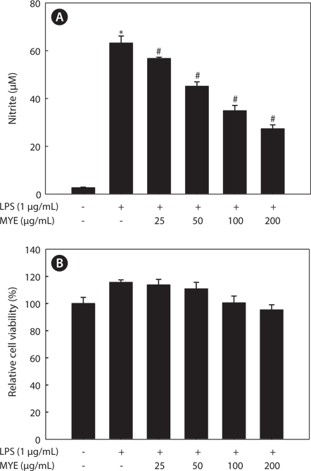
      >  MYE suppresses NO production in LPS-stimulated BV-2 cells

    Whether MYE inhibits neuroinflammation was determined by measuring NO production by LPS-stimulated BV-2 cells. Cells pretreated with various concentrations of MYE were stimulated with LPS (1 μg/mL) or treated with DMSO for 24 h. As shown in Fig. 1A, MYE significantly suppressed LPS-induced NO production in a dose-dependent manner. MTS assay revealed that MYE causes no cytotoxicity at concentrations of up to 200 μg/mL in BV-2 cells (Fig. 1B). Taken together, MYE inhibits LPS-induced NO secretion in BV-2 cells with no cytotoxicity.

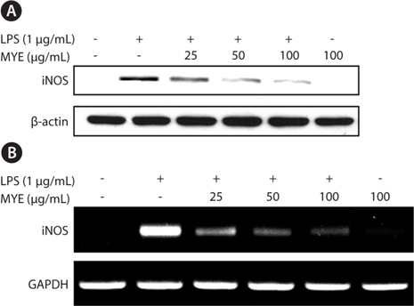
      >  MYE inhibits inflammation-triggered expression of iNOS in BV-2 cells

    We investigated whether MYE’s suppression of NO production is related to expression of iNOS by Western blotting. As shown in Fig. 2A, LPS treatment of BV-2 cells for 16 h enhanced the expression of iNOS protein. However, pretreatment with MYE led to a dose-dependent inhibition of LPS-induced iNOS protein expression in BV-2 cells; iNOS expression was almost completely suppressed at 100 μg/mL of MYE. To further examine the effect of MYE on the transcriptional regulation of iNOS expression, reverse transcription-polymerase chain reaction analysis was performed. Fig. 2B shows that MYE suppressed the expression of iNOS mRNA in a dose-dependent manner in LPS-stimulated BV-2 cells. Similar to Western blot results, 100 μg/mL of MYE strongly inhibited iNOS mRNA expression. These results indicate that MYE-mediated downregulation of iNOS expression is mainly due to its transcriptional repression in LPS-stimulated BV-2 cells.

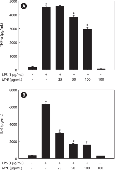
      >  MYE inhibits LPS-induced production of TNF-? and IL-6 in BV-2 cells

    Pro-inflammatory cytokines such as TNF-α and IL-6 are secreted early in the inflammatory response and are thus key markers of inflammation. We determined the effect of MYE on the levels of these cytokines in the media of LPS-stimulated BV-2 cells by ELISA following pretreatment with various concentrations of MYE for 2 h and LPS stimulation for 24 h. Stimulation of BV-2 cells with LPS induced significant increases in the levels of TNF-α (Fig. 3A) and IL-6 (Fig. 3B); pretreatment with MYE inhibited these effects in a dose-dependent manner.

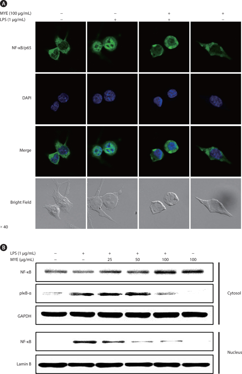
      >  MYE suppresses nuclear translocation of NF-κB in LPS-stimulated BV-2 cells

    To analyze the effect of MYE on the regulation of transcription factors during the inflammatory response, the effect of MYE on the intracellular localization of NF-κB/p65 was assessed by confocal microscopy and Western blot analysis in LPS-stimulated BV-2 cells. Immunocytochemical analysis (Fig. 4A) showed that NF-κB protein was mainly localized in the cytoplasm in unstimulated cells and was translocated into the nucleus upon stimulation with LPS. Pretreatment with MYE remarkably suppressed translocation of NF-κB regardless of LPS treatment in BV-2 cells. Subcellular fractionation and Western blot analysis confirmed LPS-induced nuclear translocation of NF-κB via degradation and phosphorylation of IκB-α, as well as MYE’s suppression of this effect (Fig. 4B). These results suggest that MYE attenuates the production of NO and pro-inflammatory cytokines at least in part through inactivation of the NF-κB pathway.

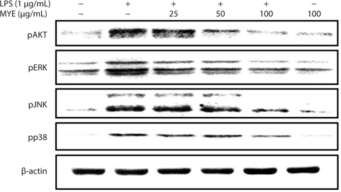
      >  MYE prevents activation of MAPKs and AKT in LPS-stimulated BV-2 cells

    To evaluate the effect of MYE on signaling associated with NF-κB activation, changes in phosphorylation of intracellular signaling proteins, including ERK, JNK, p38 MAPK, and AKT, in BV-2 cells were analyzed by Western blotting. As shown in Fig. 5, MYE inhibited phosphorylation of JNK, ERK, p38 MAPK, and AKT in a dose-dependent manner in LPS-stimulated BV-2 cells. These data suggest that inactivation of the NF-κB pathway by MYE is mediated by inhibition of MAPKs and AKT phosphorylation.

    Discussion

    Various studies have examined the biological activities of natural compounds or extracts to develop dietary supplements or therapeutic agents due to their easy availability and low side effects. Various phytochemicals from terrestrial plants have shown a wide range of anti-inflammatory activities; moreover, many studies have elucidated the molecular mechanisms of the anti-inflammatory action of extracts from marine brown algae, such as Myagropsis myagroides, Ecklonia stolonifera, and Sargassum fulvellum (Joung et al., 2012a, 2012b; Lee et al., 2012; Gwon et al., 2013; Kim et al., 2013b). The present study was designed to investigate the anti-inflammatory effect of MYE on LPS-stimulated murine BV-2 microglia cells. To further understand the molecular mechanism by which MYE reduces inflammation, the effects of MYE on the secretion of NO, TNF-α, and IL-6, the expression of iNOS and the activation of NF-κB were investigated. Our results collectively show that MYE inhibits secretion of NO, TNF-α, and IL-6 via blockade of the NF-κB pathway by inhibiting ERK, JNK, p38 MAPK, and AKT phosphorylation in LPS-stimulated BV-2 microglia cells. The inhibitory effect of MYE on the expression of inflammatory mediators suggests a potential mechanism for its anti-inflammatory action and its promise for use as a dietary supplement to reduce the risk for neuroinflammatory diseases.

    NO is a key inflammatory and neurotoxic mediator in neuroinflammation responsible for detrimental effects on the central nervous system in ischemia, Alzheimer's disease, and neuronal injury (Meda et al., 1995). Many studies have revealed that abnormally high levels of NO are found in various types of brain injuries and neurodegenerative diseases, which are caused by enhanced expression of iNOS (Teismann et al., 2003; Brown and Neher, 2010; Cunningham, 2012). Overexpression of iNOS was found to occur in microglial cells in rodent brains treated with LPS (Minghetti et al., 1999). Treatment with inhibitors of iNOS results in neuroprotection against LPS-induced neurotoxicity, suggesting that NO is an important mediator in neurotoxicity (Teismann et al., 2003;Oh et al., 2009). The present study demonstrated that MYE inhibits the production of NO in LPS-stimulated BV-2 cells. In addition, we determined that inhibition of NO production by MYE results from the suppression of both mRNA and protein expression of iNOS in these cells, indicating that inhibition of NO by MYE is associated with downregulation of iNOS at the transcriptional level. These data suggest that MYE may have a protective effect against the neuroinflammatory response in neurodegenerative diseases.

    Activated microglia produce high levels of pro-inflammatory cytokines including IL-6 and TNF-α, which are involved in the pathogenesis of several neurodegenerative disorders (Brown and Neher, 2010; Kim et al., 2013a, 2013b). Also, excessive production of pro-inflammatory cytokines such as TNF-α and IL-6 play a critical role in acute inflammatory responses as well as chronic inflammatory diseases (Jang et al., 2005; Kim et al., 2013a). Our observation that MYE suppresses secretion of these pro-inflammatory cytokines in LPSstimulated BV-2 cells further supports the potential of MYE as a neuroprotective agent against neuroinflammation.

    Transcription of iNOS and pro-inflammatory cytokine genes are mainly controlled by NF-κB, which binds to respective cis-acting elements in the iNOS promoter (Xie et al., 1993; Marks-Konczalik et al., 1998; Kim et al., 2011). A variety of external stimuli induce the phosphorylation of IκB in the NF-κB–IκB-α complex, resulting in proteasomal degradation of IκB-α and subsequent translocation of NF-κB from the cytosol into the nucleus (Janssen-Heininger et al., 2000; Jang et al., 2005). In this study, we demonstrated that MYE inhibits LPS-induced translocation of the NF-κB p65 subunit, suggesting that transcriptional repression of iNOS and pro-inflammatory cytokines by MYE primarily results from inactivation of NF-κB in LPS-stimulated microglia cells. NF-κB thus appears to be an important target transcription factor mediating the anti-inflammatory effects of MYE.

    Intracellular signaling proteins such as MAPKs and AKT are activated by LPS, leading to enhanced transcription of iNOS and pro-inflammatory cytokine genes in microglia (Herlaar and Brown, 1999; Skaper, 2007; Jung et al., 2010). Phosphorylation of ERK, JNK, p38 MAPK, and AKT is decreased by MYE treatment in a dose-dependent manner in LPS-stimulated BV-2 cells. Given the roles of ERK, JNK, p38 MAPK, and AKT in inflammatory processes, these kinases may be another target of MYE’s anti-inflammatory activity in LPS-stimulated BV-2 cells. These data indicate that MYE’s anti-inflammatory effects result at least in part from inhibition of the NF-κB pathway via suppressed phosphorylation of MAPKs and AKT.

    In conclusion, we found that MYE inhibits the secretion of inflammatory mediators such as NO and pro-inflammatory cytokines, including TNF-α IL-6, in LPS-stimulated BV-2 microglia. Moreover, the inhibitory effect of MYE was associated with inactivation of the NF-κB pathway via reduced phosphorylation of MAPKs and AKT. However, the in vitro nature of our study limits conclusions regarding MYE's effects on the in vivo neuroinflammatory response. Further studies are required to investigate the anti-inflammatory and neuroprotective effects of MYE in animal models of neurodegenerative disease. Taken together, our results suggest that MYE is a therapeutic candidate for the treatment or prevention of neuroinflammation in neurodegenerative diseases.

참고문헌
  • 1. Bozinovski S, Jones JE, Vlahos R, Hamilton JA, Anderson GP 2002 Granulocyte/macrophage-colony-stimulating factor (GM-CSF) regulates lung innate immunity to lipopolysaccharide through Akt/Erk activation of NF-κB and AP-1 in vivo [J Biol Chem] Vol.277 P.42808-42814 google
  • 2. Brown GC, Neher JJ 2010 Inflammatory neurodegeneration and mechanisms of microglial killing of neurons [Mol Neurobiol] Vol.41 P.242-247 google
  • 3. Cantley LC 2002 The phosphoinositide 3-kinase pathway [Science] Vol.296 P.1655-1657 google
  • 4. Cunningham C 2012 Microglia and neurodegeneration: the role of systemic inflammation [Glia] Vol.61 P.71-90 google
  • 5. Gwon WG, Lee MS, Kim JS, Kim JI, Lim CW, Kim NG, Kim HR 2013 Hexane fraction from Sargassum fulvellum inhibits lipopolysaccharide-induced inducible nitric oxide synthase expression in RAW 264.7 cells via NF-κB pathways [Am J Chin Med] Vol.41 P.565-584 google
  • 6. Herlaar E, Brown Z 1999 p38 MAPK signalling cascades in inflammatory disease [Mol Med Today] Vol.5 P.439-447 google
  • 7. Jana M, Jana A, Liu X, Ghosh S, Pahan K 2007 Involvement of phosphatidylinositol 3-kinase-mediated up-regulation of IκB? in anti-inflammatory effect of gemfibrozil in microglia [J Immunol] Vol.179 P.4142-4152 google
  • 8. Jang BC, Paik JH, Kim SP, Shin DH, Song DK, Park JG, Suh MH, Park JW, Suh SI 2005 Catalase induced expression of inflammatory mediators via activation of NF-NF-κB, PI3K/AKT, p70S6K, and JNKs in BV2 microglia [Cell Signal] Vol.17 P.625-633 google
  • 9. Janssen-Heininger YMW, Poynter ME, Baeuerle PA 2000 Recent advances towards understanding redox mechanisms in the activation of nuclear factor kappaB [Free Radic Biol Med] Vol.28 P.1317-1327 google
  • 10. Joung EJ, Lee MS, Choi JW, Kim JS, Shin T, Jung BM, Kim JI, Kim HR 2012 Anti-inflammatory effects of phlorofucofuroeckol B-rich ethyl acetate fraction obtained from Myagropsis myagroides on lipopolysaccharide-stimulated RAW 264.7 cells and mouse edema [Int Immunopharmacol] Vol.14 P.471-480 google
  • 11. Joung EJ, Lee MS, Choi JW, Kim JS, Shin T, Jung BM, Yoon NY, Lim CW, Kim JI, Kim HR 2012 Anti-inflammatory effect of ethanolic extract from Myagropsis myagroides on murine macrophages and mouse ear edema [BMC Complement Altern Med] Vol.12 P.171 google
  • 12. Jung WK, Lee DY, Park C, Choi YH, Choi I, Park SG, Seo SK, Lee SW, Yea SS, Ahn SC, Lee CM, Park WS, Ko JH, Choi IW 2010 Cilostazol is anti-inflammatory in BV2 microglial cells by inactivating nuclear factor-kappaB and inhibiting mitogen-activated protein kinases [Br J Pharmacol] Vol.159 P.1274-1285 google
  • 13. Kang GJ, Han SC, Yoon WJ, Koh YS, Hyun JW, Kang HK, Youl Cho J, Yoo ES 2013 Sargaquinoic acid isolated from Sargassum siliquastrum inhibits lipopolysaccharide-induced nitric oxide production in macrophages via modulation of nuclear factor-κB and c-Jun N-terminal kinase pathways [Immunopharmacol Immunotoxicol] Vol.35 P.80-87 google
  • 14. Kim AR, Lee MS, Shin TS, Hua H, Jang BC, Choi JS, Byun DS, Utsuki T, Ingram D, Kim HR 2011 Phlorofucofuroeckol A inhibits the LPS-stimulated iNOS and COX-2 expressions in macrophages via inhibition of NF-κB, Akt, and p38 MAPK [Toxicol in Vitro] Vol.25 P.1789-1795 google
  • 15. Kim AR, Lee MS, Choi JW, Utsuki T, Kim JI, Jang BC, Kim HR 2013 Phlorofucofuroeckol A suppresses expression of inducible nitric oxide synthase, cyclooxygenase-2, and pro-inflammatory cytokines via inhibition of nuclear factor-κB, c-Jun NH2-terminal kinases, and Akt in microglial cells [Inflammation] Vol.36 P.259-271 google
  • 16. Kim H, Lee HS, Chang KT, Ko TH, Baek KJ, Kwon NS 1995 Chloromethyl ketones block induction of nitric oxide synthase in murine macrophages by preventing activation of nuclear factor-kappa B [J Immunol] Vol.154 P.4741-4748 google
  • 17. Kim S, Kim JI, Choi JW, Kim M, Yoon NY, Choi CG, Choi JS, Kim HR 2013 Anti-inflammatory effect of hexane fraction from Myagropsis myagroides ethanolic extract in lipopolysaccharide-stimulated BV-2 microglial cells [J Pharm Pharmacol] Vol.65 P.895-906 google
  • 18. Kim SN, Choi HY, Lee W, Park GM, Shin WS, Kim YK 2008 Sargaquinoic acid and sargahydroquinoic acid from Sargassum yezoense stimulate adipocyte differentiation through PPAR?/γ activation in 3T3-L1 cells [FEBS Lett] Vol.582 P.3465-3472 google
  • 19. Lee MS, Kwon MS, Choi JW, Shin T, No HK, Choi JS, Byun DS, Kim JI, Kim HR 2012 Anti-inflammatory activities of an ethanol extract of Ecklonia stolonifera in lipopolysaccharide-stimulated RAW 264.7 murine macrophage cells [J. Agric Food Chem] Vol.60 P.9120-9129 google
  • 20. Makarov SS 2001 NF-kappa B in rheumatoid arthritis: a pivotal regulator of inflammation, hyperplasia, and tissue destruction [Arthritis Res] Vol.3 P.200-206 google
  • 21. Marks-Konczalik J, Chu SC, Moss J 1998 Cytokine-mediated transcriptional induction of the human inducible nitric oxide synthase gene requires both activator protein 1 and nuclear factor κB-binding sites [J Biol Chem] Vol.273 P.22201-22208 google
  • 22. Meda L, Cassatella MA, Szendrei GI, Otvos L, Baron P, Villalba M, Ferrari D, Rossi F 1995 Activation of microglial cells by β-amyloid protein and interferon-γ [Nature] Vol.374 P.647-650 google
  • 23. Minghetti L, Walsh DT, Levi G, Perry VH 1999 In vivo expression of cyclooxygenase-2 in rat brain following intraparenchymal injection of bacterial endotoxin and inflammatory cytokines [J Neuropathol Exp Neurol] Vol.58 P.1184-1191 google
  • 24. Oh YT, Lee JY, Lee J, Kim H, Yoon KS, Choe W, Kang I 2009 Oleic acid reduces lipopolysaccharide-induced expression of iNOS and COX-2 in BV2 murine microglial cells: possible involvement of reactive oxygen species, p38 MAPK, and IKK/NF-κB signaling pathways [Neurosci Lett] Vol.464 P.93-97 google
  • 25. Packard RRS, Libby P 2008 Inflammation in atherosclerosis: from vascular biology to biomarker discovery and risk prediction [Clin Chem] Vol.54 P.24-38 google
  • 26. Pahl HL 1999 Activators and target genes of Rel/NF-κB transcription factors [Oncogene] Vol.18 P.6853-6866 google
  • 27. Silva AR, Pinheiro AM, Souza CS, Freitas SRVB, Vasconcellos V, Freire SM, Velozo ES, Tardy M, El-Bacha RS, Costa MFD, Costa SL 2008 The flavonoid rutin induces astrocyte and microglia activation and regulates TNF-alpha and NO release in primary glial cell cultures [Cell Biol Toxicol] Vol.24 P.75-86 google
  • 28. Skaper SD 2007 The brain as a target for inflammatory processes and neuroprotective strategies [Ann N Y Acad Sci] Vol.1122 P.23-34 google
  • 29. Teismann P, Tieu K, Cohen O, Choi DK, Wu DC, Marks D, Vila M, Jackson-Lewis V, Przedborski S 2003 Pathogenic role of glial cells in Parkinson’s disease [Mov Disord] Vol.18 P.121-129 google
  • 30. Xie Q, Whisnant R, Nathan C 1993 Promoter of the mouse gene encoding calcium-independent nitric oxide synthase confers inducibility by interferon gamma and bacterial lipopolysaccharide [J Exp Med] Vol.177 P.1779-1784 google
  • 31. Yang EJ, Ham YM, Yang KW, Lee NH, Hyun CG 2013 Sargachromenol from Sargassum micracanthum inhibits the lipopolysaccharide-induced production of inflammatory mediators in RAW 264.7 macrophages [ScientificWorldJournal] google
이미지 / 테이블
  • [ Fig. 1. ]  Effect of Myagropsis yendoi ethanolic extracts (MYE) on cell viability and on the inhibition of nitric oxide (NO) production in lipopolysaccharide (LPS)-stimulated BV-2 cells. Cells pretreated with various concentrations of MYE for 2 h were stimulated with LPS (1 μg/mL) for 24 h. The culture media of the treated cells were used to measure the amount of nitrite to evaluate NO level (A). Cell viability was analyzed by MTS assay (B). All data are presented as means ± SDs of three independent experiments. *P < 0.05 indicates significant differences from the control group. #P < 0.05 indicates significant differences from the control group.
    Effect of Myagropsis yendoi ethanolic extracts (MYE) on cell viability and on the inhibition of nitric oxide (NO) production in lipopolysaccharide (LPS)-stimulated BV-2 cells. Cells pretreated with various concentrations of MYE for 2 h were stimulated with LPS (1 μg/mL) for 24 h. The culture media of the treated cells were used to measure the amount of nitrite to evaluate NO level (A). Cell viability was analyzed by MTS assay (B). All data are presented as means ± SDs of three independent experiments. *P < 0.05 indicates significant differences from the control group. #P < 0.05 indicates significant differences from the control group.
  • [ Fig. 2. ]  Effect of Myagropsis yendoi ethanolic extracts (MYE) on LPS-induced inducible nitric oxide synthase (iNOS) expression in BV-2 cells. Cells pretreated with various concentrations of MYE for 2 h and were stimulated with or without lipopolysaccharide (LPS; 1 μg/mL) for 16 h. Whole proteins were separated with sodium dodecyl sulfate polyacrylamide gel electrophoresis and analyzed with Western blot (A). Cells were incubated with various concentration of MYE for 2 h, and then stimulated with LPS (1 mg/mL) for 6 h. mRNA levels of iNOS and glyceraldehyde 3-phosphate dehydrogenase (GAPDH) were determined by reverse transcription-polymerase chain reaction analysis using corresponding gene-specific primers (B). The results presented are representatives of three independent experiments.
    Effect of Myagropsis yendoi ethanolic extracts (MYE) on LPS-induced inducible nitric oxide synthase (iNOS) expression in BV-2 cells. Cells pretreated with various concentrations of MYE for 2 h and were stimulated with or without lipopolysaccharide (LPS; 1 μg/mL) for 16 h. Whole proteins were separated with sodium dodecyl sulfate polyacrylamide gel electrophoresis and analyzed with Western blot (A). Cells were incubated with various concentration of MYE for 2 h, and then stimulated with LPS (1 mg/mL) for 6 h. mRNA levels of iNOS and glyceraldehyde 3-phosphate dehydrogenase (GAPDH) were determined by reverse transcription-polymerase chain reaction analysis using corresponding gene-specific primers (B). The results presented are representatives of three independent experiments.
  • [ Fig. 3. ]  Effect of Myagropsis yendoi ethanolic extracts (MYE) on the secretion of pro-inflammatory cytokines in lipopolysaccharide (LPS)- stimulated BV-2 cells. Cells pretreated with various concentrations of MYE were stimulated with or without LPS (1 μg/mL) for 24 h. Tumor necrosis factor-α (TNF-α) (A) and interleukin-6 (IL-6) (B) in the culture media were measured by ELISA. Data are means ± SDs of three independent experiments. *P < 0.05 indicates significant differences from the control group. #P < 0.05 indicates significant differences from the control group.
    Effect of Myagropsis yendoi ethanolic extracts (MYE) on the secretion of pro-inflammatory cytokines in lipopolysaccharide (LPS)- stimulated BV-2 cells. Cells pretreated with various concentrations of MYE were stimulated with or without LPS (1 μg/mL) for 24 h. Tumor necrosis factor-α (TNF-α) (A) and interleukin-6 (IL-6) (B) in the culture media were measured by ELISA. Data are means ± SDs of three independent experiments. *P < 0.05 indicates significant differences from the control group. #P < 0.05 indicates significant differences from the control group.
  • [ Fig. 4. ]  Effect of Myagropsis yendoi ethanolic extracts (MYE) on the nuclear translocation of nuclear factor-κB (NF-κB) in lipopolysaccharide (LPS)-stimulated BV-2 cells. Cells were pretreated with MYE (100 μg/mL) for 2 h, followed by LPS stimulation for 30 min. DAPI (blue) was used for staining nuclei (A). Cells pretreated with MYE for 2 h were stimulated with LPS for 30 min and levels of NF-κB in nucleus were analyzed with Western blot (B). The results presented are representative of three independent experiments.
    Effect of Myagropsis yendoi ethanolic extracts (MYE) on the nuclear translocation of nuclear factor-κB (NF-κB) in lipopolysaccharide (LPS)-stimulated BV-2 cells. Cells were pretreated with MYE (100 μg/mL) for 2 h, followed by LPS stimulation for 30 min. DAPI (blue) was used for staining nuclei (A). Cells pretreated with MYE for 2 h were stimulated with LPS for 30 min and levels of NF-κB in nucleus were analyzed with Western blot (B). The results presented are representative of three independent experiments.
  • [ Fig. 5. ]  Effect of Myagropsis yendoi ethanolic extracts (MYE) on the phosphorylations of mitogen-activated protein kinases (MAPKs) and AKT in lipopolysaccharide (LPS)-stimulated BV-2 cells. Cells pretreated with indicated concentrations of MYE for 2 h were stimulated with LPS (1 μg/mL) for 30 min. Whole cell lysates (40 μg) were analyzed by Western blotting using corresponding antibodies.
    Effect of Myagropsis yendoi ethanolic extracts (MYE) on the phosphorylations of mitogen-activated protein kinases (MAPKs) and AKT in lipopolysaccharide (LPS)-stimulated BV-2 cells. Cells pretreated with indicated concentrations of MYE for 2 h were stimulated with LPS (1 μg/mL) for 30 min. Whole cell lysates (40 μg) were analyzed by Western blotting using corresponding antibodies.
(우)06579 서울시 서초구 반포대로 201(반포동)
Tel. 02-537-6389 | Fax. 02-590-0571 | 문의 : oak2014@korea.kr
Copyright(c) National Library of Korea. All rights reserved.