검색 전체 메뉴
PDF
맨 위로
OA 학술지
Dendrodasys duplus, a New Gastrotrich Species (Macrodasyida: Dactylopodolidae) from South Korea
  • 비영리 CC BY-NC
  • 비영리 CC BY-NC
ABSTRACT
Dendrodasys duplus, a New Gastrotrich Species (Macrodasyida: Dactylopodolidae) from South Korea
KEYWORD
taxonomy , Gastrotricha , description , East Asia , new species
  • INTRODUCTION

    Dactylopodolid gastrotrichs have been considered the most basal group within macrodasyid gastrotrichs, because they show the characteristic combination of having cross-striated body muscles and monociliated epidermal cells ventrally (Hummon, 1982; Hochberg and Litvaitis, 2001). Among three genera of the family Dactylopodolidae currently recognized, the genus Dendrodasys Wilke, 1954 is characterized by a head with crenulated lateral lobes (Hummon, 1982; Todaro and Hummon, 2008).

    In the genus Dendrodasys, five species and two unidentified species are recognized: D. affinis Wilke, 1954 from the Mediterranean Sea (Naples, Tuscan Archipelago, the Ionian Sea, and the Tyrrhenian Sea); D. gracilis Wilke, 1954 from the north Atlantic Ocean (Florida and Scotland), the Pacific Ocean (Ecuador) and the Mediterranean Sea (Naples, the Ionian Sea, and the Tyrrhenian Sea); D. ponticus Valkanov, 1957 from the Black Sea (Bulgaria and Rumania) and the Mediterranean Sea (the Adriatic Sea); D. pacificus Schmidt, 1974 from the Pacific Ocean (Galapagos islands, Hawaii and Australia); D. rubomarinus Hummon, 2011 from the Red Sea (Egypt); Dendrodasys sp. Valbonesi and Luporini, 1984 from Somalia and Dendrodasys sp. Todaro and Rocha, 2004 (only photo) from Brazil (Wilke, 1954; Valkanov, 1957; Rudescu, 1967; Schmidt, 1974; Valbonesi and Luporini, 1984; Hummon et al., 1998; Todaro and Rocha, 2004; Hummon, 2009, 2011).

    Recently we discovered a new species from sublittoral fine sandy bottoms of Uljin and Jeju Island, South Korea. This is the first record of Dendrodasys from the northwest of the Pacific. Herein, we describe a new species of the genus Dendrodasys, and discuss the taxonomic affinities based on the morphological characters.

    MATERIALS AND METHODS

    Specimens were collected from shallow sublittoral fine sandy bottoms, at about 1-2m in water depth at Gujwa, the northeastern coast of Jeju Island, and at about 5-6 m deep at Uljin, the east coast of Korea (East Sea or Sea of Japan).

    Fine sands were taken by scooping the surface sediments of several centimeters using a long-handled bowl at Gujwa, and using the Smith-Mclntyre grab at Uljin. Samples were extracted through a 63 μm mesh sieve after performing the anesthetization-decantation method with 7% MgCl2 for 5-10 min, and were fixed in 4% buffered formalin. Living specimens from Uljin were video-captured using a CCD camera (eXcope X3; DIXI Optics Co., Daejeon, Korea). Methods of sample preparation, observation, and measurements follow those of our previous papers (Lee and Chang, 2003; Lee et al., 2009).

    Type specimens have been deposited at the National Institute of Biological Resources (NIBR), Incheon, Korea and the Korea Institute of Ocean Science & Technology (KIOST), Ansan, Korea.

    Terminology mostly followed that of Hummon et al. (1992) and Clausen (2000). Abbreviations used in the text are as follows: Lt, total length, from anterior tip of head to posterior tip of pedicles including adhesive tubes; PhJIn, junction between pharynx and intestine; TbA, anterior adhesive tubes; TbD, dorsal adhesive tubes; TbL, lateral adhesive tubes; TbP, posterior adhesive tubes; U, percentage unit of Lt, used for the location (U-) from anterior to posterior, or for the relative length (-U).

    SYSTEMATIC ACCOUNTS

    Order Macrodasyida Remane, 1925 [Rao & Clausen, 1970] 1*Family Dactylopodolidae Strand, 1929 2*Genus Dendrodasys Wilke, 1954

    3*Dendrodasys duplus n. sp. (Figs. 1, 2)

    Type locality. Korea: Jeju Island, Jongdal-ri, Gujwa, 33° 29′ 12′′N, 126° 54′36′′E; 2-3m depth.

    Type material. Holotype (NIBRIV0000279613) and 11 paratypes (KIOSTG0401-0409, NIBRIV0000279614, NIBRIV 0000279615) mounted in glycerin on H-S slides, 23 Apr 2006, leg. Lee J, Chang CY.

    Additional material examined: Korea: Two individuals, Uljin, Hupo, 36° 40′51′′N, 129° 28′08′′E; 5-6 m depth, 27 Aug 2013, leg. Kang TW, Shin AY.

    Etymology. The proposed specific name, duplus (L. double), alludes to the two pairs of pestle organs, the decisive characteristic of the present new species.

    Diagnosis. A slender Dendrodasys, body up to 350 μm long, pharynx length to 74 μm; head triangular, protruding anteriorly, with small triangular lateral lobes on each lateralcorner, followed by two pairs of pestle organs, separate basally; TbA one per side; TbD and TbL absent; TbP three per side, consisting of a large tube arising from anterior third of caudal peduncle and two short tubes arising terminally from bifurcate caudum; a pair of testes with different lengths; copulatory organ ellipsoidal.

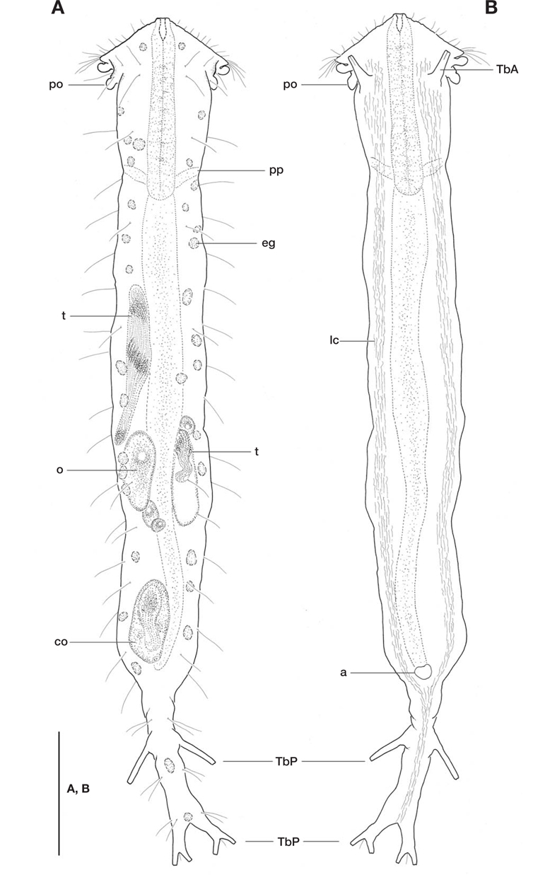
    Description of holotype. Body slender, Lt 345 μm; broadest at level of lateral lobes of head, and slightly constricted at pharyngeal pores; both lateral sides of trunk nearly parallel, before tapering into long pedunculate caudum; caudum narrow, well defined, 70 μm long (20U), about one-fifth of body length, bifurcated distally at U95. Widths of head/neck/PhJIn/ trunk/caudal base 52/30/33/38/15 μm at U05/U18/U21/ U44/U79 respectively.

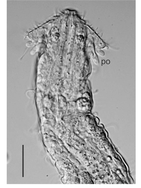
    Head triangular, with mouth slightly protruding anteriorly; armed with 8-9 cephalic sensory hairs along anterior margin. Lateral lobes small, triangular with a ciliary tuft apically, consisting of 4-5 cilia. Two pairs of pestle organs emerging between lateral lobes and TbA, each pair of pestle organs separate, not confluent at base; fore one issuing beneath lateral lobe at U06, rear one arising dorsally, split from basis of lateral lobe at U08. Sensory hairs, two per side implanted on dorsolateral side near rear pestle organ; about 9-10 dorsolateral hairs and 13-14 lateral hairs per side aligned, evenly spaced between anterior pharyngeal region and caudal peduncle (U06-U78); three paired hairs per side situated along lateral margin of caudal peduncle at regular intervals.

    Epidermal glands small, generally circular or ovoid, ~6 μm in diameter; foremost one located mid-dorsally near anterior pharyngeal region at U03; 15 glands per side, aligned asymmetrically along lateral side from anterior pharyngeal region to caudal base at U10-U77; two glands located on caudal peduncle, anterior one placed slightly behind TbP at U87, posterior one near bifurcated base of caudum at U94.

    Adhesive tubes: TbA, one per side, 13 μm long, with enlarged base, occurring slightly behind pestle organs, at U07. TbD, TbL and TbV absent. TbP, three per side, consisting of a large tube, 17 μm long, arising from anterior third of lateral margin of caudal peduncle at U85 and two short ones, 6 μm long, forming bifurcate caudum with a bristle between them.

    Ventral locomotor cilia showing paired longitudinal bands from front of TbA to caudal base at U05-U78, joining into a narrow column behind anus at U79, extending to bifurcated caudum at U95.

    Digestive tract: mouth terminal, small, 3 μm wide; buccal cavity small, U-shaped, somewhat cuticularized; pharynx 72 μm long with pharyngeal pores opened at its posterior end (U18); PhJIn at U21; intestine straight anteriorly, narrowing and slightly curved toward posterior end; anus opened at U76.

    Reproductive system: simultaneous hermaphrodite; paired testes asymmetrical, left testis longer than right one, in dorsal view; left testis reaching to anterior intestinal region at U31, and right one to mid-intestinal region at U48; one large ovum located at U48-U56 on left side in dorsal view, following two small oocytes; another small oocyte in front of right testis at U47; copulatory organ ellipsoidal, located at posterior trunk region.

    Remarks. In the genus Dendrodasys Wilke, 1954, five species are recognized: D. affinis Wilke, 1954, D. gracilis Wilke, 1954, D. ponticus Valkanov, 1957, D. pacificus Schmidt, 1974, and D. rubomarinus Hummon, 2011.

    The five congeneric species can be divided into two groups, depending on the presence or absence of pestle organs. With the pestle organs present, the new species is more closely related to D. affinis, D. gracilis, and D. rubomarinus, but it differs from them by bearing two pairs of pestle organs arising distally and dorso-distally posterior to cephalic lateral lobes while the other species have only a pair of pestle organs arising ventrolaterally. Moreover, D. duplus n. sp. is distinguished from D. gracilis due to the occurrence of large TbP, that is, larger TbP arising from the anterior third of the caudal peduncle in the new species instead of from the caudal base as in D. gracilis. Dendrodasys affinis has single testis on the right side in dorsal view, but the new species has a pair of testes with different lengths. Dendrodasys rubomarinus has a crescent-shaped anterior margin of the head, while the new species shows a triangular head.

    Aside from the absence of pestle organs, D. ponticus is different from D. duplus n. sp. due to the crescent-shaped anterior margin of head, and D. pacificus differs from the new species due to the absence of lateral lobes. Furthermore, both of them have a large TbP occurring at the base of caudal peduncle (instead of at the anterior third of the caudal peduncle in D. duplus n. sp.).

참고문헌
  • 1. Clausen C 2000 Gastrotricha Macrodasyida from the Tromsø region, northern Norway [Sarsia] Vol.85 P.357-384 google
  • 2. Hochberg R, Litvaitis MK 2001 Macrodasyida (Gastrotricha): a cladistic analysis of morphology [Invertebrate Biology] Vol.120 P.124-135 google
  • 3. Hummon WD, Parker SP 1982 Gastrotricha;Synopsis and classification of living organisms. Vol. 1 P.857-863 google
  • 4. Hummon WD 2009 Global database for marine Gastrotricha [Internet]. Taxonomic, Geographic, Bibliographic, and Video google
  • 5. Hummon WD 2011 Marine Gastrotricha of the Near East: 1. Fourteen new species of Macrodasyida and a redescription of Dactylopodola agadasys Hochberg, 2003 [Zookeys] Vol.94 P.1-59 google
  • 6. Hummon WD, Balsamo M, Todaro MA 1992 Italian marine Gastrotricha: I. Six new and one redescribed species of Chaetonotida [Bollettino di Zoologia] Vol.59 P.499-516 google
  • 7. Hummon WD, Todaro MA, Tongiorgi P, Balsamo M 1998 Italian marine Gastrotricha: V. Four new and one redescribed species of Macrodasyida in the Dactylopodolidae and Thaumastodermatidae [Italian Journal of Zoology] Vol.65 P.109-119 google
  • 8. Lee JM, Chang CY 2003 Two new marine gastrotrichs of the genus Ptychostomella (Macrodasyida, Thaumastodermatidae) from South Korea [Zoological Science] Vol.20 P.481-489 google
  • 9. Lee JM, Hwang UW, Chang CY 2009 A new gastrotrich spe- cies of the genus Ptychostomella (Macrodasyida: Thaumastodermatidae) from South Korea [Animal Cells and Systems] Vol.13 P.25-30 google
  • 10. Rudescu L 1967 Gastrotricha [Fauna Republicii Socialiste Romania] Vol.2 P.1-289 google
  • 11. Schmidt P 1974 Interstitielle Fauna von Galapagos. IV. Gastrotricha [Mikrofauna des Meeresbodens] Vol.26 P.1-76 google
  • 12. Todaro MA, Hummon WD 2008 An overview and a dichotomous key to genera of the phylum Gastrotricha [Meiofauna Marina] Vol.16 P.3-20 google
  • 13. Todaro MA, Rocha CEF 2004 Diversity and distribution of marine Gastrotricha along the northern beaches of the state of Sao Paulo (Brazil), with description of a new species of Macrodasys (Macrodasyida, Macrodasyidae) [Journal of Natural History] Vol.38 P.1605-1634 google
  • 14. Valbonesi A, Luporini P 1984 Researches on the coast of Somalia. Gastrotricha Macrodasyoidea [Monitore Zoologico Italiano Supplemento] Vol.1 P.1-34 google
  • 15. Valkanov A 1957 Erster versuch zur erforschung der Gastrotrichen des Schwarzen Meeres [Ann de L’Universite Sofia Faculte Biologie] Vol.50 P.383-399 google
  • 16. Wilke U 1954 Mediterrane Gastrotrichen [Zoologische Jahrbuecher Abteilung fuer Systematik] Vol.82 P.497-550 google
이미지 / 테이블
  • [ Fig. 1. ]  Dendrodasys duplus n. sp. A, Habitus, dorsal view; B, Habitus, ventral view. a, anus; co, copulatory organ; eg, epidermal gland; lc, locomotor cilia; o, ovum; po, pestle organ; pp, pharyngeal pore; t, testis; TbA, anterior adhesive tubes; TbP, posterior adhesive tubes. Scale bar=50 μm.
    Dendrodasys duplus n. sp. A, Habitus, dorsal view; B, Habitus, ventral view. a, anus; co, copulatory organ; eg, epidermal gland; lc, locomotor cilia; o, ovum; po, pestle organ; pp, pharyngeal pore; t, testis; TbA, anterior adhesive tubes; TbP, posterior adhesive tubes. Scale bar=50 μm.
  • [ Fig. 2. ]  Dendrodasys duplus n. sp., head and the anterior intestinal region, DIC micrographs. po, pestle organ. Scale bar=20 μm.
    Dendrodasys duplus n. sp., head and the anterior intestinal region, DIC micrographs. po, pestle organ. Scale bar=20 μm.
(우)06579 서울시 서초구 반포대로 201(반포동)
Tel. 02-537-6389 | Fax. 02-590-0571 | 문의 : oak2014@korea.kr
Copyright(c) National Library of Korea. All rights reserved.