검색 전체 메뉴
PDF
맨 위로
OA 학술지
Shengmaisan Regulates Pacemaker Potentials in Interstitial Cells of Cajal in Mice
  • 비영리 CC BY-NC
  • 비영리 CC BY-NC
ABSTRACT
Shengmaisan Regulates Pacemaker Potentials in Interstitial Cells of Cajal in Mice
KEYWORD
interstitial cells of Cajal , Shengmaisan , traditional Chinese medicine , gastrointestinal motility
  • 1. Introduction

    Traditional Chinese medicine (TCM) has become an increasingly popular alternative for the treatment of a variety of physiological disorders [1]. Shengmaisan (SMS), one of the most famous Chinese herbal formula, consists of Panax ginseng, Ophiopogon japonicas, and Schisandra chinensis, has been used for the treatment of cardiovascular and neurological diseases over a long period of time [2]. SMS prevents apoptosis through the enhancement of anti-oxidant enzymes and the inhibition of the c-jun amino-terminal kinase (JNK) signaling pathway with the promotion of nerve cell maturation. Therefore, SMS has benefits for treating of neurodegenerative diseases [3]. Also, SMS can protect against heat stroke-induced arterial hypotension and cerebral ischemia by the inhibition of inducible nitric oxide synthase (iNOS)-dependent NO overproduction in the brain and excessive accumulation of several inflammatory cytokines in the peripheral blood stream [4]. Therefore, SMS can protect against heat stroke by reducing the formations of inflammatory cytokines and nitric oxide. Moreover, in neurological disorders, SMS plays an active role in regenerative responses and facilitates axonal regrowth after spinal cord injury [4]. However, little is known about the effect of SMS on gastrointestinal (GI) motility regulation.

    Interstitial cells of Cajal (ICCs) play an important role in the generation of coordinated GI peristalsis [5-8]. The major function of the ICCs is the generation of slow waves that allow these cells to act as specialized pacemaker cells [6-8], and they have additional functions in the gastrointestinal tract as regulators of mechanical activity and neurotransmission [7-9]. Due to the central role of ICCs in GI motility, it has been suggested that defects of ICCs may contribute to motility dysfunction in several GI motility disorders [10]. Therefore, experimental observations of the ICCs should be a good tool to investigate the GI motility functions. However, the effects of SMS on small intestinal ICCs in a mouse have not yet been investigated. Therefore, we undertook this research to investigate the effects of SMS in mouse small intestinal ICCs.

    2. Materials and Methods

       2.1. Preparation of cells and cell cultures

    Balb/c mice (3-7 days old) of either sex were anesthetized with ether and sacrificed by cervical dislocation. All animal procedures were approved by the Institutional Animal Care and Use Committee of Pusan National University (Busan, South Korea) and carried out in accordance with the Guide for the Care and Use of Laboratory Animals of Pusan National University. All efforts were undertaken according to the “3R principles” of the directive to reduce the number of animals used in this study and to optimize the experimental protocols to acquire the maximum amount of data from each tested animal. The small intestines from 1 cm below the pyloric ring to the cecum were removed and opened along the mesenteric border. Luminal contents were removed by washing with Krebs-Ringer bicarbonate solution. The tissues were pinned to the base of a Sylgard dish and the mucosa were removed by sharp dissection. Small tissue strips of the intestine muscle (consisting of both circular and longitudinal muscles) were equilibrated in Ca2+-free Hanks solution (containing in mmol/L: KCl 5.36, NaCl 125, NaOH 0.34, Na2HCO3 0.44, glucose 10, sucrose 2.9, and HEPES 11) for 30 min. Then, the cells were dispersed using an enzyme solution containing collagenase (Worthington Biochemical Co., Lakewood, NJ, USA) 1.3 mg/mL, bovine serum albumin (Sigma Chemical Co., St. Louis, MO, USA) 2 mg/mL, trypsin inhibitor (Sigma) 2 mg/mL and ATP 0.27 mg/mL. Cells were plated onto sterile glass coverslips coated with murine collagen (2.5 μg/mL, Falcon/BD, Franklin Lakes, NJ,USA) in a 35-mm culture dish and then cultured at 37 °C in a 95% O2, 50 mL/L CO2 incubator in a smooth muscle growth medium (Clonetics Corp., San Diego, CA, USA) supplemented with 2% antibiotics/antimycotics (Gibco,Grand Island, NY, USA) and murine stem cell factor (SCF, 5 ng/mL, Sigma). ICCs were identified immunologically with anti-c-kit antibody (phycoerythrin-conjugated rat anti-mouse c-kit monoclonal antibody; eBioscience, San Diego,CA, USA) at a dilution of 1:50 for 20 min. ICCs were morphologically distinct from other cell types in the culture; thus, it was possible to identify the cells by using phase contrast microscopy once they had been verified with the anti c-kit antibody.

       2.2. Patch-clamp experiments

    The whole-cell patch-clamp configuration was used to record membrane potentials (current clamp) from cultured ICCs. An axopatch ID (Axon Instruments, Foster, CA, USA) was used to amplify membrane currents and potentials. The command pulse was applied using an IBM-compatible personal computer and pClamp software (version 6.1; Axon Instruments). Data obtained were filtered at 5 kHz and displayed on an oscilloscope, a computer monitor, and a pen recorder (Gould 2200, Gould, Valley View, OH, USA). Results were analyzed using pClamp and Origin (version 6.0) software. All experiments were performed at 30-32℃.

       2.3. Solutions and drugs

    The physiological salt solution used to bathe cells (Na+-Tyrode) contained (mmol/L): KCl 5, NaCl 135, CaCl2 2, glucose 10, MgCl2 1.2 and 4-(2-hydroxyethyl)-1-piperazineethanesulfonic acid (HEPES) 10, adjusted to pH 7.4 with NaOH. The pipette solution contained (mmol/L): KCl 140, MgCl2 5, K2 Adenosine triphosphate (ATP) 2.7, Na Guanosine triphosphate (GTP) 0.1, creatine phosphate disodium 2.5, HEPES 5 and ethylene glycol tetraacetic acid (EGTA) 0.1, adjusted to pH 7.2 with KOH.

    SMS was purchased from I-WORLD Pharmaceuticals (South Korea). The dosage for an adult human is 10-15 g (crude materials) per day. More information about SMS can be found on I-WORLD Pharmaceuticals Homepage (http://i-pharm.koreasme.com). The SMS was dissolved in distilled water at a concentration of 0.5 g (crude drug)/ml and stored in a refrigerator. SMS is a powder preparation and contains lactose and corn starch as an excipient. The lactose and corn starch can affect the ICCs and the GI motility. However, because SMS containing lactose and corn starch is eaten, we think that any investigation of its effects should use SMS containing that excipient.

    All other drugs were obtained from Sigma (Sigma Chemical Co., USA). The drugs were dissolved in distilled water, and just prior to use were added to the bath solution to make the desired concentrations. Addition of these chemicals to the bath solution did not alter the pH of the solution. Thapsigargin, U-73122, U-73343, chelerythrine and calphostin C were dissolved in dimethyl sulfoxide (DMSO) for the 50 or 100 mmol/L stock solution and were added (1000 times dilution) to the bathing solution on the day of the experiment. The final concentration of DMSO in the bath solution was always < 0.1%, and we confirmed that this concentration of DMSO did not affect the results that were recorded.

       2.4. Statistics

    All data are expressed as means ± S.E.’s. The student’s  t-test for unpaired data was used to compare the control and the experimental groups. A P value of less than 0.05 was considered statistically significant.

    3. Results

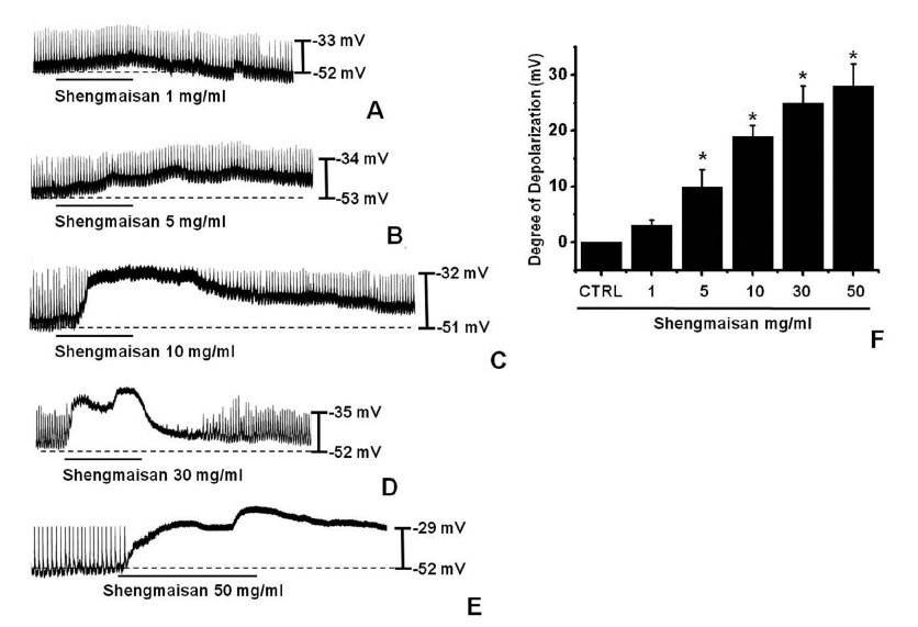
    3.1. Effect of SMS on pacemaker potentials in cultured ICCs

    The patch-clamp technique was used to investigate the ICCs that formed network-like structures in culture (2-4 days). Spontaneous rhythms were routinely recorded from cultured ICCs under current and voltage-clamp conditions, and the ICCs within the networks displayed more robust electrical rhythms. Tissue-like spontaneous slow waves have been recorded from these cells [11]. To understand the relationship between SMS and the modulation of pacemaker activity in ICCs, we examined the effects of SMS on pacemaker potentials. Recordings from cultured ICCs under current clamp conditions (I = 0) showed spontaneous pacemaker potentials. The resting membrane potential was -52.1 ± 2.1 mV and the amplitude was 20 ±  4 mV. In the presence of SMS (1 - 50 mg/ml), the membrane potentials were depolarized to 3.0 ± 1.1 mV at 1 mg/ ml, 11.2 ± 3.2 mV at 5 mg/ml, 19.4 ± 2.4 mV at 10 mg/ml, 25.2 ± 3.2 mV at 30 mg/ml and 28.1 ± 4.1 mV at 50 mg/ml (Fig 1)A-1E). The summarized values and the bar graph of SMS’s effects on pacemaker potentials are indicated in Fig. 1F (n = 4).

       3.2. Effects of an external Ca2+-free solution and a Ca2+-ATPase inhibitor of the endoplasmic reticulum on SMS-induced pacemaker potentials in cultured ICCs

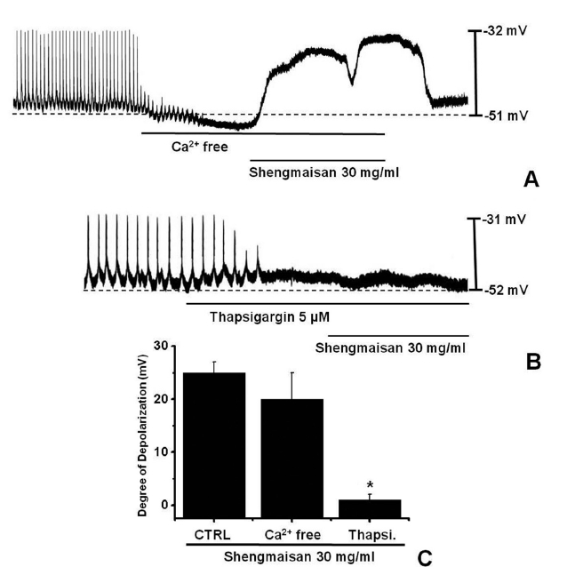
    The external Ca2+ influx is necessary for GI smooth muscle contractions and is essential for generating pacemaker potentials in the ICCs. The generation of pacemaker currents has been shown to depend on the intracellular Ca2+ oscillation [12]. To investigate the role of external Ca2+ or internal Ca2+, we tested SMS under external Ca2+-free conditions and in the presence of thapsigargin, a Ca2+-ATPase inhibitor of the endoplasmic reticulum [11, 13]. The pacemaker potentials completely disappeared when an external Ca2+-free solution was used. In this condition, SMS induced membrane depolarizations (n = 5; (Fig 2)A). Under external Ca2+-free conditions, the membrane depolarizations with SMS (30 mg/ml) were not significantly different from those with SMS (30 mg/ml) in normal Ca2+ solution (n = 5, (Fig 2)C). In addition, SMS-induced membrane depolarizations were inhibited by pretreatment with thapsigargin (n = 4, (Fig 2)B). In the presence of thapsigargin (5 μM), the membrane depolarizations with SMS were significantly different from those with SMS in the absence of thapsigargin (n = 4, (Fig 2)C).

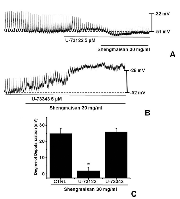
       3.3. Effects of a phospholipase C inhibitor on SMS-induced pacemaker potentials in cultured ICCs

    Because the membrane depolarizations cause by SMS were related to intracellular Ca2+ mobilization, we examined whether its effects on pacemaker potentials required phospholipase C (PLC) activation. To test this possibility, we measured SMS-induced membrane depolarizations in the absence and the presence of U-73122, an active PLC inhibitor [14]. The pacemaker membrane depolarizations completely disappeared when U-73122 (5 μM) was used. Under these conditions, SMS (30 mg/ml)-induced membrane depolarizations were not produced (n = 4; (Fig 3)A). In the presence of U-73122, the membrane depolarization produced by SMS was 2.3 ± 2.1 mV. The value of membrane depolarization caused by SMS in the presence of U-73122 was significantly different from that caused by SMS in the absence of U-73122 (n = 4, (Fig 3)C). The treatment with U-73343 (5 μM), an inactive analog of U-73122, had no influence on the SMS-induced pacemaker potentials. Under these conditions, SMS (30 mg/ml)-induced membrane depolarizations were not suppressed by using U-73343 (n = 4, (Fig 3)B).

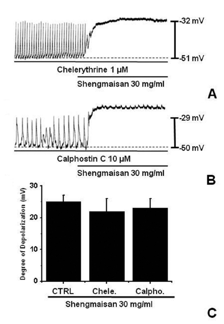
       3.4. Effect of protein kinase C inhibitor on SMS-induced pacemaker potentials in cultured ICCs

    We tested the effects of chelerythrine or calphostin C, an inhibitor of protein kinase C (PKC) [13, 15], to investigate whether the SMS-induced responses of the pacemaker potentials were mediated by the activation of PKC. Chelerythrine (1 μM) or calphostin C (10 μM), did not have any effect on the membrane depolarizations caused by SMS (30 mg/ml; (Fig 4) and the value was not significantly different from the membrane depolarization caused by SMS in the absence of chelerythrine or calphostin C (n = 4; (Fig 4)C).

    4. Discussion

    SMS is a traditional Chinese herbal medicine that has long been used for the treatment of heart failure and ischemic heart disease. It is a mixture of three herbal components. Constituent herbs of SMS include Panax ginseng, Ophiopogon japonicus and Schisandra chinensis. Experimental data have demonstrated that SMS has protective effects for the prevention of tissue injury, especially in the heart and brain, caused by oxidative stress [16-19]. SMS is useful for preventing the progression of injury in cerebral infarction after stroke [16, 17], and has a function not only as a simple antioxidant but also as a modulator of cellular antioxidant defense [18]. Also, SMS reduces adriamycin-induced cardiomyopathy in rats [19]. SMS has a function to reduce coronary arteriosclerosis [20] and to prevent the damage of myocardia and might be taken as effective therapeutic way to treat of acute viral myocarditis [21]. Although, the beneficial effects of SMS have been elucidated by antioxidant and heart and neuron diseases, the effects of SMS on GI motility and on ICCs have not been investigated yet.

    ICCs are the pacemaking cells in the GI muscles that generate the rhythmic oscillations in the membrane potential known as slow waves [5-8]. c-kit is a transmembrane protein kinase receptor that is essential for the development and function of ICCs. In the gut, c-kit is expressed only in ICCs and mast cells [5, 8]. ICCs are distributed throughout the digestive tract from the upper oesophageal sphincter to the internal sphincter of the anus [22-24]. ICCs contribute to important functions in the digestive tract due to their generation, coordination and propagation of electrical slow wave activity, their transduction of motor neural inputs from the enteric nervous system and their role in the mechanosensation of smooth muscle cells [8].

    In this study, SMS produced membrane depolarization in ICCs. Pretreatment with a Ca2+-free solution and a thapsigargin, a Ca2+-ATPase inhibitor in the endoplasmic reticulum, stopped the generation of pacemaker potentials. Under Ca2+-free solution conditions, SMS showed membrane depolarizations. However, under thapsigargin solution conditions, SMS had no effects on membrane depolarizations. Also, the pacemaker membrane depolarizations were inhibited by U-73122, an active PLC inhibitor. Furthermore, the PKC inhibitors, chelerythrine and calphostin C, did not block the SMS-induced membrane depolarizations. These results suggest that SMS might affect GI motility by the modulation of pacemaker activity in the ICCs, and that the activation takes place in an internal Ca2+ and PLC-dependent and an external Ca2+ and PKC-independent manners.

    5. Conclusion

    Our data suggest that the gastroprokinetic effects of SMS might be mediated by the induction of pacemaker potentials in the ICCs. When the effects of this drug on the ICCs are considered, further research, including finding active compound(s) and examining their action mechanisms, is clearly needed.

참고문헌
  • 1. Cheng JT 2000 Review: drug therapy in Chinese traditional medicine [J Clin Pharmacol] Vol.40 P.445-450 google cross ref
  • 2. Xu N, Qiu C, Wang W, Wang Y, Chai C, Yan Y 2011 HPLC/MS/MS for quantification of two types of neurotransmitters in rat brain and application: myocardial ischemia and protection of Sheng-Mai-San [J Pharm Biomed Anal] Vol.55 P.101-108 google cross ref
  • 3. Nishida H, Kushida M, Nakajima Y, Ogawa Y, Tatewaki N, Sato S 2007 Amyloid-β-induced cytotoxicity of PC- 12 cell was attenuated by Shengmai-san through redox regulation and outgrowth induction [J Pharmacol Sci] Vol.104 P.73-81 google cross ref
  • 4. Wang NL, Chang CK, Liou YL, Lin CL, Lin MT 2005 Shengmai San, a Chinese herbal medicine protects against rat heat stroke by reducing inflammatory cytokines and nitric oxide formation [J Pharmacol Sci] Vol.98 P.1-7 google cross ref
  • 5. Ward SM, Burns AJ, Torihashi S, Sanders KM 1994 Mutation of the proto-oncogene c-kit blocks development of interstitial cells and electrical rhythmicity in murine intestine [J Physiol] Vol.480 P.91-97 google
  • 6. Huizinga JD, Thuneberg L, Kluppel M, Malysz J, Mikkelsen HB, Bernstein A 1995 W/kit gene required for interstitial cells of Cajal and for intestinal pacemaker activity [Nature] Vol.373 P.347-349 google
  • 7. Sanders KM 1996 A case for interstitial cells of Cajal as pacemakers and mediators of neurotransmission in the gastrointestinal tract [Gastroenterology] Vol.111 P.492-515 google cross ref
  • 8. Gfroerer S, Rolle U 2013 Interstitial cells of Cajal in the normal human gut and in Hirschsprung disease [Pediatr Surg Int] Vol.29 P.889-897 google cross ref
  • 9. Won KJ, Sanders KM, Ward SM 2005 Interstitial cells of Cajal mediate mechanosensitive responses in the stomach [Proc Natl Acad Sci U S A] Vol.102 P.14913-14918 google cross ref
  • 10. Kim BJ, Lim HH, Yang DK, Jun JY, Chang IY, Park CS 2005 Melastatin-type transient receptor potential channel 7 is required for intestinal pacemaking activity [Gastroenterology] Vol.129 P.1504-1517 google cross ref
  • 11. Koh SD, Sanders KM, Ward SM 1998 Spontaneous electrical rhythmicity in cultured interstitial cells of Cajal from the murine small intestine [J Physiol] Vol.513 P.203-213 google cross ref
  • 12. Ward SM, Ordog T, Koh SD, Baker SA, Jun JY, Amberg G 2000 Pacemaking in interstitial cells of Cajal depends upon calcium handling by endoplasmic reticulum and mitochondria [J Physiol] Vol.525 P.355-361 google cross ref
  • 13. Choi S, Choi JJ, Jun JY, Koh JW, Kim SH, Kim DH 2009 Induction of pacemaker currents by DA-9701, a prokinetic agent, in interstitial cells of Cajal from murine small intestine [Mol Cells] Vol.27 P.307-312 google cross ref
  • 14. Sakamoto T, Unno T, Matsuyama H, Uchiyama M, Hattori M, Nishimura M 2006 Characterization of muscarinic receptor-mediated cationic currents in longitudinal smooth muscle cells of mouse small intestine [J Pharmacol Sci] Vol.100 P.215-226 google cross ref
  • 15. Aiello EA, Clement-Chomienne O, Sontag DP, Walsh MP, Cole WC 1996 Protein kinase C inhibits delayed rectifier K+ current in rabbit vascular smooth muscle cells [Am J Physiol] Vol.271 P.H109-19 google
  • 16. Ichikawa H, Wang L, Konishi T 2006 Prevention of cerebral oxidative injury by post-ischemic intravenous administration of Shengmai San [Am J Chin Med] Vol.34 P.591-600 google cross ref
  • 17. Ichikawa H, Wang X, Konishi T 2003 Role of component herbs in antioxidant activity of shengmai San-a traditional Chinese medicine formula preventing cerebral oxidative damage in rat [Am J Chin Med] Vol.31 P.509-521 google cross ref
  • 18. Wang L, Nishida H, Ogawa Y, Konishi T 2003 Prevention of oxidative injury in PC12 cells by a traditional Chinese medicine, Shengmai San, as a model of an antioxidant- based composite formula [Biol Pharm Bull] Vol.26 P.1000-1004 google cross ref
  • 19. You JS, Huang HF, Chang YL, Lee YS 2006 Sheng-Mai-San reduces adriamycin-induced cardiomyopathy in rats [Am J Chin Med] Vol.34 P.295-305 google cross ref
  • 20. Fang J, Jiang J, Luo DC 1987 Effect of Shengmai San oral fluid on coronary arteriosclerotic [Chin Med J] Vol.26 P.403-409 google
  • 21. Zhao MH, Rong YZ, Lu BJ 1996 Effect of Shengmai San on serum lipid peroxidation in acute viral myocarditis [Zhongguo Zhong Xi Yi Jie He Za Zhi] Vol.16 P.142-145 google
  • 22. Hagger R, Gharaie S, Finlayson C, Kumar D 1998 Distribution of the interstitial cells of Cajal in the human anorectum [J Auton Nerv Syst] Vol.73 P.75-79 google cross ref
  • 23. Faussone-Pellegrini MS, Cortesini C 1985 Ultrastructural features and localization of the interstitial cells of Cajal in the smooth muscle coat of human esophagus [J Submicrosc Cytol] Vol.17 P.187-197 google
  • 24. Torihashi S, Horisawa M, Watanabe Y 1999 C-kit immunoreactive interstitial cells in the human gastrointestinal tract [J Auton Nerv Syst] Vol.75 P.38-50 google cross ref
이미지 / 테이블
  • [ Fig. 1 ]  Effects of SMS on pacemaker potentials in cultured ICCs from murine small intestine. A-E: After the primary ICCs culture in the mouse small intestine, various dose of SMS (1 to 50 mg/mL) were applied in a bath and the membrane potentials in current clamp mode (I = 0) were estimated. Responses to SMS are summarized in F. Bars represent mean values ± S.E. *P < 0.01, indicating a significant difference from the untreated control. CTRL: Control.
    Effects of SMS on pacemaker potentials in cultured ICCs from murine small intestine. A-E: After the primary ICCs culture in
the mouse small intestine, various dose of SMS (1 to 50 mg/mL) were applied in a bath and the membrane potentials in current clamp
mode (I = 0) were estimated. Responses to SMS are summarized in F. Bars represent mean values ± S.E. *P < 0.01, indicating a significant
difference from the untreated control. CTRL: Control.
  • [ Fig. 2 ]  Effects of an external Ca2+-free solution or thapsigargin, a Ca2+-ATPase inhibitor of endoplasmic reticulum on SMS-induced pacemaker potentials in cultured ICCs. A: The external Ca2+-free solution stopped the generation of pacemaker potentials. Under these conditions, SMS (30 mg/ml)-induced membrane depolarizations were produced. B: Thapsigargin (5 μM) stopped the generation of pacemaker potentials and thapsigargin blocked the SMS (30 mg/ml)-induced membrane depolarizations. C: The responses to SMS in the external Ca2+-free solution and in the presence of thapsigargin are summarized. Bars represent the mean values ± S.E. *P < 0.01, indicating a significant difference from the untreated control. CTRL: Control.
    Effects of an external Ca2+-free solution or thapsigargin, a Ca2+-ATPase inhibitor of endoplasmic reticulum on SMS-induced
pacemaker potentials in cultured ICCs. A: The external Ca2+-free solution stopped the generation of pacemaker potentials. Under these
conditions, SMS (30 mg/ml)-induced membrane depolarizations were produced. B: Thapsigargin (5 μM) stopped the generation of
pacemaker potentials and thapsigargin blocked the SMS (30 mg/ml)-induced membrane depolarizations. C: The responses to SMS
in the external Ca2+-free solution and in the presence of thapsigargin are summarized. Bars represent the mean values ± S.E. *P < 0.01,
indicating a significant difference from the untreated control. CTRL: Control.
  • [ Fig. 3 ]  Effects of SMS on PLC inhibitors in cultured ICCs. A: U-73122 (5 μM), a PLC inhibitor, stopped the generation of pacemaker potentials. U-73122 blocked the SMS (30 mg/ml)-induced membrane depolarizations. B: The application of U-73343 (5 μM), a inactive PLC inhibitor did not show any influence on the generation of pacemaker currents. Also, U-73343 did not block the SMS-induced (30 mg/ml) membrane depolarizations. C: The responses to SMS in PLC inhibitors are summarized. Bars represent the mean values ± S.E. *P < 0.01, indicating a significant difference from the untreated control. CTRL: Control.
    Effects of SMS on PLC inhibitors in cultured ICCs. A: U-73122 (5 μM), a PLC inhibitor, stopped the generation of pacemaker potentials.
U-73122 blocked the SMS (30 mg/ml)-induced membrane depolarizations. B: The application of U-73343 (5 μM), a inactive PLC inhibitor did
not show any influence on the generation of pacemaker currents. Also, U-73343 did not block the SMS-induced (30 mg/ml) membrane depolarizations.
C: The responses to SMS in PLC inhibitors are summarized. Bars represent the mean values ± S.E. *P < 0.01, indicating a significant
difference from the untreated control. CTRL: Control.
  • [ Fig. 4 ]  Effects of chelerythrine or calphostin C, an inhibitor of PKC, upon SMS-induced pacemaker potentials in cultured ICCs of the murine small intestine. A, B: Chelerythrine (1 μM) or calphostin C (10 μM), did not have an effect on membrane depolarizations by SMS (30 mg/ml). C: Responses to PWS in the presence of chelerythrine or calphostin C are summarized. The value was not significant different when compared with the membrane depolarizations caused by SMS obtained in the absence of chelerythrine or calphostin C. Bars represent the mean values ± S.E. CTRL: Control.
    Effects of chelerythrine or calphostin C, an inhibitor of PKC,
upon SMS-induced pacemaker potentials in cultured ICCs of the murine
small intestine. A, B: Chelerythrine (1 μM) or calphostin C (10 
μM), did not have an effect on membrane depolarizations by SMS
(30 mg/ml). C: Responses to PWS in the presence of chelerythrine or
calphostin C are summarized. The value was not significant different
when compared with the membrane depolarizations caused by SMS
obtained in the absence of chelerythrine or calphostin C. Bars represent
the mean values ± S.E. CTRL: Control.
(우)06579 서울시 서초구 반포대로 201(반포동)
Tel. 02-537-6389 | Fax. 02-590-0571 | 문의 : oak2014@korea.kr
Copyright(c) National Library of Korea. All rights reserved.