Dioscin, isolated from
Due to its selectivity, sensitivity, robustness, and sample throughput, liquid chromatography/tandem mass spectrometry (LC-MS/MS) is recognized as a powerful tool for use in quantification of active compounds of herbal drugs in biological samples.11-14 There were few reports on bioanalytical methods for the determination of dioscin in rat biological fluids using LC-MS/MS method with protein precipitation method as sample clean-up procedure. 15-17 Sample preparation was important to rem-ove the endogenous interferences from herbal drug and plasma.
The purpose of this paper was to develop a rapid, selective, and sensitive LC-MS/MS assay using liquidliquid extraction as sample preparation procedure for quantification of dioscin in rat plasma. This method was applied successfully to the pharmacokinetic study of dioscin after oral administration of dioscin in male Sprague-Dawley rats.
Dioscin (purity, 92%) and chlorzoxazone (purity, 99.0%; internal standard) were obtained from PhytoLab GmbH & Co. KG (Vestenbergsgreuth, Germany) and Sigma-Aldrich Chemical (St. Louis, MO, USA), respectively. Acetonitrile and ethyl acetate (MS grade) were obtained from Fisher Scientific Co. (Pittsburgh, PA, USA). Other chemicals used were of the highest quality available. Drug-free rat plasma containing sodium heparin as the anticoagulant was obtained from male Sprague-Dawley rats.
>
Preparation of calibration standards and quality control samples
Primary stock solutions of dioscin and chlorzoxazone (1 mg/mL) were prepared in dimethylsulfoxide. Working standard solutions of dioscin were prepared by dilution of primary stock solution with acetonitrile. The internal standard working solution (500 ng/mL chlorzoxazone) was prepared by dilution of an aliquot of stock solution with acetonitrile. All standard solutions were stored at 4℃ for 4 weeks in 1.5 mL polypropylene tubes in the dark when not in use. Rat plasma calibration standards of dioscin, i.e., 1, 2, 4, 20, 40, 80, and 100 ng/mL, were prepared by addition of 2.5 μL of the working standard solutions (0.02, 0.04, 0.08, 0.4, 0.8, 1.6, and 2 μg/mL) to 50 μL of drug-free rat plasma. Quality control (QC) samples at 1, 3.5, 35, and 75 ng/mL were prepared in bulk by addition of 25 μL of the appropriate working standard solutions (0.2, 0.7, 7, and 15 μg/mL) to drug-free rat plasma (4950 μL). Bulk samples were aliquoted (50 μL) into polypropylene tubes and stored at ?80℃ until analysis.
Fifty μL of rat blank plasma, calibration standards, and QC samples were mixed with 200 μL of 50 mM ammonium formate buffer (pH 3.0), 5 μL of chlorzoxazone in acetonitrile (I.S., 500 ng/mL), and 1000 μL of ethyl acetate by using vortex mixer for 2 min. After centrifugation at 13000 rpm for 5 min, the supernatants (900 μL) were transferred in new polypropylene tubes. The aqueous layers were extracted once more with 1000 μL of ethyl acetate and the supernatants (900 μL) were transferred after centrifugation. The organic layer was evaporated to dryness at 35℃ for 30 min by using vacuum evaporator. The residues were dissolved in 60 μL of 55% acetonitrile and were sonicated for 2 min. After centrifugation, the aliquot (2 μL) was injected on UPLC and analyzed by LC-MS/MS system.
The LC-MS/MS system consisted of an Agilent 1200 series (Agilent Technologies, Wilmington, DE, USA) and a 6490 triple quadrupole mass spectrometer (Agilent Technologies). Separation was performed on a Halo C18 (2.7 μm, 2.1 mm i.d. x 50 mm, Advanced materials technology, Wilmington, DE, USA) using gradient elution of 5% acetonitrile 0.1% formic acid (mobile phase A) and 95% acetonitrile in 0.1% formic acid (mobile phase B) at a flow rate of 0.40 mL/min: 10% mobile phase B for 0.5 min, 10% to 80% mobile phase B for 1.5 min, 80% mobile phase B for 1.0 min, 80% to 10% mobile phase B for 0.1 min, 10% mobile phase B for 1.9 min. The column and autosampler were maintained at 40℃ and 10℃, respectively. ESI source settings for ionization of dioscin and chlorzoxazone in the negative mode were as follows: capillary voltage, ?3000 V; gas temperature, 200℃; heater temperature, 360℃; sheath gas flow, 11 L/min. Fragmentation of mole-cular ion for dioscin and chlorzoxazone was performed at a collision energy of 25 eV and 17 eV, respectively, using nitrogen gas as a collision gas at a pressure of 2 bar on the instrument. Selected reaction monitoring (SRM) mode was employed for the quantification:
In order to complete the method validation, batches, consisting of triplicate calibration standards at each concentration, were analyzed on three different days. In each batch, QC samples at 1, 3.5, 35, and 75 ng/mL were assayed in sets of five replicates for evaluation of intra- and inter-day precision and accuracy. The relative error (RE), percentage deviation of the mean from true values, serves as a measure of the accuracy and the coefficient of variation (CV) indicates the precision.
Recovery of dioscin and chlorzoxazone was determined by comparison of mean peak areas of the analytes spiked before extraction into plasma sources with those of the analytes spiked post-extraction into blank plasma extracts at three concentrations, i.e., 3.5, 35, and 75 ng/mL.
For evaluation of room temperature storage and long-term storage at ?80℃ stability, triplicates of QC samples at low and high concentrations (3.5 and 75 ng/mL) were subjected to storage at room temperature for 4 h or at ?80℃ for 28 days before processing. Post-extraction batch integrity was determined by batch reinjection after 12 h storage at 10℃ in the autosampler.
>
Pharmacokinetic study of dioscin in rats
Sprague-Dawley rats (body weight 235-255 g, Samtako Co. Osan, Korea) were housed in a temperature controlled room at 22-24℃ with a 12 h light/dark cycle and relative humidity of 55 ± 10%. Rats were cannulated with polyethylene tubing (PE-50, Clay Adams, Parsippany, NJ, USA) in jugular vein under anesthesia with intraperitoneal injection of zoletil (50 mg/kg). Each rat was housed individually in a rat metabolic cage and allowed to recover from anesthesia for 2 day. The rats were not restrained at any time during the study. Heparinized isotonic saline (10 U/mL) was used to flush the catheters to prevent blood clotting. The rats were fasted for more than 12 h before oral administration of drugs.
Dioscin was dissolved in a mixture of dimethylsulfoxide: propylene glycol: deionzied water (1:7:2,
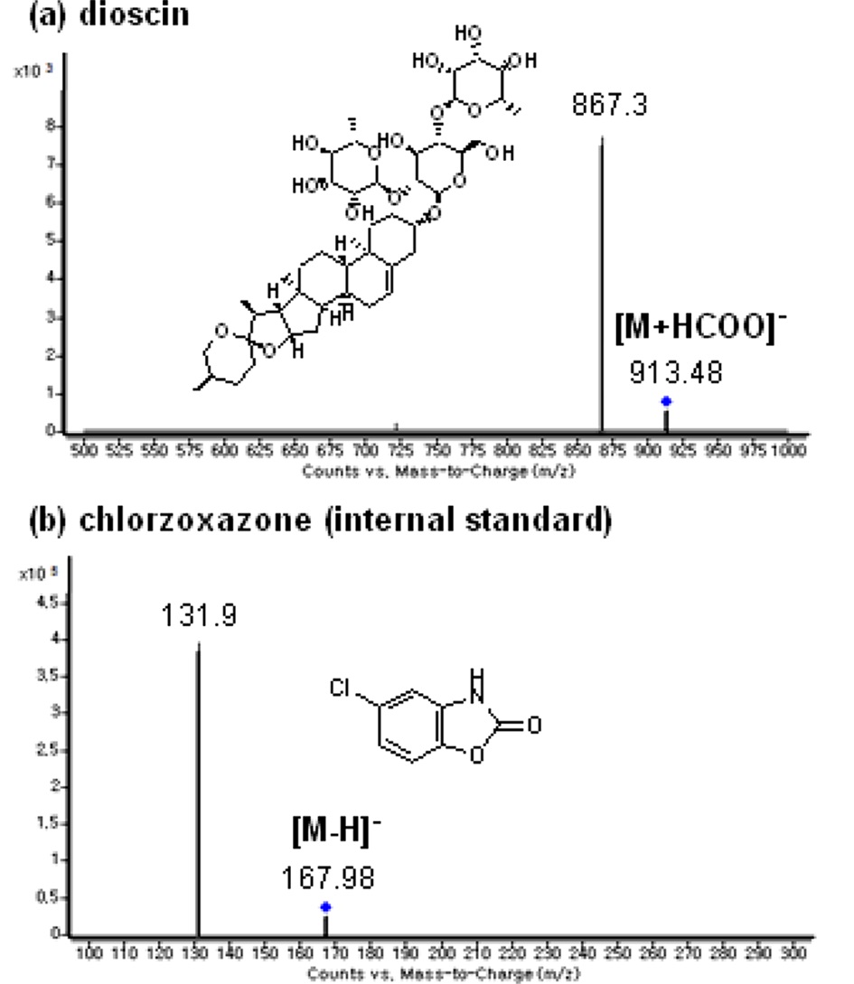
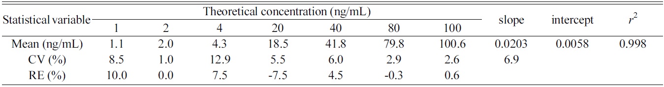
Dioscin and chlorzoxazone (internal standard) produced the molecular ions at
After many trials with different columns and mobile phase combinations, dioscin was well separated from the interferences on Halo C18 column compared to RP amide, Cadenza C18, and Unison UKC8 column. Gradient elution of acetonitrile and 0.1% formic acid as mobile phase resulted in better sensitivity compared to the use of other buffers and methanol.
In the analysis of blank plasma samples obtained from fifteen different rats, no interference peak was observed at the retention times of dioscin (3.2 min) and chlorzoxazone (2.5 min), confirming the selectivity of the present method (Figure 2a). Sample carryover effect was not observed.
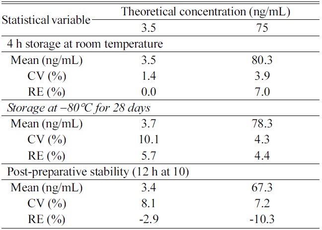
Calibration curves were obtained over the concentration range of 1 to 100 ng/mL of dioscin in rat plasma. Linear regression analysis with a weighting of 1/concentration gave

Calculated concentrations of dioscin in calibration standards prepared in rat plasma (n = 3)
the optimum accuracy (RE, ?7.5 to 10.0%) and precision (CV, ≤12.9%) of the corresponding calculated concentrations at each level (Table 1). The low CV value (6.9%) for the slope indicated the repeatability of the method (Table 1).
Table 2 shows a summary of intra- and inter-day precision and accuracy data for QC samples containing dioscin. Both intra-and inter-day CV values ranged from 1.3 to 8.0% at four QC levels. Intra- and inter-assay RE values were ?5.4 to 10.0% at four QC levels. These results indicated acceptable accuracy and precision of the present method. The lower limit of quantification (LLOQ) for dioscin was set at 1 ng/mL using 50 μL of rat plasma with a signal-to-noise ratio higher than 10 (Figure 2b).
Liquid-liquid extraction using ethyl acetate at acidic pH showed the acceptable recovery: 74.4 (±8.7)% at 3.5 ng/mL, 72.5 (±12.6)% at 35 ng/mL, 76.7 (±9.7)% at 75 ng/mL for dioscin, and 91.0 (±5.4)% for chlorzoxazone.
Dioscin was found to be stable in rat plasma for 4 h storage at room temperature and long-term (28 days) storage at ?80℃ (Table 3). Analysis of the reconstituted extracts stored for 12 h at 10℃ showed acceptable accuracy and precision for QC samples of dioscin (Table 3).
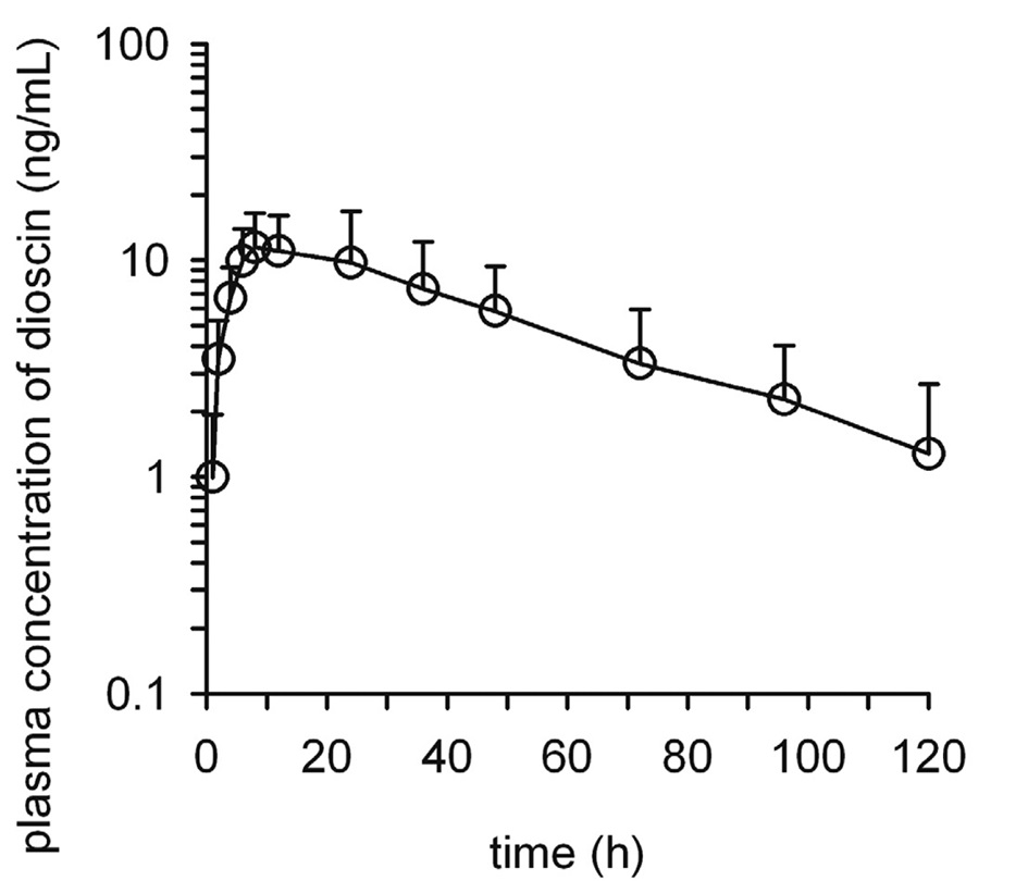
This method has been applied successfully to a pharmacokinetic study of dioscin after oral administration of dioscin at a dose of 29.2 mg/kg in seven male Sprague Dawley rats. Figure 2c shows representative SRM chrom-atograms obtained from analysis of a plasma sample obtained at 2 h after oral administration
[Table 2.] Precision and accuracy of dioscin in quality control samples
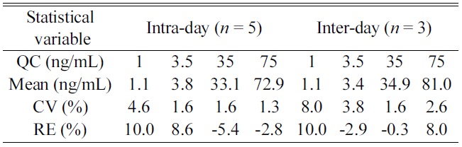
Precision and accuracy of dioscin in quality control samples
[Table 3.] Stability of QC samples (n = 3)

Stability of QC samples (n = 3)
of dioscin in a rat. The mean plasma concentrationtime plot after oral administration of dioscin in rats is shown in Figure 3. Li
A reliable, selective, and sensitive LC-MS/MS method for quantification of dioscin in rat plasma has been successfully developed. For sample preparation, dioscin and chlorzoxazone were extracted twice from plasma samples using ethyl acetate at acidic pH. Use of the present method demonstrated acceptable selectivity, sensitivity (LLOQ, 1 ng/mL), precision, accuracy, and stability. This method was applied successfully to determination of dioscin in rat plasma samples obtained after oral administration of dioscin at a dose of 29.2mg/kg in rats.