검색 전체 메뉴
PDF
맨 위로
OA 학술지
New Record of Juvenile Sigmops gracilis (Pisces: Gonostomatidae) from Jeju Island, Korea, Revealed by DNA Barcoding
  • 비영리 CC BY-NC
  • 비영리 CC BY-NC
ABSTRACT
New Record of Juvenile Sigmops gracilis (Pisces: Gonostomatidae) from Jeju Island, Korea, Revealed by DNA Barcoding
KEYWORD
Sigmops gracilis , Gonostomatidae , Pisces , DNA barcoding , Korea , First report
  • Introduction

    Worldwide, the family Gonostomatidae (order Stomiiformes) consist of 23 species in 5 genera (Banapartia, Cyclothone, Gonostoma, Margrethia, and Sigmops). These genera are distributed in the Indian, Atlantic, and Pacific oceans (Nelson, 2006), and they comprise most abundant fish group in the mesopelagic and bathypelagic zones (Nakabo, 2002). In particular, the genus Cyclothone has the greatest abundance of individuals (Froese and Pauly, 2012). Based on difficulties in the taxonomy of Gonostoma species, including non-monophyly, the genus was divided into two genera (Gonostoma and Sigmops) based on molecular phylogeny (Miya and Nishida, 2000). At present, four Sigmops species (S. bathyphilus, S. ebelingi, S. gracilis, and S. longipinnis) are considered valid (Froese and Pauly, 2012). No members of this family have previously been reported from Korean waters, possibly because of their very deep habitat. However, in this study, we report the first occurrence of a juvenile of a Gonostomatidae species in Korea, based on DNA barcoding. We describe its morphological and molecular characteristics and provide a new Korean name for the species.

    Materials and Methods

      >  Sample collection

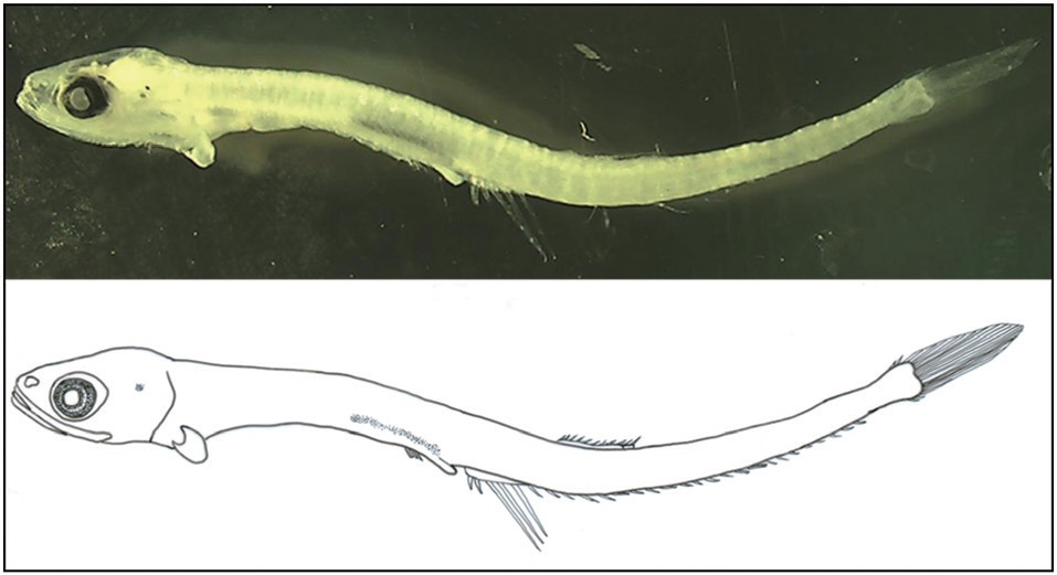
    In February 2012, a juvenile of a Gonostomatidae species was collected at a depth of 100 m at Jeju Island, Korea (32.54°N, 126.05°E) in a RN80 net (mouth opening 0.8 m, mesh size 0.33 mm). The specimen was fixed in 99% EtOH and later deposited at the National Institute of Biological Resources (NIBR-P 0000018076), Korea.

      >  DNA extraction, PCR, and sequencing

    Total DNA was extracted from the eyeball of the specimen using 10% Chelex 100 resin. Polymerase chain reaction (PCR) was used to amplify the mitochondrial DNA cytochrome oxidase subunit I (mtDNA COI) gene using the previously reported PCR primers VF2 (5′-TCA ACC AAC CAC AAA GAC ATT GGC AC-3′) and FishR1 (5′-TAC ACT TCT GGG TGG CCA AAG AAT CA-3′) (Ivanova et al., 2007). The

    PCR conditions were as follows: initial denaturation at 94℃ for 2 min, 35 cycles of denaturation at 94℃ for 30 s, annealing at 50℃ for 40 s, extension at 72℃ for 1 min, and a final extension at 72℃ for 10 min.

      >  DNA analysis

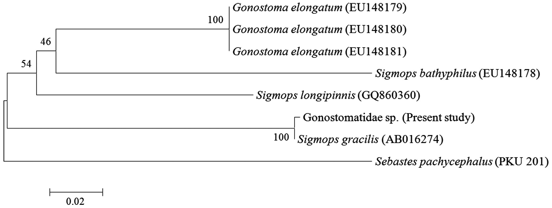
    The DNA was sequenced using an ABI 3730XL sequencer and an ABI PRISM BigDye Terminator v3.0 Ready Reaction Cycle Sequencing Kit (Applied Biosystems Inc., Foster City, CA, USA). For comparison we obtained mtDNA COI sequences for gonostomatid species from the National Center for Biological Information (NCBI) database, including Gonostoma elongatum (EU148179-81), S. bathyphilus (EU148178), S. longipinnis (GQ860360), and S. gracilis (AB016274). Sebastes pachycephalus was used as an outgroup. The mtDNA COI sequences were aligned using BioEdit version 7. Genetic distances were calculated with the Kimura 2-parameter model using the software MEGA 5. A neighbor-joining tree with 1000 bootstrap replications was constructed using MEGA 5.

      >  Morphological analysis

    Counts and measurements followed the methods of Nakabo (2002), and each body part was measured to the nearest 0.1 mm using the Active measure program (Korea). Morphological identification followed Okiyama (1988) and Ozawa and Katayama (2003).

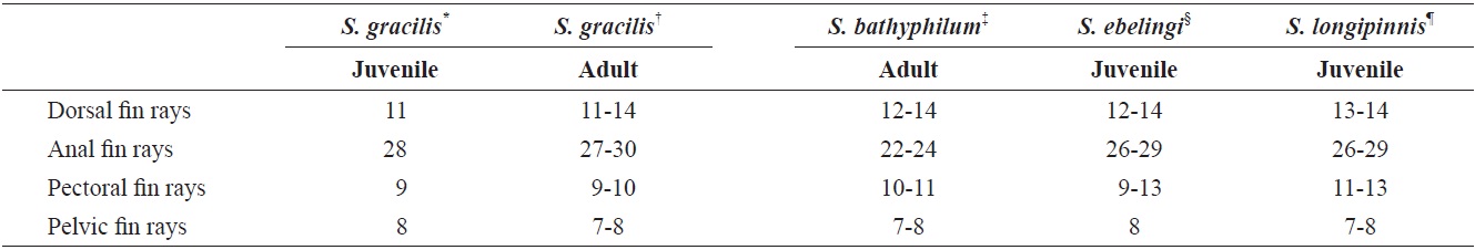
    [Table 1.] Comparison of meristic characters of Sigmops gracilis and three Sigmops species

    label

    Comparison of meristic characters of Sigmops gracilis and three Sigmops species

    Results and Discussion

    Sigmops gracilis Gunther, 1878 (Table 1, Fig. 1) (Korean name: Sol-ni-ael-tung-i)

    Gonostoma gracile Gunther, 1878 (type locality: south of Japan); Yamamoto in Okamura et al. 1982: 73; Fujii in Masuda et al. 1984: 45.

    Sigmops gracile Miya and Nishida 2000: 385; Nakabo 2002: 308; Shinohara et al. 2005: 404.

    Sigmops gracilis Mecklenburg et al. 2002: 213; Ozawa and Katayama 2003: 197; Fedorov et al. 2003: 43.

      >  Morphological description

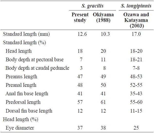
    Counts and proportional measurements for the Sigmops gracilis juvenile are presented in Tables 1 and 2, respectively. Fin rays: dorsal fin rays, 11; anal fin rays, 28; pectoral fin rays, 9; pelvic fin rays, 8. Body proportions as a percentage of standard length (SL): head length, 18; body depth at pectoral base, 7; preanus length, 47; preanal length, 48; anal fin base length, 41; location of dorsal fin origin, 57; dorsal fin base length, 12. Proportion as a percentage of head length: eye diameter, 37.

    Body moderately compressed and elongate. Head moderate and eyes round. Eye diameter longer than the snout length. Body depth uniform except at the caudal peduncle. Mouth very large with hooked teeth, with the posterior tip of the upper jaw reaching to the posterior border of the eye. Notochord flexion complete and all fins develop at their fixed locations. Pelvic and pectoral fins small. Anus not attached to anal fin base, but very close to it. Origin of dorsal fin below the 7th-8th anal fin ray. Anal fin base length much longer than the dorsal fin. Melanophores developed on lateral occipital and dorsal gut. Photophores not observed.

      >  Molecular identification

    A mtDNA COI sequence of 499 base pairs (bp) derived from the eyeball of the Gonostomatidae juvenile was compared with sequences from other gonostomatid species and an outgroup. The smallest genetic distance was between the juvenile and S. gracilis (0.002), and the distances to other members of the same genus (S. longipinnis and S. bathyphilus) were much greater (0.210 and 0.231, respectively). Therefore, the juvenile collected that was collected in the present study

    was identified as S. gracilis (Fig. 2).

      >  Distribution

    Alaska (Mecklenburg et al., 2002), China (Yang et al., 1996), Japan (Masuda et al., 1984), Kuril Islands (Mecklenburg et al., 2002), Taiwan (Shao, unpublished data), and Korea (present study).

      >  Remarks

    Juveniles of S. gracilis are distinguished from other gonostomatid juveniles by the position of the origin of the dorsal fin (Richards, 2006), which is located at the 7th-8th ray of the anal fin. Juveniles of S. gracilis are very similar to those of G. elongatum, but these species are distinguished at the photophore stage: G. elongatum has a photophore on the

    [Table 2.] Comparison with the proportional measurements of Sigmops gracilis and Sigmops longipinnis juveniles

    label

    Comparison with the proportional measurements of Sigmops gracilis and Sigmops longipinnis juveniles

    opercle at 8.0 mm SL, while S. gracilis has a photophore on the operculum at 15.5 mm SL (Okiyama, 1988). The juvenile specimen (12.6 mm SL) examined in the present study did not have a photophore, which is consistent with S. gracilis. The S. gracilis juvenile was distinguished from the congeneric species S. bathyphilus and S. longipinnis by the absence of melanophores on the caudal peduncle and the middle of the lateral body, respectively (Ozawa and Katayama, 2003). Juveniles of S. ebelingi are distinguished from those of S. gracilis by the location of the origin of the dorsal fin (Richards, 2006). According to Kawaguchi and Marumo (1967), S. gracilis is the most abundant fish group from 300 to 700 m depth in the Pacific Ocean. The water depth in the survey area in the present study was 100 m, suggesting that juveniles of S. gracilis are distributed in shallower depths than adult. The new Korean name “Sol-ni-ael-tung-i” is proposed for this species.

참고문헌
  • 1. Fedorov VV, Chereshnev IA, Mazarkin MV, Shestakov AV, Volobuev VV 2003 Catalog of Marine and Freshwater Fishes of the Northern Part of the Sea of Okhotsk. google
  • 2. Froese R, Pauly D 2012 FishBase. World Wide Web electronic publication [Internet]. google
  • 3. Gunther A 1878 Preliminary notices of deep-sea fishes collected during the voyage of H.M.S. ‘Challenger.’. [Ann Mag Nat Hist Ser] Vol.5 2 P.17-28 google
  • 4. Ivanova NV, Zemlak TS, Hanner RH, Hebert PDN 2007 Universal primer cocktails for fish DNA barcoding. [Mol Ecol Notes] Vol.7 P.544-548 google cross ref
  • 5. Kawaguchi K, Marumo R 1967 Biology of Gonostoma gracile (Gonostomatidae) I. Morphology, life history and sex reversal. In: Information Bulletin on Planktology in Japan. P.53-67 google
  • 6. Masuda H, Amaoka K, Araga C, Uyeno T, Yoshino T 1984 The Fishes of the Japanese Archipelago. Vol. 1. google
  • 7. Mecklenburg CW, Mecklenburg TA, Thorsteinson LK 2002 Fishes of Alaska. google
  • 8. Miya M, Nishida M 2000 Molecular systematics of the deep-sea fish genus Gonostoma (Stomiiformes: Gonostomatidae): two paraphyletic clades and resurrection of Sigmops. [Copeia] Vol.2000 P.378-389 google
  • 9. Nakabo T 2002 Fishes of Japan with Pictorial Keys to the Species. google
  • 10. Nelson JS 2006 Fishes of the World. google
  • 11. Okamura O, Amaoka K, Mitani F 1982 Fishes of the Kyushu-Palau Ridge and Tosa Bay: The Intensive Research of Unexploited Fishery Resources on Continental Slopes. google
  • 12. Okiyama M 1988 An Atlas of the Early Stage Fishes in Japan. google
  • 13. Ozawa T, Katayama H 2003 Early ontogeny of a South Pacific gonostomatid fish Sigmops longipinnis. [Ichthyol Res] Vol.50 P.195-197 google cross ref
  • 14. Richards WJ 2006 Gonostomatidae: Bristlemouths. In: Early stages of Atlantic Fishes: An Identification Guide for the Western Central North Atlantic. Richards WJ, ed. P.183-215 google
  • 15. Shinohara G, Sato T, Aonuma Y, Horikawa H, Matsuura K, Nakabo T, Sato K 2005 Annotated checklist of deep-sea fishes from the waters around the Ryukyu Islands, Japan. Deep-sea fauna and pollutants in the Nansei Islands. [Nat Sci Mus, Monogr] Vol.29 P.385-452 google
  • 16. Watson W 1996 Gonostomatidae: Bristlemouths. In: The Early Stages of Fishes in the California Current Region. California Cooperative Oceanic Fisheries Investigations (CalCOFI) Atlas No. 33. Moser HG, ed. P.247-267 google
  • 17. Yang J, Huang Z, Chen S, Li Q 1996 The Deep-Water Pelagic Fishes in the Area form Nansha Islands to the Northeast Part of South China Sea. google
이미지 / 테이블
  • [ Fig. 1. ]  Sigmops gracilis Gunther, 1878, NIBR-P0000018076, 12.6 mm in standard length.
    Sigmops gracilis Gunther, 1878, NIBR-P0000018076, 12.6 mm in standard length.
  • [ Table 1. ]  Comparison of meristic characters of Sigmops gracilis and three Sigmops species
    Comparison of meristic characters of Sigmops gracilis and three Sigmops species
  • [ Fig. 2. ]  Neighbor joining tree based on partial mitochondrial DNA cytochrome oxidase subunit I sequences, showing the relationships among juvenile of Gonostomatidae sp., four gonostomatids, and one outgroup (Sebastes pachycephalus). The tree was constructed using the K2P model and 1,000 bootstrap replications. The bar indicates a genetic distance of 0.02.
    Neighbor joining tree based on partial mitochondrial DNA cytochrome oxidase subunit I sequences, showing the relationships among juvenile of Gonostomatidae sp., four gonostomatids, and one outgroup (Sebastes pachycephalus). The tree was constructed using the K2P model and 1,000 bootstrap replications. The bar indicates a genetic distance of 0.02.
  • [ Table 2. ]  Comparison with the proportional measurements of Sigmops gracilis and Sigmops longipinnis juveniles
    Comparison with the proportional measurements of Sigmops gracilis and Sigmops longipinnis juveniles
(우)06579 서울시 서초구 반포대로 201(반포동)
Tel. 02-537-6389 | Fax. 02-590-0571 | 문의 : oak2014@korea.kr
Copyright(c) National Library of Korea. All rights reserved.