검색 전체 메뉴
PDF
맨 위로
OA 학술지
Evaluation of a Visible Implant Fluorescent Elastomer Tag in the Greenling Hexagrammos otakii
  • 비영리 CC BY-NC
  • 비영리 CC BY-NC
ABSTRACT
Evaluation of a Visible Implant Fluorescent Elastomer Tag in the Greenling Hexagrammos otakii
KEYWORD
Hexagrammos otakii , Tag readability , Tag retention , Visible implant fluorescent elastomer tag
  • Introduction

    Identification of individual fish is essential for growth, migration, mortality, stock identification, and gear selectivity to trace particular fish populations. Although short-term tag retention may suffice for some experiments, the effect of a tag on fish survival, behavior, growth, recognition, and cost of the marking technique should be considered. However, traditional external tags (such as spaghetti or dart tags) are often lost quickly (Crossland, 1980; Bergman et al., 1992) and may affect growth or survival (Crossland, 1976; Tong, 1978; Mc- Farlane and Beamish, 1990; Serafy et al., 1995). Furthermore, these types of tags can only be read by recapturing the fish.

    Internally situated but externally readable devices such as acoustic tags are often limited by their expense. Problems associated with the physiological characteristics of the fish, reliability of identification, fouling of the tag by algae (Jones, 1987; Barrett, 1995), tag retention (Crossland, 1976; Parker, 1990), and external visibility have reduced the confidence of in situ studies of reef fish ecology.

    Many fish species have transparent tissue suitable for tagging, including opecula, mandible, top of the head, body, and fins. However, sites to retain tags vary among species. Tagging sites in other body locations may also be used successfully. An example of a visual mark that has been successfully applied to several fish species is the visible implant fluorescent elastomer

    (VIE) tag (Northwest Marine Technology, Ltd., Shaw Island, WA, USA). It was developed primarily for tagging large batches of small or juvenile fishes. The VIE is comprised of a viscous liquid elastomer that sets into a pliable solid over a period of hours. The elastomer can be injected into transparent or translucent tissues to form a permanent, biocompatible mark. The compound fluoresces brightly when exposed to blue light (UV lamp) and viewed through an amber filter.

    Tag size can easily be varied according to the requirements of the researcher and the size of the fish to be tagged. This system has been used to identify groups or cohorts of juvenile reef fishes (Frederick, 1997) and salmonids (F. Haw personal communication) but is also proving potentially effective for controlled laboratory studies of adults (Dewey and Zigler, 1996). As an externally visible but subdermally situated marking system, VIE tags are potentially able to eliminate many of the problems experienced with other methods and can be used to trace individual fish.

    Greenling Hexagrammos otakii which is a benthic species in the family Hexagrammidae, is commonly found in coastal areas throughout the year in Korea and is a favorite seawater food fish due to its taste and tender meat. In this study, we determined the tag colors that resulted in the highest rates of detection, as well as the likelihood of tag or handling-related mortality and retention rates of VIE tags at particular body sites.

    Materials and Methods

    The fish used in this experiment were sub-adult greenling Hexagrammos otakii (mean body length ± standard deviation [SD], 21.0 ± 1.4 cm; mean body weight ± SD, 154.4 ± 13.8 g). The VIE tags were applied to treated and control fish on April 16, 2009. According to the method of Park et al. (2003) all fish were anaesthetized in 800 ppm lidocaine-hydrochloride/ NaHCO3 at a water temperature of 18℃. Fish were sedated until they were completely immobile, individually removed from the anesthetic solution, rinsed in fresh water, and placed on a flat surface for tagging.

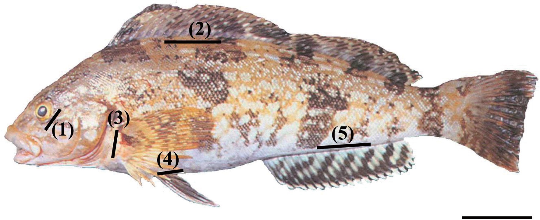
    Fifty fish in triplicate groups were individually marked with orange, yellow, red, or green elastomer at five body locations (Fig. 1): 1) the adipose eyelid, 2) the surface of the dorsal fin base, 3) the inside surface of the pectoral fin base, 4) the inside surface of the pelvic fin base, and 5) the surface of the anal fin base. Control fish (n = 50) were anesthetized but not marked. We used the VIE Hand Injection Master Kit (Northwest Marine Technology, Ltd.). The elastomer and curing agent were properly mixed at a 10:1 ratio, and the elastomer was injected as a liquid. Each site of the injection volume was 4 mL. Tagged fish were placed in tanks according to tag color and body part marked.

    The fish were held in 18 flow-through fiberglass-reinforced plastic tanks (2 × 2 × 0.5 m; water volume, 2,000 L) supplied with filtered seawater. The bottom of the tanks was coated with a black sheet to facilitate decoding of the tags. Flow rate was 100 L min-1 tank-1, and mean water temperature was 20 ± 2.5℃ respectively. A day-night cycle was established, and all tanks were covered with mesh to prevent fish jumping out. The fish were fed daily to satiation with dry commercial flounder feed (Agribrand Furina Korea Co., Seoul, Korea) throughout the 20-month trial.

    Growth of fish and retention, underwater visibility, and readability of the tags were determined at 2-month intervals, and dead fish were removed daily. Only individuals marked with color on the dorsal fin could be distinguished more than 2 m away by eye and with a blue light (UV lamp) after 2 months. Tag retention rates were calculated according to the method of Zerrenner et al. (1997). The mark-retention data recorded from dead fish were used to calculate the percent retention up to the date on which they died, but were not used in subsequent calculations.

    Data are expressed as means of triplicate experiments. Differences in survival and growth between the control and experimental groups were assessed by the t-test (Cody and Smith, 1991) and tag retention rates (%) among tagging sites were evaluated by one-way analysis of variance and Duncan’s multiple range test (Duncan, 1955). A P < 0.05 was considered to indicate statistical significance.

    Results and Discussion

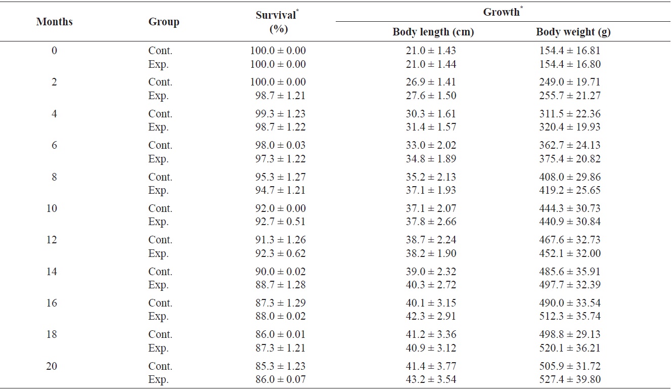
    Survival and growth of greenling Hexagrammos otakii during the 20-month trial are shown in Table 1. Tagging did not affect survival or growth of greenling. Among the 150 individuals in the control and experimental groups, 21 marked fish and 23 control fish died. After the 20-month trial, the body length of marked greenling (43.2 ± 3.5 cm, mean ± SD) did not differ from that of the control (41.4 ± 3.7 cm, mean ± SD). Additionally, the body weight of marked greenling (527.4 ± 39.8 g, mean ± SD) did not differ from that of the controls (505.9 ± 31.7 g, mean ± SD).

    The primary causes of mortality in caged snapper tagged with the same system are internal damage and infection from gas bladder rupture, or infection from anatomical trauma caused by handling (Willis and Babcock, 1998). Greenling was less fragile; however, steps should be taken to minimize the possibility of capture-related injury. Therefore, skilled application of the elastomer injection was crucial for maintaining low mortality, as suggested by the decrease in mortality of marked fish during a laboratory experiment (Frederick, 1997). Jang et al. (2007) investigated the effectiveness of passive integrated transponder (PIT) tags in a small cyprinid Pseudorasbora parva (n = 110; mean body length ± SD, 17.4 ±

    [Table 1.] Survival (%) and growth in greenling Hexagrammos otakii from 0 to 20 months after visible implant fluorenscent elastomer (VIE) tagging

    label

    Survival (%) and growth in greenling Hexagrammos otakii from 0 to 20 months after visible implant fluorenscent elastomer (VIE) tagging

    1.1 cm; mean body weight ± SD, 71.7 ± 0.2 g). Upon use of three types of tags, including small PIT tags (width, 11.0 mm; height, 2.1 mm; weight, 0.088 g), mid-sized PIT tags (width, 20.0 mm; height, 3.5 mm; weight, 0.188 g), and large PIT tags (width, 30.0 mm; height, 3.5 mm; weight, 0.298 g), survival rates of 100 individuals 30 days after tag insertion were as follows: large tag, 50.0%; middle tag, 57.5%; and small tag, 61.4%. Survival rates varied among the three tag types because death was due to surgical damage. However, we used the VIE tag which resulted in a high survival rate and no surgical damage to the fish.

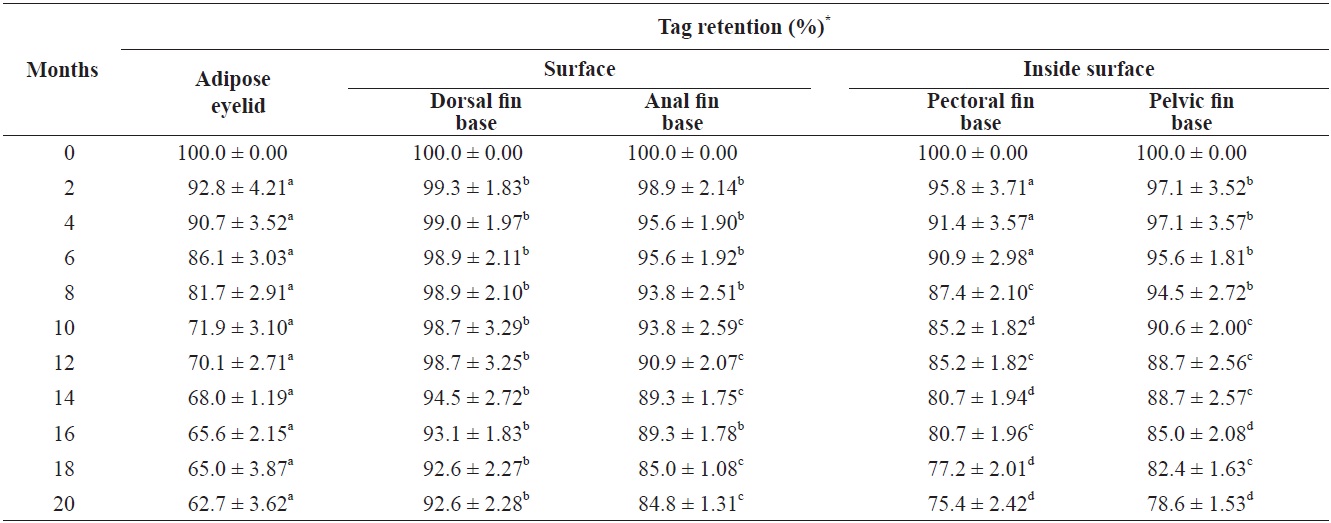
    Greenling retained > 90% of the tags at the surface of the dorsal fin base (Table 2), which is similar to previous VIE studies (Dewey and Zigler, 1996; Willis and Babcock, 1998). The anal fin base showed a higher tag retention rate than marking the inside surface of the pectoral fin and pelvic fin bases (P < 0.05). The high loss of the VIE at the inside surface of the pectoral and pelvic fin bases during tagging seemed to related to the benthic behavior of this species. Buckley et al. (1994) found that VIE tags in juvenile Sebastes spp. released on an artificial reef could be identified visually after 258 days during strip transects using ultraviolet underwater lights.

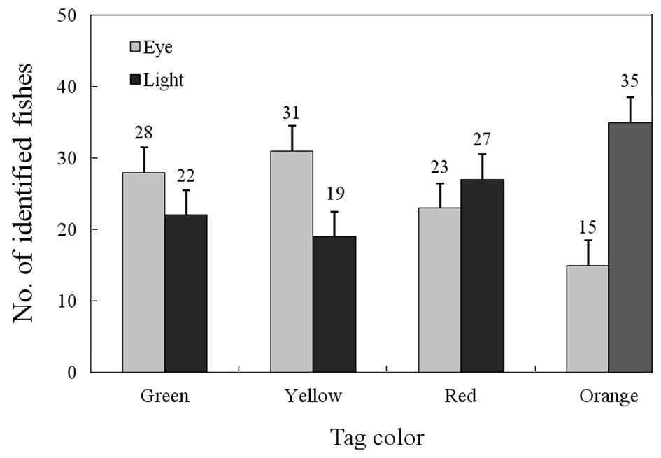
    Tags of different colors were not equally identifiable underwater (Fig. 2). Only individuals marked with color on the

    dorsal fin were distinguishable at distances > 2 m by eye or with a UV lamp. Twenty-eight fish tagged with green were identifiable with the naked eye, and 22 were with a UV lamp. Thirty-one fish tagged with yellow were identifiable with the naked eye, and 19 with a UV lamp. Twenty-three fish tagged with red were identifiable with the naked eye, and 27 with a UV lamp. Fifteen fish tagged with orange were identifiable with the naked eye, and 35 with a UV lamp. These results are similar to those of a previous study (Willis and Babcock, 1998).

    [Table 2.] Retention rate (%) of visible implant fluorenscent elastomer (VIE) tags in each sites of greenling Hexagrammos otakii from 0 to 20 months after VIE tagging

    label

    Retention rate (%) of visible implant fluorenscent elastomer (VIE) tags in each sites of greenling Hexagrammos otakii from 0 to 20 months after VIE tagging

    Significant differences were found among tag colors; red and orange were easier to detect than green and yellow using a UV lamp. Green and yellow could be detected easily with the naked eye. The method of detection (naked eye vs. UV lamp) did not affect overall recognition rates, although there was a significant interaction between method and color because the green and yellow tags were more difficult to see when the UV lamp was used (Fig. 2). This was likely because the green and yellow tags emit fluorescence in response to a smaller range of light wavelengths than that of brighter colors (G. Thomburgh personal communication).

    Buckley et al. (1994) found that red monofilament micro tags were difficult to detect underwater because of the attenuation of short wavelength light. However, we could more easily detect red compared to green or yellow (Willis and Babcock, 1998). All dives in this study were limited to 2 m. Deeper water where natural light levels are lower may result in greater attenuation of red light (Willis and Babcock, 1998). Red tags are clearly recognizable at up to 5 m in clear water under direct sunlight (Willis and Babcock, 1998).

    As mentioned by Willis and Babcock (1998), a variety of factors may influence the distance at which tags can be positively identified. It was often possible to recognize a fish as possessing a tag at > 6 m without being able to detect tag color. The direction and intensity of ambient light made a large difference; when the sun was low, and behind the observer, tag color could be correctly identified at a greater distance. The amount of suspended material was also a contributing factor, particularly in the effectiveness of fluorescence. Backscatter from large particles as well as strong ambient light decreases the effective range of a UV lamp beam, thereby decreasing the range of detection. The range of detection of orange tagged fish using a UV lamp and amber filter appears to be limited primarily by the water clarity and the power of the light used (Willis and Babcock, 1998). We found that the VIE mark was easy to apply, requiring < 1 min per fish, and was readily visible when viewed under an UV lamp in greenling, a benthic species.

참고문헌
  • 1. Barrett NS 1995 Short- and long-term movement patterns of six temperate reef fishes (Families Labridae and Monacanthidae). [Mar Freshw Res] Vol.46 P.853-860 google cross ref
  • 2. Bergman PK, Haw F, Blankenship HL, Buckley RM 1992 Perspectives on design, use, and misuse of fish tags. [Fisheries] Vol.17 P.20-25 google cross ref
  • 3. Buckley RM, West JE, Doty DC 1994 Internal micro-tag systems for marking juvenile reef fishes. [Bull Mar Sci] Vol.55 P.848-857 google
  • 4. Cody RP, Smith JK 1991 Applied Statistics and the SAS Programming Language. P.122-135 google
  • 5. Crossland J 1976 Snapper tagging in north-east New Zealand, 1974: analysis of methods, return rates, and movements. [N Z J Mar Freshw Res] Vol.10 P.675-686 google cross ref
  • 6. Crossland J 1980 Population size and exploitation rate of snapper, Chrysophrys auratus, in the Hauraki Gulf from tagging experiments, 1975-1976. [N Z J Mar Freshw Res] Vol.14 P.255-261 google
  • 7. Dewey MR, Zigler SJ 1996 An evaluation of fluorescent elastomer for marking bluegills in experimental studies. [Prog Fish-Cult] Vol.58 P.219-220 google cross ref
  • 8. Duncan DB 1955 Multiple-range and multiple F tests. [Biomentrics] Vol.11 P.1-42 google cross ref
  • 9. Frederick JL 1997 Evaluation of fluorescent elastomer injection as a method for marking small fish. [Bull Mar Sci] Vol.61 P.399-408 google
  • 10. Jang MH, Yoon JD, Do Y, Joo GJ 2007 Survival rate on the small cyprinidae by PIT tagging application. [Korean J Ichthyol] Vol.19 P.371-377 google
  • 11. Jones GP 1987 Competitive interactions among adults and juveniles in a coral reef fish. [Ecology] Vol.68 P.1534-1547 google cross ref
  • 12. McFarlane GA, Beamish RJ 1990 Effect of an external tag on growth of sablefish (Anoplopoma fimbria), and consequences to mortality and age at maturity. [Can J Fish Aquat Sci] Vol.47 P.1551-1557 google cross ref
  • 13. Park IS, Jo JH, Lee SJ, Kim YA, Park KE, KE JW, Yoo JS, Song YC 2003 Anaesthetic effect of lidocaine hydrochloride-sodium bicarbonate and MS-222 on the greenling (Hexagrammos otakii). [J Korean Fish Soc] Vol.36 P.449-453 google
  • 14. Parker RO Jr. 1990 Tagging studies and diver observations of fish populations on live-bottom reefs of the U.S. Southeastern coast. [Bull Mar Sci] Vol.46 P.749-760 google
  • 15. Serafy JE, Lutz SJ, Capo TR, Ortner PB, Lutz PL 1995 Anchor tags affect swimming performance and growth of juvenile red drum (Sciaenops ocellatus). [Mar Freshw Behav Physiol] Vol.27 P.29-35 google cross ref
  • 16. Tong LJ 1978 Tagging snapper Chrysophrys auratus by scuba divers. [N Z J Mar Freshw Res] Vol.12 P.73-76 google
  • 17. Willis TJ, Babcock RC 1998 Retention and in situ detectability of visible implant fluorescent elastomer (VIFE) tags in Pagrus auratus (Sparidae). [N Z J Mar Freshw Res] Vol.32 P.247-254 google
  • 18. Zerrenner A, Josephson DC, Krueger CC 1997 Growth, mortality, and mark retention of hatchery brook trout marked with visible implant tags, jaw tags, and adipose fin clips. [Prog Fish-Cult] Vol.59 P.241-245 google cross ref
이미지 / 테이블
  • [ Fig. 1. ]  Elastomer injection locations shown on the greenling Hexagrammos otakii: 1) the adipose eyelid, 2) the surface of the dorsal fin base, 3) the inside surface of the pectoral fin base, 4) the inside surface of the pelvic fin base, and 5) the surface of the anal fin base. Scale bar = 3 cm.
    Elastomer injection locations shown on the greenling Hexagrammos otakii: 1) the adipose eyelid, 2) the surface of the dorsal fin base, 3) the inside surface of the pectoral fin base, 4) the inside surface of the pelvic fin base, and 5) the surface of the anal fin base. Scale bar = 3 cm.
  • [ Table 1. ]  Survival (%) and growth in greenling Hexagrammos otakii from 0 to 20 months after visible implant fluorenscent elastomer (VIE) tagging
    Survival (%) and growth in greenling Hexagrammos otakii from 0 to 20 months after visible implant fluorenscent elastomer (VIE) tagging
  • [ Fig. 2. ]  Comparison of visible implant fluorescent elastomer (VIE) tag detection, identification between the four tag colors used two detection methods in greenling Hexagrammos otakii, using observations taken on 2 m above water visibility. Number of observations for each category is given above each bar. Tags site were dorsal fin base (eye, tags observed with the naked eye; light, tags observed using a blue UV lamp light to activate fluorescence. Error bars are 95% confidence).
    Comparison of visible implant fluorescent elastomer (VIE) tag detection, identification between the four tag colors used two detection methods in greenling Hexagrammos otakii, using observations taken on 2 m above water visibility. Number of observations for each category is given above each bar. Tags site were dorsal fin base (eye, tags observed with the naked eye; light, tags observed using a blue UV lamp light to activate fluorescence. Error bars are 95% confidence).
  • [ Table 2. ]  Retention rate (%) of visible implant fluorenscent elastomer (VIE) tags in each sites of greenling Hexagrammos otakii from 0 to 20 months after VIE tagging
    Retention rate (%) of visible implant fluorenscent elastomer (VIE) tags in each sites of greenling Hexagrammos otakii from 0 to 20 months after VIE tagging
(우)06579 서울시 서초구 반포대로 201(반포동)
Tel. 02-537-6389 | Fax. 02-590-0571 | 문의 : oak2014@korea.kr
Copyright(c) National Library of Korea. All rights reserved.