Suborder Calcaxonia belongs to the order Alcyonaria within the subclass Octocorallia and includes five gorgonian fam-ilies, of which two families, Primnoidae and Isididae have been introduced from King Sejong Station (62° 13′S, 058° 47′W), Korea Polar Institute (KOPRI) of Korea Ocean Rese-arch and Development Institute (KORDI) for the Korea An-tarctic Research Program on King George Island in the pre-sent study. The former Primnoidae comprising more than 233 species within 36 genera has the axis revealed undulating concentric layers of calcified material embedded in gorgonin, and the latter Isididae possesses the characteristic axis com-posed of alternating horny nodes and nonspicular calcareous internodes. In a series of taxonomic studies on anthozoans from King Sejong Station, KOPRI, three species in Primnoi-dae and one in Isididae have newly been added to the octo-corallian fauna.
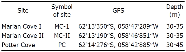
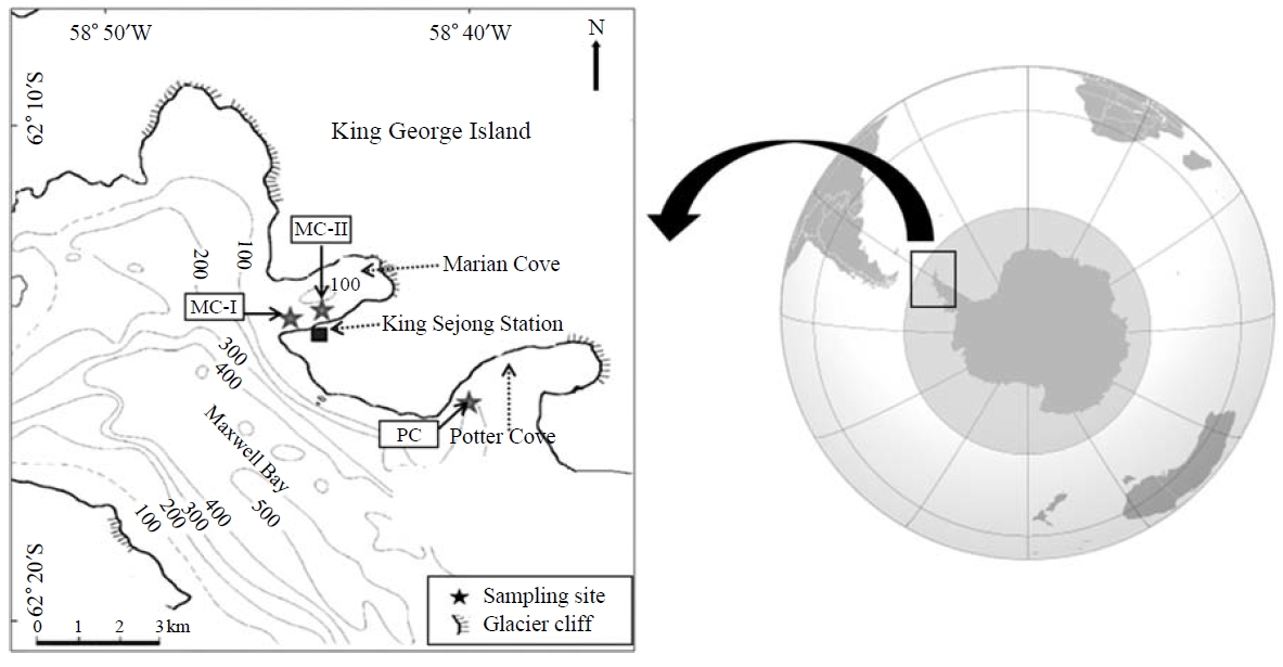
All specimens examined in this study were collected from subtidal zones in the coastal regions of King Sejong Station, KOPRI between depths of 10 and 45 m from two sites, which were the Marian Cove and a site in Potter Cove of King Geo-rge Island, by SCUBA diving from December 2009 to Jan-uary 2011 (Fig. 1). Information on the detailed collecting sites is shown in Table 1. The specimens were anesthetized with menthol for 6-8 h, and then fixed in 4-5% (v/v) formalin with seawater. After that, they were washed with tap water, and preserved in 70% alcohol (v/v).
For identification, each specimen was examined for exter-nal and internal morphological characteristics such as growth form, branching pattern and size on each part of colonies, arrangement of polyps on stem and branches, and also col-oration, anthostele, shape and size of sclerites in polyps and coenenchymes under the stereomicroscopes (Semi SV-6 and SV-11; Carl Zeiss, Jena, Germany and S8APO; Leica, Wet-zlar, Germany) and light microscope (Eclipse 80i; Nikon Co., Tokyo, Japan). The color of each part was recorded with a color code based on the color chart (New Color Cards 199b; Japan Color Research Services Company, Japan). To examine the sclerites, a small part of tissue from the each body part was dissolved in a diluted solution of the clorax for five min-utes. In addition, the white sclerites of the polyp head were
[Table 1.] Information of collection sites in King Sejong Station Korea Polar Institute (KOPRI)

Information of collection sites in King Sejong Station Korea Polar Institute (KOPRI)
stained with a mixture of methylene blue (one or two drops) and ethanol for the visualization of anthostele.
The living calcaxoian colonies under water were photogra-phed with a digital camera (5060-WZ; Olympus, Tokyo, Japan) with underwater housing (Patima-7070; Patima Uw_ Eng Co. Ltd, Seoul, Korea). The specimens were photogra-phed with a digital camera (G7; Canon Inc., Tokyo, Japan) before fixation. Photographs of the arrangement of polyps to branches and the anthostele of polyps were taken by a stere-omicroscope (Semi SV-11; Carl Zeiss) mounted with a digi-tal camera (5060-WZ; Olympus) and a second stereomicro-scope (S8APO; Leica) with another digital camera (DFC 290; Leica). Next, the size of polyps, and also the interval and angle of polyp head to branchlet were measured using an image analyzer (Motic Image Plus 2.0; Motic China Group Co., Xiamen, China and LAS Version 3.6; Leica). Images of sclerites were taken with a light microscope (Eclipse 80i; Nikon Co.) mounted with a camera (DS-5Mc; Nikon Co.), and then the size of sclerites were calculated with an image analyzer (NIS-Elements BR 3.0; Nikon Co.). In addition, an image editing program (HeliconFocus 5.1 Pro; Helicon Soft Ltd., Kharkov) was used to create one completely focused image of a sclerite from several partially focused images by combining the focused areas.
For identification and classification, Cairns and Bayer’s (2009) generic revision of the Primnoidae and also Bayer and Stefani’s (1987) classification of the Isididae were employed. The samples examined in this study have been deposited in the Korean Coral Resource Bank (KCRB), Ewha Womans University and some of them are stocked in the Korea Polar Research Institute of KORDI.
Phylum Cnidaria Hatschek, 1888
Class Anthozoa Ehrenberg, 1834
Subclass Octocorallia Haeckel, 1866
Order Alcyonacea Lamouroux, 1816
Suborder Calcaxonia Grasshoff, 1999
Diagnosis. Colonies with a solid axis without a central hol-low core. Axis composed of large amount of non-scleritic calcareous material between horny fibers or present as a cen-tral core or as solid intermodal sections alternating with nodes and pure gorgonin in a segmented axis. Solid calcareous in-ternodes white or colored. In some families, axis having a green to golden metallic or iridescent sheen.
Family Primnoidae Milne Edwards, 1857
Diagnosis. Axis reveals undulating concentric layers of cal-cified material embedded in gorgonin, resulting from a lon-gitudinal pattern of calcification, without a chambered cen-tral core. Colonies branched in a variety of manners or un-branched, usually firmly attached to substrate by a calcare-ous discoidal holdfast. Polyps non-retractile, calyces occurr-ing in a variety of arrangements and orientations.
Genus Arntzia Lopez-Gonzalez, Gili and Orejas, 2002
Diagnosis. Flagelliform, unbranched Primnoidae. Colonies not densely spiculated. Polyps tall, cylindrical, standing almost vertical from axis, arranged in whorls. Bases of polyps fused basally. Basal brooding part of polyps fused forming a common brood-chamber within axial coenenchyme. Eight distalmost opercular scales, larger than marginal or submar-ginal scales. Body completely covered by scales, except for proximal and basal parts of adaxial side of polyps.
Arntzia gracilis (Molander, 1929) (Table 2, Figs. 2A, 3, 4)
Material examined. Antarctic: Marian Cove I, 1 ind., 23 Dec 2009, 30-35 m deep; Marian Cove I, 1 ind., 24 Dec 2009, 30-35 m deep; Marian Cove I, 1 ind., 3 Jan 2011, 30 m deep.
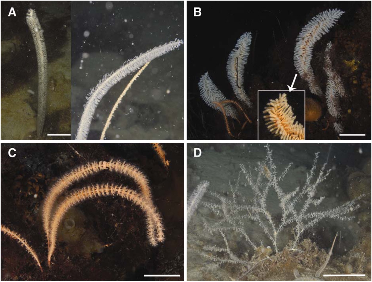
Description. Colonies unbranched, flagelliform, whip-like (Fig. 2A). Specimens with thin holdfast up to 5×4 mm in diameter, 250×3-330×4 mm (height×diameter) including stalks 10-35 mm long. Axis mostly 0.6-1.7 mm in diameter, 1.5-1.7 mm at stalk, 1.4-1.5 mm at middle part and 0.6-0.7 mm at upper part.
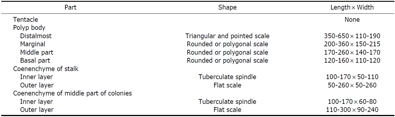
[Table 2.] Measurement (μm) and shape of sclerites of Arntzia gracilis

Measurement (μm) and shape of sclerites of Arntzia gracilis
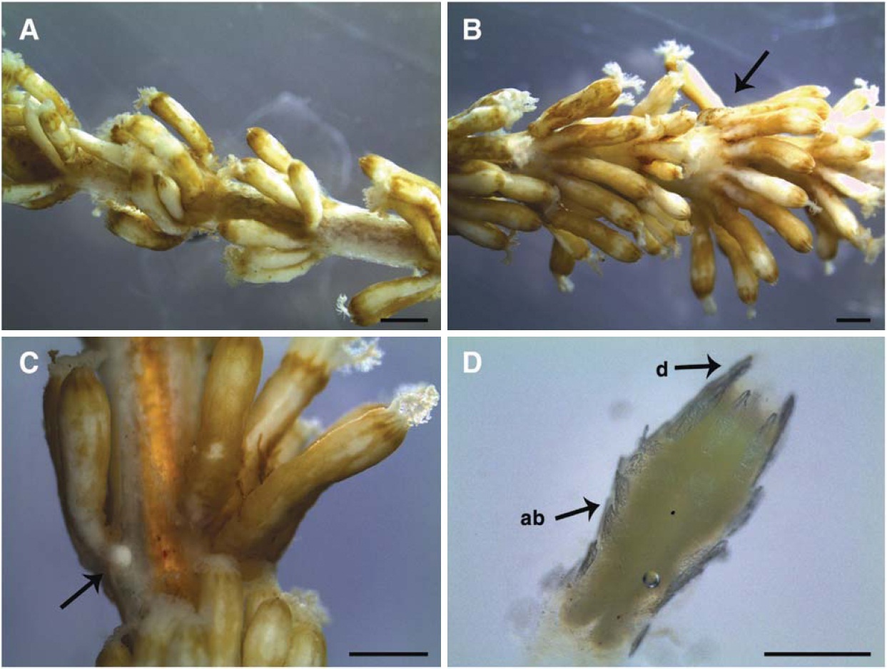
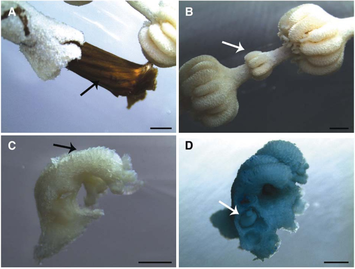
Polyps cylindrical, irregular in size, 2.0-3.0×0.5-0.7 mm (length×diameter), standing almost vertical from axis, arrang-ed in whorls, and arranged 10-16 on low annular thickenings of coenenchyme at middle part of colonies, with proximal portion partially or negligibly fused, notable mainly in repro-ductive stages (Fig. 3C). Polyps directed upwards, but not strongly adhered to coenenchyme. About 2-5 whorls per cm of axial length (Fig. 3A, B). Polyps with eight longitudinal
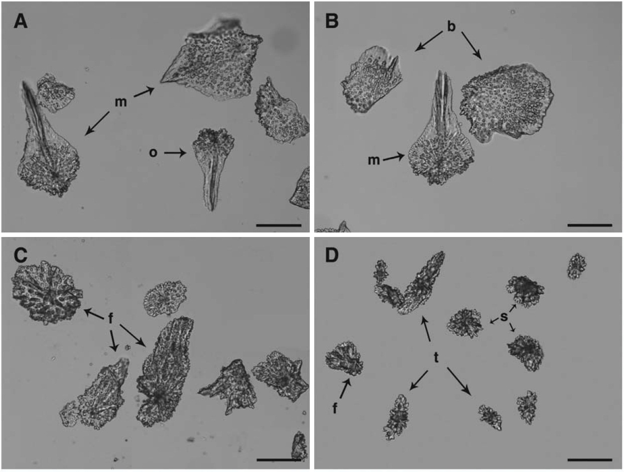
rows of scales perfectly aligned on distal half of polyps, so-metimes irregular arrangement or complete absence of scales on proximal half. Scales slightly overlapping, 5-7 (8) scales at two adaxial longitudinal rows (Fig. 3D), and 8-10 (11) scale at abaxial and lateral longitudinal rows.
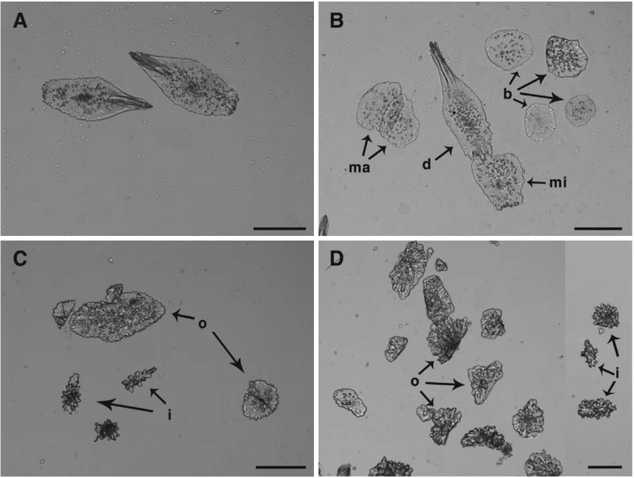
Individuals covered by a thin layer of epidermal tissue overlain by brownish cuticle. Gonads large in size, approxi-mately 280 μm in diameter, but few number within gastrovas-cular cavities of polyps at middle part of colonies (Fig. 3C). Tentacles with 12-13 pairs of pinnule, 1-2 mm long.
In coloration, colonies pinkish white (itg8) in life and white in alcohol, showing brownish by thin membranous cuticle.
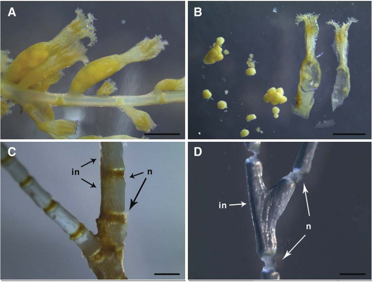
Distalmost scales (opercular) of polyp more or less triangular and pointed, 350-550×110-190 μm, two adaxial scales smaller 370-395×120-136 μm (Fig. 4A, B). Marginal scales smaller, more or less rounded or polygonal, similar to remaining body scales (Fig. 4B). Abaxial and lateral longitu-dinal rows with 8-10 (11) scales (Fig. 3D), 560-648×190-220 μm and 500-591×160-180 μm, respectively. Two adax-ial longitudinal rows with 5-7 (8) scales smaller in size, 370-392×120-136 μm, than abaxial and marginal scales. Basal and proximal portions on adaxial side of polyp body naked and markedly fleshy (Fig. 3D), Coenenchyme thick, 0.36-0.46 mm thick at stalk and 0.30-0.31 mm at middle part, with an outer layer containing sclerites like those on bodies of polyps, 50-300×50-240 μm (Fig. 4C, D). Inner layer, containing longitudinal stem canals, with more or less irregular rods covered by compound tubercles, 90-170×50-110 μm. Super-ficial coenenchymal scale-like and inner irregular sclerites, more densely packed in smaller colonies than in larger ones.
Remarks. The characteristics of our specimens mostly agree with those of Lopez-Gonzalez et al. (2002), but they differ in the number and size of scales overlapping at adaxial, lateral and abaxial longitudinal rows of polyps. This species are abundantly living at depths of 30-35 m in the Marian Cove I in front of King Sejong Station.
Distribution. Antarctic and Subantarctic (Weddell Sea, Scotia Arc, and Ross Sea).
Genus Thouarella (Thouarella) Gray, 1870
Diagnosis. Colonies of most species branch in a bottlebrush fashion, although two species have pinnate branching. Caly-ces isolated (not in pars of whorls), arranged in no apparent order on all sides of branches and stand perpendicular or in-clined upward (type species) on branch.
Well-developed operculum present. Marginal scales arrang-ed in two circles of four that alternate with one another, cir-cumference of distal polyp not being large enough to accom-modate eight adjacent marginal scales; distal margins of mar-ginals spinose, folding over lower portion of operculars, form-ing a circumoperculum. Inner face of operculars and margin-als bear ornate keels, each longitudinal keel bearing several smaller ridges oriented at right angles, altogether forming a foliate process; distal edges of same scales finely serrate. Polyps protected by six longitudinal rows of body wall scales, the adaxial face being covered by enlarged inner-lateral scal-es, resulting in complete coverage of polyp. Outer and inner faces of body wall scales radially ridged distally. Coenechy-mal scales in two layers; outer layer composed of irregular-ly shaped scales with granular outer surface; inner layer comp-osed of small spheroids.
Thouarella (Thouarella) antarctica (Valenciennes, 1846) (Table 3, Figs. 2B, 5, 6)
[Table 3.] Measurement (μm) and shape of sclerites of Thouarella (Thouarella) antarctica

Measurement (μm) and shape of sclerites of Thouarella (Thouarella) antarctica
Bayer, 2009: 34, fig. 6g-l.
Material examined. Antarctic: Potter Cove, 1 ind., 10 Jan 2010, 30-40 m deep; Potter Cove, 2 inds., 19 Jan 2010, 30-40 m deep; Potter Cove, 1 ind., 24 Jan 2011, 45 m deep.
Description. Colonies branch in a bottlebrush fashion, with one lateral branch 30-90 mm long at middle part (Fig. 2B). Branchlets generally simple, sometimes branching, 16-42 (50) mm long, arranged at interval of 10-15 per cm at stem and branch, inclined 80-85° to stem arising from 5-6 sides with longitudinal rows around stem (Fig. 2B). Specimens with thin holdfast 5×5-7×5 mm in diameter, attaching peb-ble, 330×50-450×70 mm (height×diameter) including stalks 18-24 mm long. Axis stiff, bristle-like, yellow with golden sheen on surface, 1.0-3.0 mm in diameter.
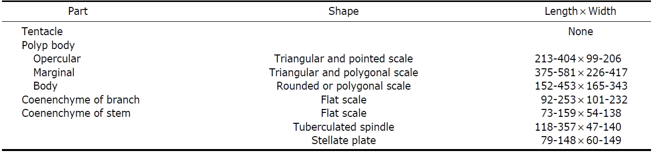
Calyces isolated (not in pars of whorls), arranged in com-pactly spiral order on all sides of branchlets, 30-36 per cm, and inclined upward on branchlets, 30-50° (Fig. 5A). Polyps club-shaped. cylindrical, irregular in size, 0.5-1.6×0.3-0.6 mm (height×diameter). Polyps with six longitudinal rows of scales perfectly aligned on distal half of polyps, sometimes irregular arrangement or complete absence of scales on prox-imal half at adaxial side. Scales slightly overlapping, 4-5 scales at two adaxial longitudinal rows and 6-7 scales at abax-ial longitudinal rows (Fig. 5B-D).
Individuals covered by a thin layer of epidermal tissue overlain by brownish cuticle. In coloration, colonies pinkish yellow (PI-10) in life and white in alcohol, showing brownish by thin membranous cuticle.
Opercular scales of polyps higher than broad, triangular and pointed with strong prominence, and a median keel run-ning out into a shot spine, 430-650×200-400 μm (Fig. 6A, B). Succeeding scales from marginal to body broader than high, with small serrated upper edge, often interspersed with stronger teeth (Fig. 6A, B). Scales of coenenchyme irregular triangular to four-corned with unequal sides, 73-253×54-232 μm (Fig. 6C, D).
Remarks. The characteristics of our specimens mostly agree with those of Wright and Studer (1889), but they differ in the number of longitudinal rows of branchlets around the stem, and also in the number of scales overlapping at the adaxial and abaxial longitudinal rows of polyps. This species are abundantly living at a depth of 30 m in front of King Sejong Station.
Distribution. Antarctic (Faulkland and Crozets).
Genus Onogorgia Cairns and Bayer, 2009
Diagnosis. Colonies unbranched (flagelliform). Calyces ap-pressed facing upwards, slightly fused basally within a whorl, arranged in whorls of up to 24. Well-developed operculum present, inner face of operculars bear multiple longitudinal
[Table 4.] Measurement (μm) and shape of sclerites of Onogorgia nodosa

Measurement (μm) and shape of sclerites of Onogorgia nodosa
ridges but no single keel. Eight marginal scales fold over operculars, forming a circumoperculum, distal edges of mar-ginals elongate and pointed but not spinose. Polyps protected by eight longitudinal rows of body wall scales. Two abaxial rows consisting of up to 14 scales, scales on lateral rows equal in number and size to abaxials, but adaxial scales smal-ler and randomly arranged. Body wall scales ascus-types. Coenenchymal scales in two layers, outer layer composed of flat or ascus-types scales, inner layer composed of irregular tuberculate sclerites.
Onogorgia nodosa (Molander, 1929) (Table 4, Figs. 2C, 7, 8)
Material examined. Antarctic: Marian Cove II, 1 ind., 24 Dec 2009, 30-35 m deep; Potter Cove, 2 inds., 10 Jan 2010, 30-40 m deep; Potter Cove, 3 inds., 25 Jan 2010, 30-40 m deep; Potter Cove, 1 ind., 22 Jan 2011, 40 m deep.
Description. Colonies unbranched, flagelliform, whip-like (Fig. 2C). Specimens with thin holdfast 5×2-5×4 mm in diameter, 270×2-400×6 mm (height×diameter) including stalks 25-90 mm long. Axis having longitudinal grooves, 1.5-2.2 mm in diameter at basal part, 1.3-1.6 mm at middle, and 0.7-1.0 mm at top. Coenenchyme 0.34 mm in thickness (Fig.7A).
Polyps cylindrical, irregular in size, larger ones 2.5-3.9×0.9-1.1 mm (height×diameter), and smaller ones 1.0-2.3×0.5-0.9 mm, directed upwards, but not strongly adhered to coenenchyme. New smaller polyps inserted between mature individuals within whorl, and also new whorl added between old whorls (Fig. 7B). About 2-3 whorls per cm of axial leng-th, mostly at intervals of 1.5-2.1 mm and up to 4.5 mm at middle part of colonies. Polyps arranged in one row at each
whorl, 15-20 per whorl on low annular thickenings of coen-enchyme at middle part of colonies, and 5 per whorl at lower part. Gonads large in size, 0.75×0.59-1.05×0.78 mm in diameter, similar like immature stage of embryos, but few number, approximately 1-2 per polyp within gastrovascular cavities of polyps at middle part of colonies (Fig. 7D). Scales of abaxial and lateral rows longitudinally aligned in all but basalmost part of body, circumopercular scales overreach operculars (Fig. 7C, D). Adaxial surface of polyps covered by scales in rows shorter and more or less irregular than those of abaxial surface, and they decrease toward adaxial side of polyp in size. Scales slightly overlapping, up to 20 scales at two abaxial and lateral longitudinal rows. Tentacles with 14 pairs of pinnule, 1.0-1.5 mm long.
In coloration, colonies pinkish yellow (PI-10) in life and white in alcohol, showing brownish by a thin layer of mem-branous cuticle. Axis dark brown.
Opercular scales of polyp more or less triangular and point-ed, 327-417×65-121 μm (Fig. 8A), Circumopercular pointed ascus-scale, 458-522×156-253 μm (Fig. 8B). Scales decrease in size toward adaxial side of polyp, adaxial almost half size of abaxial (Fig. 8C). Coenenchyme thick, with an outer layer containing flat tuberculate scale like those on bodies of polyps, 118-288×69-215 μm (Fig. 8D). Inner layer, with more or less irregular rods covered by compound tuberculate spindle, 66-204×30-96 μm.
Remarks. The characteristics of our specimens mostly agree with those of
Distribution. Antarctic (Scotia Sea, Ross Sea, South Shetl-and Islands, South Georgia, and Antarctic Peninsula).
Family Isididae Lamourous, 1812
Diagnosis. Axis composed of alternating horny nodes and
non-spicular calcareous internodes. Isidids branched from internodes, in several planes or all sides in form of bottle-brush. Polyps cylindrical or clavate, standing straight or in-clined from axis, sometimes recurved toward axis, scattered on all sides of branches. Sclerites in form of serrated scales or plates, sometimes narrow and fusiform, transversely place in polyps. Bases of tentacles armed with converging scales.
Genus Tenuisis Bayer and Stefani, 1987
Diagnosis. Very slender isidids sparsely dichotomously bran-ched from internodes, occasionally from nodes. Internodes cylindrical, solid, slightly enlarged at each end, smooth or with only faint longitudinal striation confined to larger bran-ches and main stem. Polyps tall, narrowly campanulate, usu-ally 1 or 2 per internodes. Sclerites of polyps flat scales never projecting between tentacle bases, spindles or rods. Sclerites of coenenchyme small, thin scales with dentate margins.
Tenuisis microspiculata (Molander, 1929) (Table 5, Figs. 2D, 9, 10)
Material examined. Antarctic: Potter Cove, 1 ind., 10 Jan 2010, 30-35 m deep; Marian Cove II, 1 ind., 15 Jan 2010, 30-35 m deep; Marian Cove II, 1 ind., 18 Jan 2010, 30-35 m deep; Potter Cove, 1 ind., 25 Jan 2010, 35 m deep.
Description. Specimens with thin holdfast 3×4-7×5 mm in diameter, almost one plane, 80×50×20-190×120×30 mm (height×width×thickness) including stalks 10-22 mm long (Fig. 2D). Colonies dichotomously branched from mostly internodes and sometimes nodes at upper part of colonies (Fig. 9A). 1st branches 12-15 mm long, longer and longer towards top of colonies, 8th 20-40 mm long. At angles of
[Table 5.] Measurement (μm) and shape of sclerites of Tenuisis microspiculata
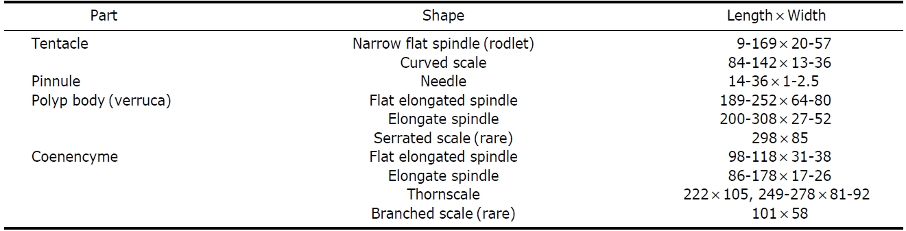
Measurement (μm) and shape of sclerites of Tenuisis microspiculata
branching, 1st 60-90° and narrower and narrower towards 8th 30°. Nodes brownish, 0.06-0.32×0.01-0.91mm (length×diameter), in detail 0.20-0.32×0.72-0.91 mm at lower part of colonies, 0.20-0.22×0.34-0.53 mm at middle, and 0.06-0.12×0.01 mm at upper. Internodes whitish, sculptured lower longitudinal groove (Fig. 9D), 0.50-1.83×0.17-1.00 mm long (length×diameter), in detail 0.50-1.50×0.55-1.00 mm at lower part of colonies, 1.30-1.83×0.50-0.60 mm at middle, and 1.26-1.31×0.17-0.18 mm at upper (Fig. 9C, D).
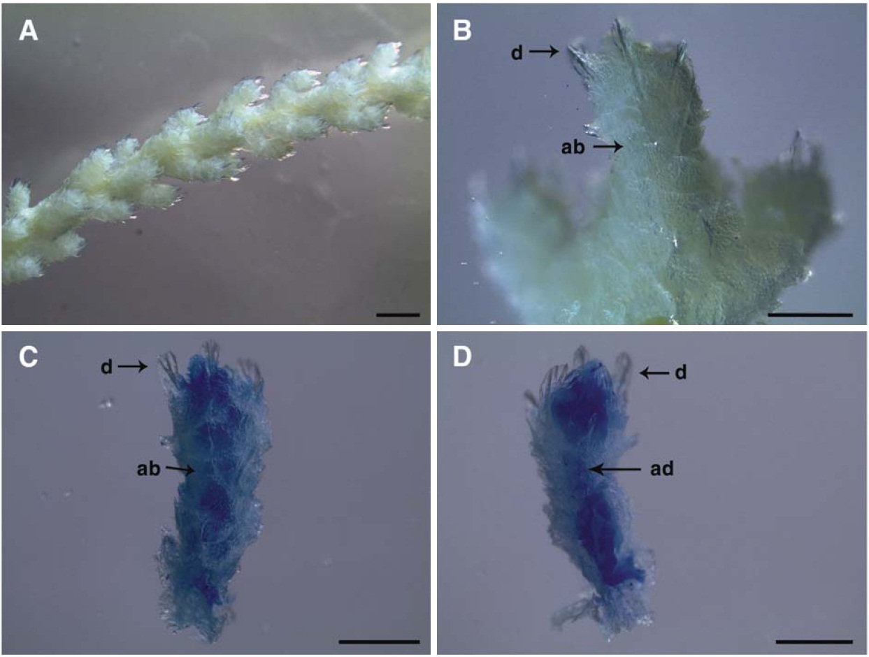
Polyps irregular in size, 0.4-3.0×0.2-0.7 mm (height×diameter), smaller ones 0.4-0.8×0.2-0.3 mm and larger ones 1.6-3.0×0.5-0.7 mm, trumpet-shaped, standing 40-50° to
axis, and arranged alternately at intervals of 0.5-2.0 mm, 11-16 per cm in number of polyps, usually one or occasionally two per internode, all around branches (Fig. 9A). Larger polyps usually arising from every node and one or two smal-ler polyps between larger ones. Individuals covered by a thin layer of epidermal tissue overlain by cuticle. Gonads 50-286μm in diameter, up to 35 per polyp, well developed within gastrovascular cavities of polyps at upper part of colonies (Fig. 9A, B). Tentacles with 10-11 pairs of pinnules, 0.5-1.0 mm long. Coenenchyme very thin membranous, 117-120 μm in thickness (Fig. 9C).
In coloration, colonies white, polyps showing yellowish orange by thin membranous cuticle.
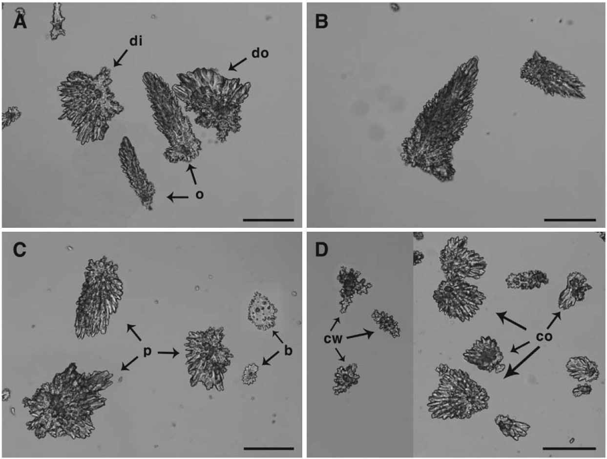
Distal half of polyp body, beneath tentacle, contains a sin-gle layer of flat, elongated scales longitudinally arranged, di-stalmost sclerites weakly converging in 8 indistinct points. Polyp sclerites short, flat elongated spindles, cylinders and clubs, sparsely thorned, 190-308 μm long, narrow curved scales up to 0.3 mm long (Fig. 10A, B). Pinnules minute rod-lets 14-36 μm long, and larger rodlets flattened and marginally lobate. At distal part of polyp body, a single layer of flat. Scl-erites of coenenchyme flat elongated spindles and thornscales, sparsely visualized in membranous tissue (Fig. 10C, D).
Remarks. The characteristics of our specimens mostly agree with those of Bayer and Stefani (1987), but they differ in the size and shape of sclerites of the verrucal wall.
Distribution. South Atlantic (South Shetland Islands), An-tarctic Peninsula (Graham Land, Palmer Land, Viecennes Bay, and Wikes Land).