검색 전체 메뉴
PDF
맨 위로
OA 학술지
Effect of Egg White Combined with Chalcanthite on Lipopolysaccharide induced Inflammatory Cytokine Expression in RAW 264.7 cells
  • 비영리 CC BY-NC
  • 비영리 CC BY-NC
ABSTRACT
Effect of Egg White Combined with Chalcanthite on Lipopolysaccharide induced Inflammatory Cytokine Expression in RAW 264.7 cells
KEYWORD
cytokine , GM-CSF , IL-1β , IL-6 , lipopolysaccharide , TNF?α
  • 1. Introduction

    Chalcanthite derived from the old Latin name Chalcanthum meaning “flowers of copper” is a richly-colored, blue, water-soluble sulfate mineral commonly found in the late-stage oxidation zones of copper deposits [1]. Copper is the main component of chalcanthite and the third most common mineral in the body, being essential in human growth and development [1-3]. Copper is found throughout the muscul-o-skeletal system, with largest amount being found in the brain and the liver. It helps to release energy in cells and contributes to the production of melanin; several key enzym-es, neurotransmitters and neuroactive compounds; collagen; and red blood cells. It also helps in the functioning of antioxidants, the formation and regulation of melatonin, and the absorption and transport of iron [4]. Copper toxicities caused by inadequate diet or surgical interventions may lead to cardiac hypertrophy, poor neuronal myelination, blood vessel abnormalities, and impaired immune responses [2, 3]. Elevated copper levels are associated with morphological and metabolic changes in the liver and in several other tissues and show a wide spectrum of hepatic abnormalities, including hepatitis, cirrhosis, acute liver failure and/or neurological and psychiatric diseases that may progress to death if left untreated [3]. Recent reports indicate that changes in the expression of copper transpor-ters alter the sensitivity of cancer cells to major chemothe-rapeutic agents, such as cisplatin, although the mechanism behind this phenomenon remains unclear [5]. Regulation of human copper homeostasis has significant therapeutic potential and requires detailed understanding of copper transport mechanisms [6]. Normally, most of the copper entering the blood in ionic form goes directly to the liver and the kidneys before reappearing in blood plasma in newly

    formed ceruloplasmin. Copper is transported by albumin and transcuprein, a high-affinity copper carrier in the plasma involved in the initial distribution of copper entering blood. Studies have discovered that transcuprein is a macroglobulin regulated by copper and iron availability [3, 7]. The main ingredient of chalcanthite is CuSO4, which exists as a series of compounds that differ in their degrees of hydration [1]. The anhydrous form occurs as a rare mineral known as chalcocya-nite. The hydrated copper sulfate occurs in nature as chalcanth-ite (CuSO4? 5H2O), and two of more rare ones: bonattite (CuSO4 ? 3H2O) and boothite (CuSO4? 7H2O) [1]. Macrophages are known to play an important role in immune reactions, allergies, and inflammation. During inflammation response, macrophag-es release many different kinds of cytokines, such as interleuk-in-1β (IL-1β), interleukin-6 (IL-6), tumor necrosis factor-α (TNF-α) and granulocyte-macrophage colony-stimulating factor (GM-CSF), initiating and maintaining specific immune responses as part of the immune/inflammatory cascade [8]. Lipopolysacch-aride (LPS) is a major outer membrane component of gram-negative bacteria and is frequently used as an inflammatory model due to its ability to activate macrophages. LPS stimulates immune responses by interacting with the membrane receptor CD14 to induce cytokine release through nuclear factor kappa-light-chain-enhancer of activated B cells (NF-κB) activation [7-11]. Therefore, the modulation of macrophage-mediated inflammatory responses is important in order to formulate a new therapeutic approach against these inflammatory diseases. Minerals have been traditionally used as part of oriental herbal medicine treatment, especially for immune-related diseases, in Korea, China, and other Asian countries. According to oriental medicine, chalcanthite has sour, acrid, cold and toxic properti-es, and is affiliated with the liver meridian. Its main function is to eliminate toxins. Internally, it functions as a purgative and helps the body get rid of excess phlegm and toxins. Chalcanthite can also be applied externally for skin abscesses, inflammation, and the removal of necrotic tissue [12]. The traditionally used dose of chalcanthite is between 0.3 to 0.6 grams of powdered mine-ral. Larger amounts can be used if chalcanthite is being applied externally [12]. However, chalcanthite is poisonous and should be used with extreme caution. To date, due to safety issues con-cerning their cytotoxical mechanisms, mineral medicines have rarely been used. Previously, we reported that egg white combined with chalcanthite (IS4) inhibited the growth of NCI-H460 human lung cancer cells by inducing apoptotic cell death via caspase-3 activation. Mixing egg white with mineral drugs is a method to enhance pharmaceutical properties and to reduce toxicity [13]. In this study, the anti-inflammatory effects of IS4 on LPS-stimulated cytokines in mouse RAW 264.7 macrophage-like cells were investigated.

    2. Materials and methods

       2.1. IS4

    Egg white combined with chalcanthite (IS4) was used as the main experimental material. The eggs were from Hamyang, Korea, and the chalcanthite was obtained from Manila, The Phil-ippines. The chalcanthite was heated and dehydrated. During the heating process, chalcanthite uniformly received heat while being carefully watched for color change every 3 to 5 hours until it turned fully grey. The heating duration varied with the amount and the quality of the mineral used and was in the range of 10 to 24 hours. Once the chalcanthite had been dehydrated, it was cooled until the remaining heat was completely released. The moisture content of the chalcanthite ranged from 0% to 5%. After it had been cooled, the chalcanthite was finely pulverized. The pulverized chalcanthite should be stored in a concealed airtight container in a dry place to prevent any moisture from entering. The prepared chalcanthite powder was rapidly mixed with egg whites in a ratio of 30:13. The egg whites had to be ho-mogeneously mixed with a wooden spatula in a ceramic vessel so that the utensils would not interfere with or cause any chem-ical reactions during the mixing process. After the chalcanthite and egg whites had been sufficiently mixed, the mixture was cooled until the reaction heat was completely released. One-hundred mg of the attained substance was then dissolved in 1 ml of distilled water and was centrifuged at 6,000 rpm for 10 minutes. The upper level was filtered in 0.8㎛ pore sizes, and the filtered substance diluted in distilled water was used for treatment. The treatment doses of IS4 (12.5, 25, 50 ㎍/ml) were based on our previous IS4 [13].

       2.2. Cell culture

    The RAW 264.7 macrophage-like cell lines obtained from ATCC® were cultured in Dulbecco’s modified Eagle’s medium (DMEM) with 10% fetal bovine serum (FBS) and penicillin-streptomycin at 37 °C under 5% CO2.

       2.3. Reagents

    The fetal bovine serum (FBS) was purchased from HyClone(Logan, UT) and high-glucose DMEM, L-glutamine, penicillin, and streptomycin were purchased from GIBCO BRL (Life Technologies, Grand Island, NY). Purified LPS derived from E.coli serotype 055:B5, Allo, Tris, sodium dodecyl sulfate (SDS),Triton X-100, Tween 20, phenylmethylsulfonyl fluoride, leupe-ptin, aprotinin, proteinase K, and other chemicals were purcha-sed from Sigma-Aldrich (St. Louis, MO).

       2.4. Measurement of cytokines

    RAW 264.7 cells were seeded into 24 well plates at 1 x 10⁵ cells/well and were cultured in DMEM containing 10% FBS at 37°C for 24 hours. After treatment with LPS (1 ㎍/ml) for 24 hours, the concentrations of IL-1β and IL-6 in the supernatant were measured by using a commercial enzyme-linked imm-unosorbent assay (ELISA) kit (BD Biosciences, San Diego, CA). The concentrations of TNF-α and GM-CSF in the supernatant were measured by using an ELISA kit (Millipore, Billerica, MA). The concentrations were calculated using linear-regression equations obtained from the standard absorbance values.

       2.5. Statistical analysis

    Numerical data from each experiment were averages from triplicate samples. Experiments were repeated three times to avoid any experimental errors and to ensure the repeatability of the used methods. Data were expressed as means ± SD. Statistical differences were analyzed using the Mann?Whitney U-test in SPSS ver. 10.0 (statistical software package, Chicago, IL). P-values less than 0.05 were considered statistically significant.

    3. Results

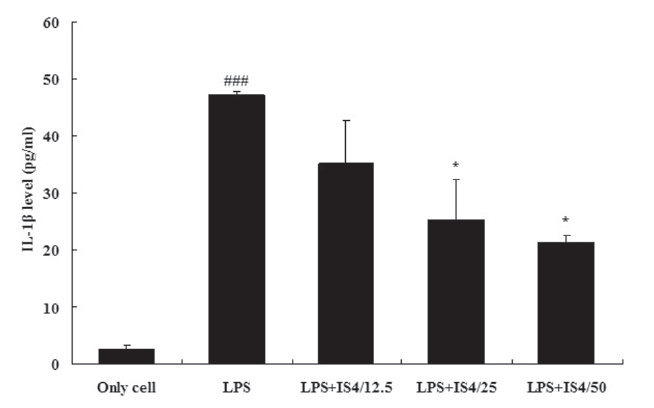
       3.1. IL-1βexpression in RAW 264.7 cells

    To test the effect of IS4 on LPS-induced IL-1β production, we cultured RAW 264.7 cells with LPS (1 ㎍/ml) and various conce-ntrations of IS4 (12.5, 25, 50 ㎍/ml) for 24 hours. The IL-1β concentration in the supernatants was determined by using an ELISA. As shown in Fig. 1, stimulation with LPS significantly inc-reased the expression of IL-1β in RAW 264.7 cells when com-pared to the untreated control (P < 0.001). Interestingly, the cells treated with IS4 inhibited expression of IL-1β in a concentration- dependent manner, which showed 25% inhibition with 12.5 ㎍/ml, 46% inhibition with 25 ㎍/ml (P<0.05), and 55% inhibition with 50 ㎍/ml (P<0.05).

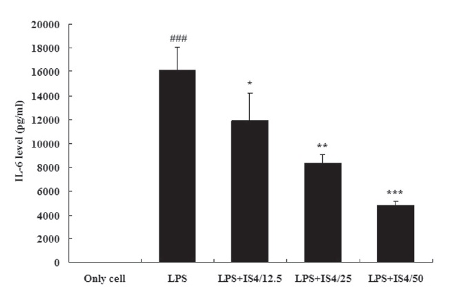
       3.2. IL-6 expression in RAW 264.7 cells

    To test the effect of IS4 on LPS-induced IL-6 production, we cultured RAW 264.7 cells with LPS (1 ㎍/ml) and various conce-ntrations of IS4 (12.5, 25, 50 ㎍/ml) for 24 hours. The IL-6 conce-ntration in the supernatants was determined by using an ELISA. As shown in Fig. 2, stimulation with LPS showed 99.9% increase in the expression of IL-6 in RAW 264.7 cells when compared to the untreated control (P〈 0.001). Moreover, the cells treated with IS4 inhibited IL-6 expression in a concentration-dependent manner, which showed 26% inhibition with 12.5 ㎍/ml (P〈 0.05), 48% inhibition with 25 ㎍/ml (P〈 0.01), and 70% inhibition with 50㎍/ml (P〈 0.001).

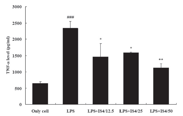
       3.3. TNF-α expression in RAW 264.7 cells

    To test the effect of IS4 on LPS-induced TNF-α production, we cultured RAW 264.7 cells with LPS (1 ㎍/ml) and various conc-entrations of IS4 (12.5, 25, 50 ㎍/ml) for 24 hours. The TNF-α concentration in the supernatants was determined by using an ELISA. As shown in Fig. 3, stimulation with LPS significantly increased the expression of TNF-α in RAW 264.7 cells when compared to the untreated control (P〈 0.001). Moreover, the cells treated with high doses of IS4 (25 and 50 ㎍/ml) inhibited TNF-α expression in a concentration-dependent manner, which showed 38% inhibition with 12.5 ㎍/ml (P〈 0.05), 32% inhibition with 25 ㎍/ml (P〈 0.05), and 52% inhibition with 50 ㎍/ml (P〈 0.01).

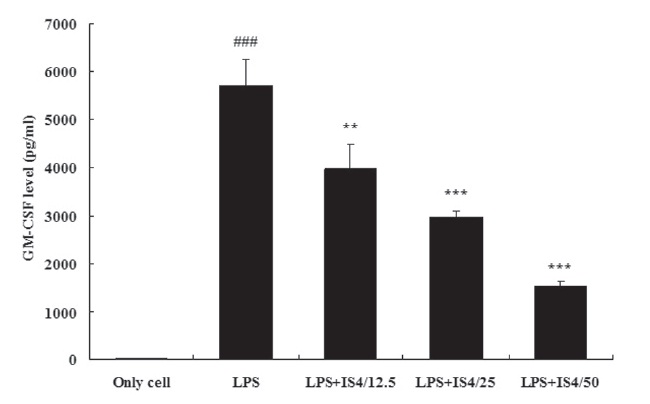
       3.4. GM-CSF expression in RAW 264.7 cells

    To test the effect of IS4 on LPS-induced GM-CSF production, we cultured RAW 264.7 cells with LPS and various concentrations of IS4 (12.5, 25, 50 ㎍/ml) for 24 hours. The GM-CSF concentration in the supernatants was determined by using

    an ELISA. As shown in Fig. 4, stimulation with LPS (1 ㎍/ml) remarkably increased the expression of GM-CSF in RAW 264.7 cells when compared to the untreated control (P〈 0.001). Moreover, the cells treated with IS4 inhibited of GM-CSF expression in a concentration-dependent manner, which showed 30% inhibition with 12.5 ㎍/ml (P〈 0.01), 48% inhibition with 25 ㎍/ml (P〈 0.001), and 73% inhibition with 50 ㎍/ml (P〈 0.001).

    The results were analyzed using Korea Basic Science Institute (KBSI)’s EAS (Expression Analysis System).

    4. Discussion

    Previously, we had tested the efficacy of IS3 (heat treated chalcanthite), IS4 (egg white combined with chalcanthite), and IS5 (raw chalcanthite) on NCI-H460 human lung cancer cells. Among them, the egg-white treated IS4 most successfully inhibited the growth of cells by inducing apoptotic cell death via caspase-3 activation [13]. Other than being a quality protein source, egg whites contain many essential minerals, including potassium, magnesium, calcium, phosphorus, copper, zinc, and iron. They are also a good source of riboflavin and selenium,along with essential vitamins such as folate, B12, niacin betaine, and choline [14]. Historically, in medieval times in the West, egg whites were used for medicinal purposes, such as treating wounds, skin disorders, and mending broken bones [15]. Today, egg whites are considered to be one of the most nutrition-dense foods and are widely recommended for children, the elderly, athletes, and patients. Copper has been used in France since the 18th century to stabilize egg foams. Reactive sulfur items, such as egg whites beaten in a copper bowl, create tighter bo-nds. The bonds created are so tight that the sulfur atoms from the egg whites are prevented from reacting with any other materials [15]. Avidin is a tetrameric biotin-binding glycoprotein found in raw egg white. In chicken egg white, avidin makes up approximately 0.05% of the total protein (approximately 1.8 mg per egg) [16]. The tetrameric protein contains four identical subunits (homotetramers), each of which can bind to biotin (vitamin B7) with a high degree of affinity and specificity [16]. Research in the 1970s helped establish the avidin-biotin system as a powerful tool in biological sciences [17]. Avidin|s affinity for biotin is applied in wide-ranging biochemical assays, including western blot, ELISA, ELISPOT and pull-down assays. Avidin immobilized onto solid supports is also used as decontaminati-on medium to capture biotin-labeled proteins or nucleic acid molecules [17]. For example, lectins, proteins that bind specific sugar molecules on glycoproteins and glycolipids, are expressed at various levels on the surfaces of tumor cells. Conjugation of cytotoxic agents to glycoproteins recognized by lectins could be useful in the treatment of tumors. Avidin, a highly glyco-positively charged protein, contains terminal N-acetylglucosamine and mannose residues that bind to some lectins [18].

    In a 1998 study, the ability of avidin targeting diffe-rent tumor models was tested through radioactively-labeled biotin conjugation. Avidin pre-targeting, followed by injection of radioactive biotin led to avidin-biotin conjugation in-vivo. As a result, radio-labeled avidin were highly and rapidly localized in the tumors, and the uptakes of radioactivity in normal tissues and other organs were low, yielding high tumor-to-non tumor activity ratios [18]. Previously, functional avidin was thought to be found only in raw eggs as the biotin avidity of the protein is destroyed by cooking. A 1966 study claimed that the structure of avidin remained stable at temperatures below 70℃. At temperatures above 70℃, avidin|s structure was rapidly disrup-ted and by 85℃, extensive loss of structure and ability to bind biotin was found [19]. However, a 1992 study disagreed, concluding that protein denaturation was not equivalent to loss of biotin-binding activity [20]. Based on these findings, we examined the effect of IS4 on LPS-stimulated inflammatory cytokines in mouse RAW 264.7 macrophages. Concentrations of pro-inflammatory cytokines, such as IL-1β, IL-6, TNF-α, and GM-CSF, were determined by using an ELISA. As shown in the results, stimulation with LPS significantly increased the expressions of cytokines in RAW 264.7 cells when compared to the untreated control. Moreover, cells treated with IS4 (12.5, 25 and 50 ㎍/ml) inhibited expressions of IL-1β, IL-6 and GM-CSF in concentration-dependent manner. In addition, high doses of IS4 (25 and 50 ㎍/ml) inhibited TNF-α expression, also in a concentration-dependent manner.

    5. Conclusion

    Egg white combined with chalcanthite may be considered as a possible future candidate for an anti-inflammatory agent. Furt-her studies are needed to evaluate the inhibitory mechanisms toward pro-inflammatory cytokines and should focus on in-vivo biological activity assessment of egg white combined with chalcanthite. It would also be interesting to biotinylate the apoptotic, anti-inflammatory cytokine-stimulating agent chalcanthite and have avidin from the egg whites work as a pre-targeting vehicle using avidin-biotin conjugation mechanisms.

      >  Appendix A. Supplementary data

    An online supplementary data associated with this article can be found at doi:10.1016/j.cyto.2008.03.010.

      >  Appendix B. Toxicity data

    Single and repeated oral dose toxicity tests of IS4 were performed and confirmed its safety. The results are currently under review and will be published in the near future.

참고문헌
  • 1. Nordstrom DK, Alpers CN, Negative pH 1999 efflorescent mine-ralogy, and consequences for environmental restoration at the Iron Mountain Superfund site, California. [Proc Natl Acad Sci USA] Vol.96 P.3455-62 google cross ref
  • 2. Gupta A, Chattopadhyay I, Mukherjee S, Sengupta M, Das SK, Ray K 2010 A novel COMMD1 mutation Thr174Met associated with elevated urinary copper and signs of enhanced apopt-otic cell death in a Wilson Disease patient. [Behavioral and Brain Functions] Vol.15 P.33 google
  • 3. Gupta A, Lutsenko S 2009 Human copper transporters: mec-hanism, role in human diseases and therapeutic potential. [Future Medicinal Chemistry] Vol.1 P.1125-42 google cross ref
  • 4. 1984 Technical Note 34, Copper and Human Health - A Review. google
  • 5. Blair BG, Larson CA, Safaei R, Howell SB 2009 Copper transp-orter 2 regulates the cellular accumulation and cytotoxicity of cisplatin and carboplatin. [Clin Cancer Res.] Vol.15 P.4312-21 google cross ref
  • 6. Ishida S, Lee JK, Thiele DJ, Herskowitz I 2002 Uptake of the antic-ancer drug cisplatin mediated by the copper transporter Ctr1 in yeast and mammals. [Proc Natl Acad Sci USA.] Vol.99 P.14298-302 google cross ref
  • 7. Hong YH, Chao WW, Chen ML, Lin BF 2009 Ethyl acetate extracts of alfalfa (Medicago sativa L.) sprouts inhibit lipopolysaccha-rideinduced inflammation in vitro and in vivo. [Journal of Bio-medical Science] Vol.146 P.64 google
  • 8. Tsai JT, Liu HC, Chen YH 2010 Suppression of inflammatory mediators by cruciferous vegetable-derived indole-3-carbinol and phenylethyl isothiocyanate in lipopolysaccha-ride-activated macrophages. [Mediators of Inflammation] P.2936-42 google cross ref
  • 9. Shin SL, Lee SW, Kwon JH, Moon SH, Lee SJ, Lee CK 2009 Cordyce-pin Suppresses Expression of Diabetes Regulating Genes by Inhibition of Lipopolysaccharide-induced Inflamm-ation in Macrophages. [Immune Network] Vol.9 P.98-105 google cross ref
  • 10. Kimura A, Naka T, Nakahama T, Chinen I, Masuda K, Nohara K 2009 Aryl hydrocarbon receptor in combination with Stat1 regulates LPS-induced inflammatory responses. [J Exp Med] Vol.206 P.2027-35 google cross ref
  • 11. Liu N, Lo LS, Askary SH, Jones L, Kidane TZ, Trang T 2007 Transcuprein is a macroglobulin regulated by copper and iron availability. [The Journal of Nutritional Biochemistry] Vol.18 P.597-608 google cross ref
  • 12. 2011 Herbs & Botanicals, Chalcanthite (danfan). google
  • 13. Choi EA, Kim KH, Yoo BC, Yoo HS 2009 Induction of Apoptotic Cell Death by Egg white combined-Chalcanthite on NCI-H460 Human Lung Cancer Cells. [Journal of Pharmacopuncture] Vol.12 P.49-59 google cross ref
  • 14. Lunsford P 2008 The Health Benefits and Nutritional Value ofEgg Whites. google
  • 15. McGee Harold, Vinay 2004 On Food and Cooking: The Science and Lore of the Kitchen. P.102-3 google
  • 16. 2010 Wikipedia Meaning and Defini-tion on Avidin. google
  • 17. Eakin RE, McKinley WA, Williams RJ 1940 Egg-white injury in chi-cks and its relationship to a deficiency of vitamin H (biotin). [Science] Vol.92 P.224-5 google cross ref
  • 18. Yao Z, Zhang M, Sakahara H, Saga T, Arano Y, Konishi J 1998 Avi-din Targeting of Intraperitoneal Tumor Xenografts. [J Natl Cancer Inst] Vol.90 P.25-9 google cross ref
  • 19. Pritchard AB, McCormick DB, Wright LD 1966 Optical rotatory dispersion studies of the heat denaturation of avidin and the avidin-biotin complex. [Biochem Biophys Res Commun] Vol.25 P.524-8 google cross ref
  • 20. Durance TD, Wong NS 1992 Kinetics of thermal inactivation of avidin. [Food Research International] Vol.25 P.89-92 google cross ref
이미지 / 테이블
  • [ Figure 1 ]  Inhibitory effect of IS4 on IL-1βproduction in RAW 264.7cells. Cells were incubated with LPS (1 ㎍/ml) for 24 hr.Data are expressed as percentages of the control values.Values shown are means ± SD of three independentexperiments, each run in triplicate. ### Statisticallysignificant difference from the only cell at P<0.001. *Statistically significant difference from the LPS-treatedcontrol at P<0.05.
    Inhibitory effect of IS4 on IL-1βproduction in RAW 264.7cells. Cells were incubated with LPS (1 ㎍/ml) for 24 hr.Data are expressed as percentages of the control values.Values shown are means ± SD of three independentexperiments, each run in triplicate. ### Statisticallysignificant difference from the only cell at P<0.001. *Statistically significant difference from the LPS-treatedcontrol at P<0.05.
  • [ Figure 2 ]  Inhibitory effect of IS4 on IL-6 production in RAW 264.7cells. Cells were incubated with LPS (1 ㎍/ml) for 24 hr. Data are expressed as percentages of the control values.Values shown are means ± SD of three independentexperiments, each run in triplicate. ### Statisticallysignificant difference from the only cell at P<0.001. *,**,***Statistically significant difference from the LPS-treatedcontrol at P<0.05, P<0.01, and P<0.001, respectively. Means± SD were calculated from five individual results.
    Inhibitory effect of IS4 on IL-6 production in RAW 264.7cells. Cells were incubated with LPS (1 ㎍/ml) for 24 hr. Data are expressed as percentages of the control values.Values shown are means ± SD of three independentexperiments, each run in triplicate. ### Statisticallysignificant difference from the only cell at P<0.001. *,**,***Statistically significant difference from the LPS-treatedcontrol at P<0.05, P<0.01, and P<0.001, respectively. Means± SD were calculated from five individual results.
  • [ Figure 3 ]  Inhibitory effect of IS4 on TNF-α production in RAW 264.7cells. Cells were incubated with LPS (1 ㎍/ml) for 24 hr. Data are expressed as percentages of the control values.Values shown are means ± SD of three independentexperiments, each run in triplicate. ### Statisticallysignificant difference from the only cell at P<0.001. *,**Statistically significant difference from the LPS-treatedcontrol at P<0.05 and P< 0.01, respectively.
    Inhibitory effect of IS4 on TNF-α production in RAW 264.7cells. Cells were incubated with LPS (1 ㎍/ml) for 24 hr. Data are expressed as percentages of the control values.Values shown are means ± SD of three independentexperiments, each run in triplicate. ### Statisticallysignificant difference from the only cell at P<0.001. *,**Statistically significant difference from the LPS-treatedcontrol at P<0.05 and P< 0.01, respectively.
  • [ Figure 4 ]  Inhibitory effect of IS4 on GM-CSF production in RAW 264.7cells. Cells we-re incubated with LPS (1 ㎍/ml) for 24hr. Data are expressed as percentag-es of the control values.Values shown are means ± SD of three independe-ntexperiments each run in triplicate. ### Statistically significant differen-ce from the only cell at P<0.001. ***** Statistically significant difference from the LPS-treatedcontrol at P〈 0.01 and P〈 0.001 respectively.
    Inhibitory effect of IS4 on GM-CSF production in RAW 264.7cells. Cells we-re incubated with LPS (1 ㎍/ml) for 24hr. Data are expressed as percentag-es of the control values.Values shown are means ± SD of three independe-ntexperiments each run in triplicate. ### Statistically significant differen-ce from the only cell at P<0.001. ***** Statistically significant difference from the LPS-treatedcontrol at P〈 0.01 and P〈 0.001 respectively.
(우)06579 서울시 서초구 반포대로 201(반포동)
Tel. 02-537-6389 | Fax. 02-590-0571 | 문의 : oak2014@korea.kr
Copyright(c) National Library of Korea. All rights reserved.