검색 전체 메뉴
PDF
맨 위로
OA 학술지
바지락(Ruditapes philippinarum) 아가미로부터 항균 펩타이드의 탐색 및 정제 Screening and Purification of an Antimicrobial Peptide from the Gill of the Manila Clam Ruditapes philippinarum
  • 비영리 CC BY-NC
  • 비영리 CC BY-NC
ABSTRACT
바지락(Ruditapes philippinarum) 아가미로부터 항균 펩타이드의 탐색 및 정제
KEYWORD
Manila clam , Ruditapes philippinarum , Antimicrobial peptide , Innate defense , Purification
  • 서 론

    대부분의 척추동물 및 무척추동물들은 세균, 바이러스, 곰팡이, 또는 기생충 등과 같은 외부 미생물의 침입으로부터 자신을 방어하기 위한 효과적인 면역 체계를 각각 포함하고 있다(Hancock and Diamond, 2000). 선천면역과 적응면역이 각각 잘 발달되어 있는 척추동물과는 달리 무척추동물은 선천면역 만을 포함하고 있으므로 외부 환경에 대한 방어 작용은 오로지 선천면역에만 의존하고 있다. 일반적으로 선천면역은 외부 침입물질에 대한 초기방어 및 적응 면역의 유도를 담당하므로 그 기능과 역할에 대한 관심이 최근 들어서 증대되고 있는 실정이다. 선천면역을 담당하는 대표적인 물질로는 lectin, prophenoloxidase activating factor, phagocytosis, antimicrobial peptide 등이 알려져 있으며, 이들은 외부로부터 생체 내로의 이물질 침입에 대한 1차 방어 역할을 담당 한다고 알려져 있다(Relf et al., 1999; Tincu and Taylor, 2004). 최근 들어서 여러 가지의 선천 면역 요소들 중에서 비 특이적이고 빠른 반응성을 가지는 자연 친화적인 항균 펩타이드에 대한 관심이 증가되고 있는 실정이다(Zasloff, 2002).

    항균 펩타이드는 유전자에 의해 암호화되어 있는 양친매성의 α-helix나 β-sheet 구조를 취하는 positive net charge를 가지는 12-100개의 아미노산으로 구성된 선천면역에 관여하는 물질로서, 미생물뿐만 아니라 무척추동물에서 척추동물에 이르는 넓은 범위에서 분포하고 있는 단백질성 항균물질이다(van’t Hof et al., 2001; Stark et al., 2002; Izadpanah and Gallo, 2005). 또한 항균 펩타이드는 막을 주요 target site로 삼고 있으므로 내성균 유발의 가능성이 거의 없는 특징을 가지고 있어서 차세대 항생제 개발을 위한 주요 후보물질로서 각광을 받고 있다(Hancock REW and Diamond G, 2000; Zasloff M, 2002).

    항균 펩타이드는 구조에 따라서 크게 4가지 종류로 나눌 수 있다(Andreu and Rivas, 1998): i) Cys을 포함하지 않는 linear α-helical peptide, ii) Cys을 포함하는 looped peptide, iii) 특이 구조를 취하지 않는 random extended peptide, iv) 특이 아미노산을 많이 포함한 peptide 등이다. 항균 펩타이드는 이온 결합과 소수성 상호작용을 이용한 barrel-stave model, toroidal model 또는 carpet model을 통해서 membrane pore를 형성하거나 세포내부로 들어가 세포 내 기관이나 대사과정의 저해를 통해서 항균 효과를 나타낸다고 알려져 있다(Shai, 2002; Li, 2009). 현재까지 해양무척추동물 유래 항균 peptide는 갯지렁이(Ovchinnikova et al., 2004), 군소(Iijima et al., 2003), 투구게(Miyata et al., 1989; Saito et al., 1995), 꽃게(Khoo et al., 1999), 흰 다리 새우(Destoumieux et al., 1997), 홍합(Charlet et al., 1996; Mitta et al., 1999; Mitta et al., 2000), 굴(Seo et al., 2005; 2012; 2013; Nam et al., 2015; Gueguen et al., 2006), 전복(De Zoysa et al., 2009) 등이 보고되어 있다.

    바지락은 우리나라의 패류산업에서 큰 비중을 차지하는 패류 양식 자원중의 하나로서 중요성이 크게 증대되고 있으나, 최근 들어서 다양한 병원성 미생물 감염원에 의한 대량 폐사가 일어나고 있어서 경제적, 환경적으로 큰 문제가 되고 있다(Park et al., 2006). 따라서 이러한 문제점을 해결하기 위한 기초자료로서 바지락의 host defense mechanism을 이해하고, 질병 감염원의 종류 또는 분포와 바지락의 선천면역 요소들 간의 상호연관성 연구가 절실히 요구되고 있는 실정이다. 특히, 다양한 해양무척추동물에 있어서 선천면역요소 중의 하나인 항균 펩타이드의 질병감염에 대한 효과적인 방어기능이 보고되면서 질병감염에 대한 연구가 활발히 진행되고 있는 반면, 바지락과 연관된 항균 펩타이드에 대한 연구는 극히 제한적으로 진행되고 있는 실정이다. 현재까지 보고된 바지락 유래 항균 펩타이드는 defensin(Zhang et al., 2015), big defensin (Zhao et al., 2010) 또는 mussel defensin MGD-1 유사 peptide (Kang et al., 2006) 등과 같은 defensin 류에 한정되어 있으므로 선천면역을 담당하는 다양한 항균 펩타이드에 대한 연구가 필요한 실정이다.

    따라서 본 연구는 바지락(Manila clam, Ruditapes philippinarum)의 면역관련 조직인 아가미 추출물을 이용해서 항균활성의 탐색 및 항균물질의 정제과정을 수행하고 항균물질의 구조 규명 과정을 수행함으로써 바지락의 선천면역에 대한 기초자료를 제공하고자 수행되었다.

    재료 및 방법

      >  실험동물

    본 실험에 사용한 바지락(Manila clam, Ruditapes philippinarum)은 충청남도 곰소만에서 구입하였으며 바지락의 아가미와 외투막 조직을 추출에 사용하였다. 모든 추출물은 사용하기 전까지 −70℃에 보관하였다.

      >  시약 및 재료

    항균활성 측정을 위한 tryptic soy broth (TSB)와 agarose type I (Low EEO Agar)은 Merck사(Merck, Darmstadt, Germany)와 Sigma사(St. Louis, MO, USA)에서 각각 구입하여 사용하였다. 정제 과정에서 사용된 HPLC용 water와 acetonitrile (CH3CN)은 Tedia사(Ohio, USA)로부터 구입하였고, 그 이외의 모든 시약들은 특급을 사용하였다.

      >  추출방법

    바지락의 패각을 흐르는 수돗물에 씻어서 패각 오염물을 제거한 후, 아가미와 외투막 조직을 취하여 5 mL volume이 될 때까지 얼음 속에서 모았다. 모아진 아가미와 외투막 조직에 끓고 있는 4 배량의 1% acetic acid(v/v)를 첨가하여 100℃에서 5분 동안 끓인 후, 얼음에 보관하여 충분히 냉각 시켰다. 충분히 냉각된 조직은 homogenizer (T10 basic ULTRA-TURRAX, IKA, USA)를 사용하여 완전히 파쇄 시켰다. 조직 파쇄액은 4℃에서 20분 동안 8,000 g로 원심분리(VS-21SMT, Vision Scientific, Buchon, Korea)를 행한 후에 상층액을 취해서 항균활성 탐색 및 항균물질의 정제과정에 사용하였다.

      >  항균활성 측정방법 및 사용균주

    항균활성 측정에는 그람 양성균인 Bacillus subtilis KCTC 1021과 그람 음성균인 Escherichia coli D31 (미국 North Carolina 주립대 수의과대학 Noga 교수가 제공)을 사용하였다. 활성측정 방법으로는 서로 다른 농도를 포함한 두 층의 배지를 사용하는 ultrasensitive radial diffusion assay (URDA)법을 이용하였다(Lehrer at al., 1991). 항균활성 측정에 사용된 균주는 우선 tryptic soy broth (TSB)에 접종한 후 18시간 동안 37℃에서 pre-culture를 수행한 후 colorimeter (Product No. 52-1210, BioMerieux, Inc., USA)를 사용하여 균 농도를 84%T(≒ 1×10⁸ CFU/mL)가 되도록 조정하였다. 그 후, 9.5 mL의 0.03% TSB, 1% Type I agarose 및 10 mM phosphate buffer (PB) (pH 6.5)를 포함하는 underlay gel에 각각의 농도로 희석된 균액 0.5 mL를 넣고 잘 섞은 후에 plate에 편평하게 부어 굳혔다. 굳은 plate에 punch를 사용하여 직경 2.5 mm의 well을 뚫은 후에 5 μL의 각 추출물 또는 정제 단계의 분획물을 도입시켰다. 추출물 또는 분획물이 배지에 스며들면 3시간 동안 37℃에서 1차 배양한 후, 그 위에 10 mL의 6% TSB, 1% TypeⅠ agarose 및 10 mM phosphate buffer (pH 6.5)를 포함하는 overlay gel을 붓고 굳힌 후에 동일한 온도에서 18시간 동안 2차 배양하였다. 다음날 well 주위에 생긴 clear zone의 크기를 측정하여 항균활성을 확인하였다. 항균활성 측정 동안 positive control로는 미국산 잡종 농어(Morone saxatilis×Morone chrysops)의 mast cell에서 정제된 항균 펩타이드인 piscidin 1을 사용하였고 negative control로는 1% acetic acid 또는 0.01% acetic acid를 사용하였다(Silphaduang and Noga, 2001).

      >  Protease 처리에 의한 단백질성 확인

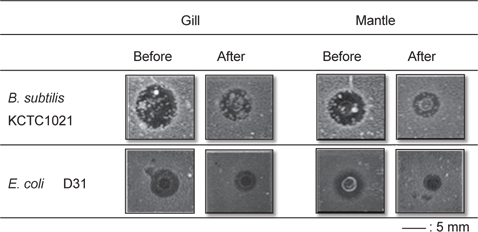
    추출물 또는 정제된 항균물질의 단백질성 확인을 위해서 trypsin 처리 전·후의 항균활성 변화를 확인하였다. 바지락 추출물 또는 정제된 물질 4 μL에 trypsin (1,000 μg/mL) 1 μL를 첨가하고 37℃에서 60분간 반응시킨 뒤, URDA법으로 B. subtilis KCTC1021에 대한 항균활성 변화를 측정하였다.

      >  Hemolysis

    추출물에 존재하는 독성물질의 유무를 확인하기 위해서 인간의 적혈구에 대한 용혈활성을 측정하였다(Seo et al., 2014). 사람의 혈액(Blood type: B)에 동량의 phosphate buffered saline (PBS, 50 mM sodium phosphate, 150 mM NaCl, pH 7.4)을 넣고 혼합한 후 800 rpm에서 15분간 원심 분리하여 상등액은 버리고 침전물을 얻었다. 이러한 세척과정을 3회 반복한 후 얻어진 적혈구를 1% 혼합용액이 되도록 PBS를 첨가한 후 사용하였다. 얻어진 적혈구 90 μL를 96-well microplate에 도입시키고 바지락 추출물 또는 정제물질 10 μL를 첨가한 다음 37℃에서 60분간 반응시켰다. 이후 반응액을 원심분리(1,500 rpm, 10분, 4℃) 하여 얻어진 상등액 80 μL를 새로운 microtiter plate로 옮긴 후 헤모글로빈 유출 정도를 확인하기 위해서 490 nm에서 흡광도를 측정하였다. 적혈구의 100% 용혈을 위한 비교물질로는 triton X-100 (0.1%)을 사용하였다.

      >  Acid-Urea PAGE와 bug-blot

    바지락 추출물 내의 양이온성 단백질/펩타이드의 조성분포와 활성 band를 확인하기 위해서 acid-urea PAGE와 bug-blot을 E. coli D31에 대해서 수행하였다(Seo et al., 2005). Bug-blot에 사용된 E. coli D31은 TSB에서 37℃로 18시간 동안 배양한 후 colorimeter (Product No. 52-1210, BioMerieux, Inc., USA)를 사용하여 균 농도를 84%T (≒ 1×10⁸ CFU/mL)가 되게 조정하였다. 그 후, 9.5 mL의 LB, 1% TypeⅠ agarose, 10 mM phosphate buffer (PB) (pH 6.7) 및 NaCl를 포함하는 gel에 희석된 균액 0.5 mL를 넣고 잘 섞은 후에 plate에 편평하게 부어 굳혔다

    AU-PAGE를 위한 gel은 urea 3.6 g, D.W 3.4 mL, 30% acrylamide/1.6% bis-acrylamide solution 5 mL, glacial HAc 0.65 mL를 비커에 넣고 stirring (10min, RT)과 degassing (20 min, RT, desiccator)을 한 후 TEMED 0.06 mL, 10% APS 0.275 mL를 첨가한 후 stirring (1 min, RT)하고, gel액을 붓고 유출 유무를 확인한 뒤 comb를 꽂은 후 충분히 실온에서 굳혔다. 이후 장치에 5% HAc를 붓고 pre-run (150 v, 60 min, RT)을 하였다. Well에 도입할 각 시료는 20 µL의 2×sample buffer와 1:1(v/v)로 혼합해서 사용하였고, standard는 histone H1 (∼21 kDa), lysozyme (∼11 kDa), aprotinin (∼6.5 kDa) 및 piscidin 1 (∼2.5 kDa)을 포함하는 20 µL의 0.01% HAc와 2×Sample buffer를 1:1 (v/v)로 혼합해서 사용하였다. Pre-run이 끝난 후 running buffer (5% HAC)를 교체하고 well에 시료를 도입시킨 후 전기영동을 실시하였다(150 v, 45 min). 전기영동 후, 1개의 gel은 CBB R-250으로 염색시켰고, 나머지 1개의 gel을 rinse buffer (10 mM PB buffer, pH 7.4)로 2회 세척을 한 후에 E. coli D31을 포함하는 LB plate에 overlay시킨 후 37℃에서 3시간 배양하였다. 배양 후에 overlay시켰던 gel을 제거한 후에 LB plate는 동일한 온도에서 18시간 동안 2차 배양하였다. 다음날 LB plate에 나타난 clear zone을 염색 및 탈색 된 gel의 band 결과와 비교하였다.

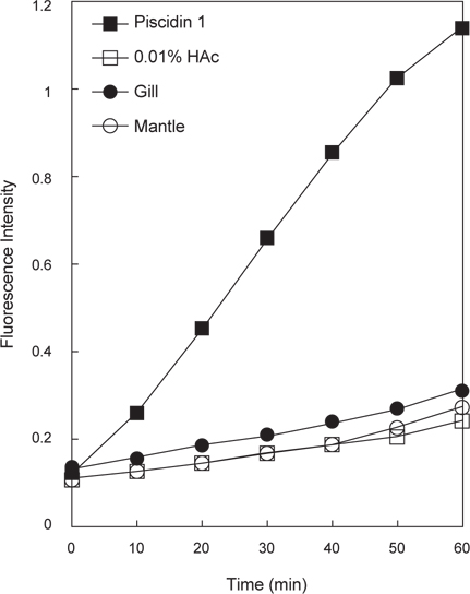
      >  추출물들의 cytoplasmic membrane permeabilizatin assay

    바지락 추출물의 세균 내막 투과성 측정을 위해서 nonmembrane-permeative chromogenic substrate인 o-nitrophenyl-β-D-galactopyranoside (ONPG)에 대한 추출물 도입 후의 E. coli ML35p의 세포질에서 유출된 β-galactosidase의 활성을 확인하는 실험을 수행하였다 (Skerlavaj et al., 1990). 배양된 mid-log phase의 E. coli ML35p를 10 mM sodium phosphate buffer (pH 7.4)로 세척을 한 후, 1.5 mM의 ONPG를 포함하는 동일 butter에 용해시켰다. 그 후, 측정할 추출물들을 첨가한 뒤 37℃에서 60분간 배양하면서 10분 간격으로 유출된 β-galactosidase에 의한 ONPG의 o-nitrophenol로의 가수분해 정도를 405 nm에서 측정하였다. 막 투과활성 측정의 표준물질로는 강한 막 투과성을 포함하는 것으로 알려진 항균 펩타이드인 piscidin 1을 사용하였다.

      >  DNA Binding Assay

    바지락 추출물에 존재하는 염기성의 DNA 결합성 물질의 존재유무를 확인하기 위해서 DNA binding에 의한 DNA band들의 agarose gel-electrophoresis에서의 이동의 저해정도를 확인하는 DNA-binding assay를 수행하였다(Hsu et al., 2003; Alfred et al., 2013). 이를 위해서 1 kb DNA marker (λ-HindIII-digested DNA, 50 ng)와 추출물을 혼합해서 37℃에서 60분 동안 반응시킨 후 0.5 μg/mL EtBr을 포함하는 1.4% agarose gel에서 전기영동을 수행한 후 DNA-binding 정도를 확인하였다.

      >  항균활성물질의 분리 및 정제

    아가미 추출물에 존재하는 항균물질의 분리 및 정제과정은 HPLC (YL9100 HPLC system, Young Lin Instrument Co., Korea)를 이용하여 수행하였다. 추출물에 존재하는 항균물질의 정제과정은 역상 및 이온 교환성을 갖는 2가지 특성의 HPLC column을 사용하여 수행되었고, 정제과정 중의 분획들에 대한 항균활성은 B. subtilis KCTC1021에 대해서 URDA법으로 측정하였다. 첫 번째 정제과정으로 CapCell-Pak C18 column (4.6 mm×250 mm, Shiseido, Japan)을 이용하여 다음과 같은 조건으로 분리과정을 수행하였다. A 용매는 0.1% Trifluoroacetic acid (TFA)를 포함하는 H2O (pH 2.2)이며 B용매는 0.1% TFA를 포함하는 100% CH3CN (pH 2.2)를 사용하였다. 분리조건은 5%에서 65%까지 60분간 B용매를 순차적으로 증가시켰으며, 유속은 1.0 mL/min, 파장은 220 nm에서 확인하였고 모든 정제과정은 실온에서 수행되었다. 항균활성을 지닌 분획들을 모아서 TSK-gel SP-5PW column (7.5 mm×75 mm, Tosoh, Japan)에 도입시키고 다음과 같은 조건으로 분리과정을 수행하였다. A buffer는 10 mM phosphate buffer (PB, pH 6.0)이며 B buffer는 1.0 M NaCl을 포함하는 10 mM PB (pH 6.0)를 사용하였다. 분리조건은 0%에서 100%까지 100분 동안 B buffer를 순차적으로 증가시켰으며, 유속은 1.0 mL/min, 파장은 220 nm에서 분리과정을 수행하였다. 활성이 확인된 분획은 CapCell-Pak C18 column (4.6 mm×250 mm)에 적용시켜 다음과 같은 조건으로 최종 분리과정을 수행하여 단일 peak를 정제하였다. A 용매는 0.1 % TFA를 포함하는 H2O (pH 2.2)이며 B 용매는 0.1% TFA를 포함하는 100% CH3CN (pH 2.2)를 사용하였다. 분리조건은 5%에서 45%까지 40분간 B용매를 순차적으로 증가시켰으며, 유속은 1.0 mL/min, 파장은 220 nm에서 확인하였다.

      >  분자량 측정

    최종 정제된 물질의 분자량은 Ultraflex III matrix-assisted laser desorption ionisation time-of-flight (MALDI-TOF) mass spectrometer (Bruker Daltonik GmbH, Bremen, Germany)를 사용하여 linear mode로 측정하였다. 정제물질을 0.1% TFA/50% CH3CN (1:1, v/v)에 녹인 후 α-cyano-4- hydroxycinnamic acid (CHCA) matrix solution (10 mg/mL CHCA in 0.1% TFA/50% CH3CN, 1:1, v/v)과 혼합하여 MALDI plate에 도입한 후에 분자량을 측정하였다. Standard로는 Calmix 2를 사용하였다.

      >  펩타이드 일차구조 분석

    정제물의 N-terminal amino acid sequence는 pulse liquid automatic sequencer (model 473A; Applied Biosystems, Foster City, CA, USA)를 사용하여 분석하였다.

    결과 및 고찰

      >  바지락 추출물의 생리활성

    바지락 추출물의 항균활성

    바지락(Manila clam, Ruditapes philippinarum)으로부터 채취한 아가미와 외투막 조직들은 1% HAC를 이용한 약산추출 과정을 수행하였으며, 각 추출물 내의 단백질성 항균물질의 존재유무를 확인하기 위하여 각 추출물(4 μL)의 trypsin 처리 전∙후의 항균활성 변화를 URDA법을 이용하여 B. subtilisE. coli D31에 대해서 측정하였다(Fig. 1). 실험 결과, 각 추출물은 B. subtilisE. coli D31에 대해서 강한 항균활성을 나타내었으며, trypsin 처리 후에는 항균활성이 현저히 감소되었다. 이러한 결과는 바지락 추출물에 단백질성 항균물질이 포함되어 있음을 의미하는 것이다. 또한 추출물 또는 균주 간의 활성 비교에서는 아가미 추출물이 외투막 추출물보다는 강한 항균활성을 나타내었으며, E. coli D31보다 B. subtilis에 더욱 효과적인 것으로 나타났다. 이러한 결과를 토대로 바지락유래 단백질성 항균물질을 정제하기 위해서 아가미 추출물을 연구과정에 사용하였다.

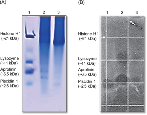
    Acid-urea PAGE와 bug-blot

    바지락 추출물내의 염기성 단백질/펩타이드의 구성분포와 그들의 항균활성 유무를 확인하기 위해서 AU-PAGE와 bug-blot를 수행하였다(Fig. 2). 그 결과, 바지락 추출물들은 저분자 물질보다는 상대적으로 고분자 물질의 농도가 높게 나타났으며, 외투막 추출물보다는 아가미 추출물에서 구성물질들의 다양성이 더 높게 나타났다. 또한, 추출물의 항균활성은 slow migration zone (SMZ) (~11 kDa)과 rapid migration zone (RMZ) (~2.5 kDa)을 포함하는 두 위치에서 나타났으며, RMZ의 항균활성이 SMZ보다는 강하게 나타났다. 이러한 결과는 바지락 추출물에는 다양한 분자량의 항균물질이 존재하고 있으며, 아마도 저분자의 항균물질이 보다 중추적인 역할을 담당하는 것으로 판단된다.

    바지락 추출물의 hemolysis

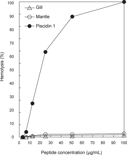
    바지락 추출물들에 포함된 독성물질의 유무를 확인하기 위해서 추출물을 인간 적혈구(혈액형: B형)에 대한 용혈활성(hemolytic activity)을 측정하였다(Fig. 3). 비교물질로는 piscidin 1을 사용하였으며 100% hemolysis의 표준물질로는 0.1% TritonX 100을 사용하였다. 측정결과 대조군으로 사용된 항균 펩타이드인 piscidin 1은 낮은 농도(12.5 μg/mL)에서도 강한 용혈활성을 나타낸 반면, 바지락 추출물들은 용혈활성을 거의 나타내지 않았다. 이러한 결과는 바지락 추출물들은 세포독성을 나타내는 물질을 거의 포함하고 있지 않다는 것을 의미하는 것이다.

    바지락 추출물에 포함된 항균물질의 작용부위 확인 실험

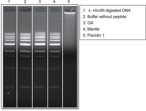
    바지락 추출물에 포함된 항균물질의 target site를 예측하기 위해서 세균 내막 투과성과 핵내 구성물인 DNA와의 상호작용 유무를 관찰하였다. 우선, 항균물질의 target site가 핵 내부의 DNA인지를 확인하기 위해서 DNA-peptide interaction에 의한 DNA migration 저해현상을 관찰할 수 있는 DNA-binding assay를 수행하였다(Fig. 4). 그 결과, 양성 대조군으로 사용한 piscidin 1은 강한 DNA-binding 활성을 나타낸 반면 바지락 추출물들은 DNA migration에 전혀 영향을 주지 않는 것으로 나타났다. 이러한 결과는 바지락 추출물에 존재하는 항균물질은 핵 내의 DNA와의 상호작용에 의해 항균작용을 나타내는 것은 아님을 의미하는 것이다. 두 번째로, 추출물에 포함된 항균물질의 target site가 세포막 자체인지를 확인하기 위해서 membrane permeabilization assay를 수행하였다(Fig. 5). 실험결과 양성 대조군으로 사용한 piscidin 1은 강한 내막 투과성을 나타낸 반면 음성 대조군으로 사용한 1% HAc와 측정에 사용된 바지락 추출물들은 막 투과활성을 거의 나타내지 않았다. 이것은 실험에 사용된 1% HAc는 막 투과활성에 거의 영향이 없음을 나타내는 것이므로 바지락 추출물들에는 막 투과 활성물질이 거의 포함하고 있지 않다는 것을 의미하는 것이다. 이러한 결과는 추출물내의 항균물질의 예상 작용 target site은 막 자체는 아니라는 것을 의미하는 것이다. 두 가지의 실험 결과 바지락 추출물에 포함된 항균물질의 작용기작은 세포막을 직접 공격하거나 핵 내부의 DNA와의 직접적인 상호작용에 의한 것보다는 세포표면 물질들과의 상호작용 또는 세포질에 존재하는 세포 내 구성 성분에 작용할 가능성이 있다는 것을 의미하는 것이다.

      >  항균활성 물질의 정제

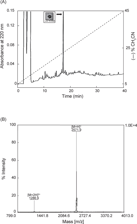
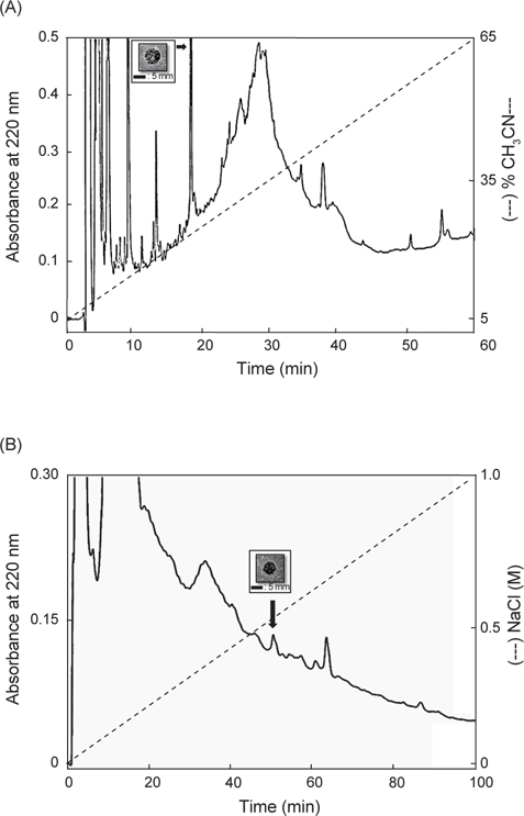
    바지락 아가미 추출물로부터 항균물질을 정제하기 위해서 역상 HPLC column 및 이온교환 column을 사용한 정제과정을 수행함으로써 순수한 항균물질을 정제하였다. 첫 번째 정제과정으로 아가미 추출물을 CapCell-Pak C18 column (4.6 mm×250 mm, Shiseido, Japan)에 도입하였으며, 분리된 각각의 분획들은 B. subtilis에 대해서 URDA법으로 항균활성을 측정하였다(Fig. 6A). 그 결과, 18분에서 19분 사이에 용출된 분획에서 항균활성이 나타났으며 활성분획을 모아서 cation-exchange column인 TSK-gel SP-5PW column (7.5 mm×75 mm, Tosoh, Japan)에 도입해 다음 분리과정을 수행하였다(Fig. 6B). 항균활성은 0.5 M NaCl 농도에서 용출된 분획에서 확인되었으며 항균활성이 확인된 양이온 교환 column으로부터 얻은 분획을 모아 최종적으로 CapCell-Pak C18 column (4.6 mm×250 mm)에 도입하여 순수한 항균물질을 정제하였다(Fig. 7A). 최종 정제된 물질에 trypsin을 처리한 후 항균활성을 확인한 결과 정제물질의 항균활성이 완전히 소실되었다(data not shown). 이러한 결과는 최종 정제된 항균물질은 단백질성 항균물질임을 의미하는 것으로써 정제된 항균물질의 일차구조 분석을 위해서 분자량 측정(MALDI-TOF MS)과 edman degradation 과정을 수행하였다.

      >  정제된 항균활성 물질의 일차구조 분석

    최종 정제된 항균물질의 분자량은 Ultraflex III matrix-assisted laser desorption ionisation time-of-flight (MALDI-TOF) mass spectrometer (Bruker Daltonik GmbH, Bremen, Germany)를 이용하여 linear mode로서 측정하였다(Fig. 7B). 분자량 측정을 위한 matrix로는 CHCA (ɑ-cyano-4-hydroxycinamic acid)를 사용하였으며, standard로는 calmix 2를 사용하였다. 분자량을 측정한 결과, 정제된 물질의 singly charged species는 2571.9 [M+H]+와doubly charged species는 1,286.9 [M+2H]2+의 mass data가 얻어졌으며, 이를 바탕으로 정제 물질의 분자량은 2,571.9 Da으로 확인되었다. 이러한 결과를 바탕으로 정제 물질의 일차구조를 분석하기 위해서 edman degradation을 통한 N-말단 서열분석을 수행하였으나 구성 아미노산에 대한 분석 결과를 얻지 못하였다. 이것은 아마도 정제된 항균물질의 N-말단이 번역 후 변형(post-translational modification) 과정에 의한 specific functional group으로 blocking된 것에 기인한 것으로 판단된다. 일반적으로 척추 또는 무척추동물 유래의 많은 항균 펩타이드들에서 분해효소에 대한 저항성 강화 등의 목적으로 번역 후 변형과정을 통해서 N-말단이 blocking되어 있는 경우가 자주 보고되고 있다(Andreu and Rivas, 1998). 본 연구에서 정제된 단백질성 항균물질의 N-terminal blocked group의 확인과 완전한 일차구조의 규명을 위한 추가적인 연구과정이 수행되어야 할 것으로 판단된다.

    본 연구의 결과 바지락의 아가미와 외투막에는 다양한 단백질성 항균물질들이 포함되어 있는 것으로 확인되었으며 특히, 아가미 추출물에서 분자량이 2571.9 Da의 단백질성 항균물질의 존재를 확인할 수 있었다. 이러한 결과는 바지락의 선천면역에 대한 이해를 증대시킬 수 있는 기초자료로 활용될 수 있을 것으로 판단된다.

참고문헌
  • 1. Alfred RL, Palombo EA, Panozzo JF, Bhave M 2013 The antimicrobial domains of wheat puroindolines are cell-penetrating peptides with possible intracellular mechanism of action [PLoS One] Vol.8 P.e75488 google cross ref
  • 2. Andreu D, Rivas L 1998 Animal antimicrobial peptides: an overview [Biopolymers] Vol.147 P.415-433 google
  • 3. Charlet M, Chernysh S, Philippe H, Hetru C, Hoffmann JA, Bulet P 1996 Innate immunity: Isolation of several cysteine-rich antimicrobial peptides from the blood of a mollusk, Mytilus edulis [J Biol Chem] Vol.271 P.21808-21813 google cross ref
  • 4. Destoumieux D, Bulet P, Loew D, Van DA, Rodriguez J, Bachere E 1997 Penaeidins, a new family of antimicrobial peptides isolated from the shrimp Penaeus vannamei (Decapoda) [J Biol Chem] Vol.272 P.28398-28406 google cross ref
  • 5. De Zoysa M, Nikapitiya C, Whang I, Lee JS, Lee J 2009 Abhisin: a potential antimicrobial peptide derived from histone H2A of disk abalone (Haliotis discus discus) [Fish Shellfish Immunol] Vol.27 P.639-646 google cross ref
  • 6. Gueguen Y, Herpin A, Aumelas A, Garnier J, Fievet J, Escoubas JM, Bulet P, Gonzalez M, Lelong C, Favrel P, Bachere E 2006 Characterization of a defensin from the oyster Crassostrea gigas. Recombinant production, folding, solution structure, antimicrobial activities, and gene expression [J Biol Chem] Vol.281 P.313-323 google cross ref
  • 7. Hancock REW, Diamond G 2000 The role of cationic antimicrobial peptides in innate host defences [Trends Microbiol] Vol.8 P.402-410 google cross ref
  • 8. Hsu CH, Chen C, Jou ML, Lee AYL, Lin YC, Yu YP, Huang WT, Wu SH 2005 Structural and DNA-binding studies on the bovine antimicrobial peptide, indolicidin, evidence for multiple conformations involved in binding to membranes and DNA [Nucl Acids Res] Vol.33 P.4053-4064 google cross ref
  • 9. Iijima R, Kisugi J, Yamazaki M 2003 A novel antimicrobial peptide from the sea hare Dolabella auricularia [Dev Comp Immunol] Vol.27 P.305-311 google cross ref
  • 10. Izadpanah A, Gallo RL 2005 Antimicrobial peptides [J Am Acad Dermatol] Vol.52 P.381-390 google cross ref
  • 11. Kang YS, Kim YM, Park KL, Cho SK, Choi KS, Cho M 2006 Analysis of EST and lectin expression in hemocytes of Manila clams (Ruditapes phylippinarum) (Bivalvia: Mollusca) infected with Perkinsus olseni [Dev Comp Immunol] Vol.30 P.1119-1131 google cross ref
  • 12. Khoo L1, Robinette DW, Noga EJ 1999 Callinectin, an antibacterial peptide from blue crab, Callinectes sapidus, hemocytes [Mar Biotechnol] Vol.1 P.44-51 google cross ref
  • 13. Lehrer RI, Rosenman M, Harwig SSL, Jackson R, Eisenhaur P 1991 Ultrasensitive assay for endogenous antimicrobial polypeptides [J Immunol Methods] Vol.137 P.167-173 google cross ref
  • 14. Li Y 2009 The role of antimicrobial peptides in cardiovascular physiology and disease [Biochem Biophys Res Commun] Vol.390 P.363-367 google cross ref
  • 15. Mitta G, Hubert F, Dyrynda EA, Boudry P, Roch P 2000 Mytilin B and MGD2, two antimicrobial peptides of marine mussels: gene structure and expression analysis [Dev Comp Immunol] Vol.24 P.381-393 google cross ref
  • 16. Mitta G, Hubert F, Noel T, Roch P 1999 Myticin, a novel cysteine-rich antimicrobial peptide isolated from haemocytes and plasma of the mussel Mytilus galloprovincialis [Eur J Biochem] Vol.265 P.71-78 google cross ref
  • 17. Miyata T, Tokunaga F, Yoneya T, Yoshikawa K, Iwanaga S, Niwa M, Takao T, Shimonishi Y 1989 Antimicrobial peptides, isolated from horseshoe crab hemocytes, tachyplesin II, and polyphemusins I and II: chemical structures and biological activity [J Biochem] Vol.106 P.663-668 google
  • 18. Nam BH, Seo JK, Lee MJ, Kim YO, Kim DG, An CM, Park NG 2015 Functional analysis of Pacific oyster (Crassostrea gigas) β-thymosin: Focus on antimicrobial activity [Fish Shellfish Immunol] Vol.45 P.167-174 google cross ref
  • 19. Ovchinnikova TV, Aleshina GM, Balandin SV, Krasnosdembskaya AD, Markelov ML, Frolova EI, Leonova YF, Tagaev AA, Krasnodembsky EG, Kokryakov VN 2004 Purification and primary structure of two isoforms of arenicin, a novel antimicrobial peptide from marine polychaeta Arenicola marina [FEBS Lett] Vol.577 P.209-214 google cross ref
  • 20. Park KI, Paillard C, Chevalier P, Choi KS 2006 Report on the occurrence of brown ring disease (BRD) in Manila clam, Ruditapes philippinarum, on the west coast of Korea [Aquaculture] Vol.255 P.610-613 google cross ref
  • 21. Relf JM, Chisholm JRS, Kemp GD, Smith VJ 1999 Purification and characterization of a cystein-rich 11.5-kDa antibacterial protein from the granular haemocytes of the shore crab, Carcinus maenas [Eur J Biochem] Vol.264 P.350-357 google cross ref
  • 22. Saito T, Kawabata S, Shigenaga T, Takayenoki Y, Cho J, Nakajima H, Hirata M, Iwanaga S 1995 A novel big defensin identified in horseshoe crab hemocytes: isolation, amino acid sequence, and antibacterial activity [J Biochem] Vol.117 P.1131-1137 google
  • 23. Seo JK, Crawford JM, Stone KL, Noga EJ 2005 Purification of a novel arthropod defensin from the American oyster, Crassostrea virginica [Biochem Biophys Res Commun] Vol.338 P.1998-2004 google cross ref
  • 24. Seo JK, Lee MJ, Go HJ, Kim GD, Jeong HD, Nam BH, Park NG 2013 Purification and antimicrobial function of ubiquitin isolated from the gill of Pacific oyster, Crassostrea gigas [Mol Immunol] Vol.53 P.88-98 google cross ref
  • 25. Seo JK, Lee MJ, Go HJ, Kim YJ, Park NG 2014 Antimicrobial function of the GAPDH-related antimicrobial peptide in the skin of skipjack tuna, Katsuwonus pelamis [Fish Shellfish Immunol] Vol.36 P.571-581 google cross ref
  • 26. Seo JK, Lee MJ, Nam BH, Park NG 2013 cgMolluscidin, a novel dibasic residue repeat rich antimicrobial peptide, purified from the gill of the Pacific oyster, Crassostrea gigas [Fish Shellfish Immunol] Vol.35 P.480-488 google cross ref
  • 27. Shai Y 2002 Mode of action of membrane active antimicrobial peptides [Biopolymers] Vol.66 P.236-248 google cross ref
  • 28. Silphaduang U, Noga EJ 2001 Peptide antibiotics in mast cells of fish [Nature] Vol.414 P.268-269 google cross ref
  • 29. Skerlavaj B, Romeo D, Gennaro R 1990 Rapid membrane permeabilization and inhibition of vital functions of Gram-negative bacteria by bactenecins [Infect Immun] Vol.58 P.3724-3730 google
  • 30. Stark M, Liu LP, Deber CM 2002 Cationic hydrophobic peptides with antimicrobial activity [Antimicrob Agents Chemother] Vol.46 P.3585-3590 google cross ref
  • 31. Tincu JA, Taylor SW 2004 Antimicrobial peptides from marine invertebrates [Antimicrob Agents Chemother] Vol.48 P.3645-3654 google cross ref
  • 32. van’’t Hof W, Veerman EC, Helmerhorst EJ, Amerongen AV 2001 Antimicrobial peptides: properties and applicability [Biol Chem] Vol.382 P.597-619 google cross ref
  • 33. Zasloff M 2002 Antimicrobial peptides of multicellular organisms [Nature] Vol.415 P.389-395 google cross ref
  • 34. Zhang L, Yang D, Wang Q, Yuan Z, Wu H5, Pei D, Cong M, Li F, Ji C, Zhao J 2015 A defensin from clam Venerupis philippinarum: Molecular characterization, localization, antibacterial activity, and mechanism of action [Dev Comp Immunol] Vol.51 P.29-38 google cross ref
  • 35. Zhao J, Li C, Chen A, Li L, Su X, Li T 2010 Molecular characterization of a novel big defensin from clam, Venerupis philippinarum [PLoS One] Vol.5 P.e13480 google cross ref
OAK XML 통계
이미지 / 테이블
  • [ Fig. 1. ]  Antimicrobial activity of the crude extracts of the Manila clam Ruditapes philippinarum. Antimicrobial activity of gill or mantle extract (before) and trypsin treated extract (after) against B. subtilis KCTC1021 and E. coli D31. Scale bar indicates 5 mm.
    Antimicrobial activity of the crude extracts of the Manila clam Ruditapes philippinarum. Antimicrobial activity of gill or mantle extract (before) and trypsin treated extract (after) against B. subtilis KCTC1021 and E. coli D31. Scale bar indicates 5 mm.
  • [ Fig. 2. ]  AU-PAGE gel overlay assay of gill and mantle extract against E. coli D31. (A) AU-PAGE run and stained with Coomassie Brilliant Blue R-250. Lane 1, molecular weight markers: 3 μg of human histone H1, 2 μg of human lysozyme, 1 μg of aprotinin, and 1 μg of piscidin 1, lane 2: 20 μL of gill extract, lane 3: 20 μL of mantle extract. (B) AU-PAGE gel overlay assay of gill and mantle extract against E. coli D31.
    AU-PAGE gel overlay assay of gill and mantle extract against E. coli D31. (A) AU-PAGE run and stained with Coomassie Brilliant Blue R-250. Lane 1, molecular weight markers: 3 μg of human histone H1, 2 μg of human lysozyme, 1 μg of aprotinin, and 1 μg of piscidin 1, lane 2: 20 μL of gill extract, lane 3: 20 μL of mantle extract. (B) AU-PAGE gel overlay assay of gill and mantle extract against E. coli D31.
  • [ Fig. 3. ]  Hemolytic activities of gill or mantle extract and piscidin 1 against human erythrocytes (blood type: B).
    Hemolytic activities of gill or mantle extract and piscidin 1 against human erythrocytes (blood type: B).
  • [ Fig. 4. ]  Gel retardation analysis or the binding of gill or mantle extract to DNA. Binding of extract to DNA was assessed by measuring the retardation of commercial molecular weight marker λ-HindIII-digested DNA (50 ng) migration through an agarose gel. Lane 1: λ-HindIII-digested DNA(50 ng), lane 2: negative control, 0.0.1% HAc, lane 3: gill extract, lane 4: mantle extract, lane 5: positive control, piscidin 1 1 μg.
    Gel retardation analysis or the binding of gill or mantle extract to DNA. Binding of extract to DNA was assessed by measuring the retardation of commercial molecular weight marker λ-HindIII-digested DNA (50 ng) migration through an agarose gel. Lane 1: λ-HindIII-digested DNA(50 ng), lane 2: negative control, 0.0.1% HAc, lane 3: gill extract, lane 4: mantle extract, lane 5: positive control, piscidin 1 1 μg.
  • [ Fig. 5. ]  Cytoplasmic membrane permeabilization of E. coli ML35p by gill or mantle extract and piscidin 1. Cytoplasmic membrane permeabilization was monitored as an increase in fluorescence intensity by the hydrolysis of the impermeable, chromogenic substrate ONPG in the presence of piscidin 1 (1 μg/mL) or extract (3 μL) for 60 min.
    Cytoplasmic membrane permeabilization of E. coli ML35p by gill or mantle extract and piscidin 1. Cytoplasmic membrane permeabilization was monitored as an increase in fluorescence intensity by the hydrolysis of the impermeable, chromogenic substrate ONPG in the presence of piscidin 1 (1 μg/mL) or extract (3 μL) for 60 min.
  • [ Fig. 6. ]  Purification of antimicrobial peptide from the gill extract. (A) Reversed-phase HPLC separation of the Manila clam gill extract. The acidic extract was fractionated by the CapCell-Pak C18 column. Elution was performed with a linear gradient of 5->65% CH3CN in 0.1% TFA for 60 min at a flow rate of 1 mL/min. The eluate was monitored at 220 nm. Fraction of the absorbance peak (indicated by the arrow) showed antimicrobial activity against B. subtilis KCTC1021 (inset). The elution point of the active peak was at 23% CH3CN. (B) Active fraction was pooled and then applied to a cation-exchange column (TSK-gel SP-5PW, 7.5×75 mm). Elution was performed with a linear gradient of buffer A (10 mM PB, pH 6.0) and buffer B (1.0 M NaCl in 10 mM PB, pH 6.0) from 0 to 1.0 M NaCl for 100 min at a flow rate of 1 mL/min. The eluate was monitored at 220 nm. The elution point of the active fraction occurred at 0.51 M NaCl (arrow). Fraction of the absorbance peak (indicated by the arrow) showed antimicrobial activity against B. subtilis KCTC1021 (inset). Scale bar indicates 5 mm.
    Purification of antimicrobial peptide from the gill extract. (A) Reversed-phase HPLC separation of the Manila clam gill extract. The acidic extract was fractionated by the CapCell-Pak C18 column. Elution was performed with a linear gradient of 5->65% CH3CN in 0.1% TFA for 60 min at a flow rate of 1 mL/min. The eluate was monitored at 220 nm. Fraction of the absorbance peak (indicated by the arrow) showed antimicrobial activity against B. subtilis KCTC1021 (inset). The elution point of the active peak was at 23% CH3CN. (B) Active fraction was pooled and then applied to a cation-exchange column (TSK-gel SP-5PW, 7.5×75 mm). Elution was performed with a linear gradient of buffer A (10 mM PB, pH 6.0) and buffer B (1.0 M NaCl in 10 mM PB, pH 6.0) from 0 to 1.0 M NaCl for 100 min at a flow rate of 1 mL/min. The eluate was monitored at 220 nm. The elution point of the active fraction occurred at 0.51 M NaCl (arrow). Fraction of the absorbance peak (indicated by the arrow) showed antimicrobial activity against B. subtilis KCTC1021 (inset). Scale bar indicates 5 mm.
  • [ Fig. 7. ]  Final purification and molecular weight determination of the purified peptide. (A) Final purification of the peak purified in Fig. 6 (B) and antimicrobial activity of the purified peak against B. subtilis KCTC1021 (inset). Scale bar indicates 5 mm. The peak was purified with a CapCell-Pak C18 column using a linear gradient of 5->45% CH3CN in 0.1% TFA for 40 min at a flow rate of 1 mL/min. The eluate was monitored at 220 nm. (B) The molecular weight of the purified peptide was determined using an UltrafleXtreme MALDI-TOF mass spectrometer equipped with a pulsed smart beam II in linear mode. MALDI-TOF mass spectrum showed the singly and doubly charged species. The molecular weight of the purified peptide is 2571.9 Da.
    Final purification and molecular weight determination of the purified peptide. (A) Final purification of the peak purified in Fig. 6 (B) and antimicrobial activity of the purified peak against B. subtilis KCTC1021 (inset). Scale bar indicates 5 mm. The peak was purified with a CapCell-Pak C18 column using a linear gradient of 5->45% CH3CN in 0.1% TFA for 40 min at a flow rate of 1 mL/min. The eluate was monitored at 220 nm. (B) The molecular weight of the purified peptide was determined using an UltrafleXtreme MALDI-TOF mass spectrometer equipped with a pulsed smart beam II in linear mode. MALDI-TOF mass spectrum showed the singly and doubly charged species. The molecular weight of the purified peptide is 2571.9 Da.
(우)06579 서울시 서초구 반포대로 201(반포동)
Tel. 02-537-6389 | Fax. 02-590-0571 | 문의 : oak2014@korea.kr
Copyright(c) National Library of Korea. All rights reserved.