검색 전체 메뉴
PDF
맨 위로
OA 학술지
Telestacicola turgipes n. sp. (Copepoda, Poecilostomatoida, Rhynchomolgidae) Associated with Basket Stars (Ophiuroidea, Euryalida) from Korea
  • 비영리 CC BY-NC
  • 비영리 CC BY-NC
ABSTRACT
Telestacicola turgipes n. sp. (Copepoda, Poecilostomatoida, Rhynchomolgidae) Associated with Basket Stars (Ophiuroidea, Euryalida) from Korea
KEYWORD
association , Astroboa , Copepoda , new species , Rhynchomolgidae , Telestacicola
  • INTRODUCTION

    Basket stars (Echinodermata, Ophiuroidea) seem to be capable of supporting large number of copepods. Humes (1973) could collect 27,209 specimens of copepods from three basket stars of Astroboa nuda (Lyman) in Madagascar. According to Humes (1986), four poecilostomatoid species and three siphonostomatoid species were known as associates of basket stars until that time. All of the four poecilostomatoid species belong to the family Rhynchomolgidae. The Rhynchomolgidae Humes and Stock, 1972 is a large family comprising 44 genera and more than 250 species. The great majority of them live in association with cnidarians, but some species are associated with sponges, mollusks and echinoderms (Boxshall and Halsey, 2004).

    Basket stars live common in the southeastern coast of Korea and are frequently caught with fishing nets. The authors have been examined the basket stars caught as fisheries bycatches for copepods, but repeatedly failed to find any, certainly due to their detachment from the hosts during the haul of nets. However, recent trials yielded some copepod material of a new species which allow them to describe it herein.

    MATERIALS AND METHODS

    Basket stars (class Ophiuroidea, order Euryalida), the hosts of the examined copepods, were collected as fisheries bycatches from fishing boats. These hosts were fixed with absolute ethanol immediately after the collection. Later, in the laboratory copepod material were obtained from washings of these hosts and preserved in 80% ethanol. Prior to microscopic observation and dissection, copepod specimens were immersed in lactic acid for at least 10 minutes. Dissection and observation were done following the reversed slide method (Humes and Gooding, 1964). All illustrations were drawn with the aid of a drawing tube mounted on microscope. The terminology for the setae of maxilla follows that of Humes and Boxshall (1996). The intact type specimens have been deposited in the National Institute of Biological Resources (NIBR), Incheon, Korea. Descriptions of species were made on the basis of the dissected and figured paratypes. In the description of species body length was measured from the anterior apex of the cephalothorax to the posterior margin of caudal rami, excluding caudal setae. In the formula for the armature of legs 1-4, Roman numerals indicate spines and Arabic numerals represent setae.

    SYSTEMATIC ACCOUNTS

      >  Telestacicola turgipes n. sp. (Figs. 1-3)

    Material examined. 9♀♀, 1♂ from external washings of 3 basket stars, Astroboa arctos Matsumura, caught with a gill net as fisheries bycatches, off Yangpo Port (35°52′51″N, 129°31′21″E), Pohang, Korea, 23 May 2014. Holotype (♀, NIBRIV0000357358) and paratypes (7♀♀, NIBRIV00003 57359) have been deposited in the NIBR. Dissected paratypes (1♀, 1♂) are retained in the collection of Kim IH.

    Other material examined. 4♀♀, 17♂♂ (1♀, 1♂ dissected) from external washings of 10 basket stars, Astroboa sp., caught with cage nets as fisheries bycatches, off Haeundae in Pusan (35°09′29″N, 129°10′17″E), Korea, 11 Dec 2014.

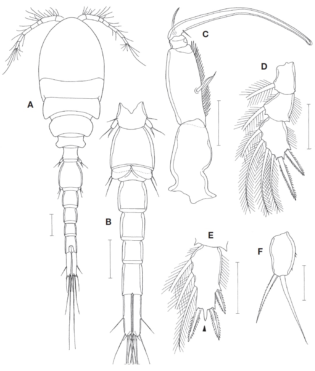
    Female. Body (Fig. 1A) slender, 1.50 mm long (1.44-1.61 mm), based on 6 specimens. Figured and described specimen 1.51 mm long. Prosome 810 μm long. Cephalothorax 513×415 μm, longer than wide, with faint dorsal suture line delimiting cephalosome and first pedigerous somite. Second pedigerous somite with slightly projected posterolateral corners. Urosome (Fig. 1B) 5-segmented. Fifth pedigerous somite 125 μm wide. Genital double-somite 221×144 μm, 1.53 times as long as wide, evenly tapering from broadest proximal 0.2 region to distal; with angular lateral apex at broadest region; genital aperture located dorsally at proximal 0.3 region of double-somite. Three abdominal somite 400×83, 75×70, and 108×67 μm, respectively. Anal somite unornamented, with smooth ventro-distal border. Caudal rami slightly divergent. Each ramus (Fig. 1C) 154×30 μm (length/width ratio 5.13 : 1), with 6 setae; outer lateral seta naked and located at 0.58 region of caudal ramus length; 4 distal setae plumose, and small dorsal seta naked.

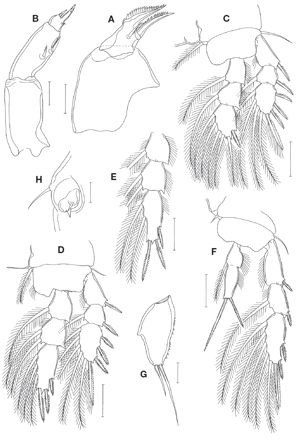
    Rostrum (Fig. 1D) tapering, slightly wider than long, with rounded distal apex. Antennule (Fig. 1E) slender, 345 μm long, 7-segmented; armature formula: 4, 13, 6, 3, 4+aesthetasc, 2+aesthetasc, and 7+aesthetasc; plumose setae 1 on fifth and sixth segments each, and 4 on terminal segment; aesthetascs on distal segments slender, confusable with setae. Antenna (Fig. 1F) 4-segmented. Coxobasis (first segment) with 1 distal seta. Endopod 3-segmented; first segment with 1 subdistal seta and ornamented with fine spinules on outer margin; short second segment with 3 setae at inner distal region, distalmost one of them being small and spiniform (Fig. 1G); terminal segment 77×28 μm, about 2.75 times as long as wide, armed with 2 slender, strongly curved spines and 5 setae, and ornamented with fine spinules on outer margin.

    Labrum (Fig. 1H) with broad, smooth posterior lobes; proximal part of lateral margin bluntly produced. Mandible (Fig. 1I) with shallow inner proximal notch; inner margin short and oblique; outer proximal part of gnathobase with 2 rows of spinules followed by more than 10 large teeth; distal lash elongate, with denticles along both margins. Maxillule (Fig. 1J) armed with 4 setae, 3 distal and 1 lateral. Maxilla (Fig. 2A) 2-segmented. Syncoxa (proximal segment) large but unarmed. Basis (distal segment) with short, stiff, tapering lash bearing about 20 denticles along convex outer margin and 2 setae; inner seta (seta I) strong, with spinules along convex outer margin; distal lash and inner seta together giving appearance of pincers; anterior seta (seta II) small and spinulose unilaterally; outer proximal seta (seta III) absent. Maxilliped (Fig. 2B) 3-segmented (syncoxa, basis, and 1-segmented endopod). Syncoxa largest but unarmed. Basis with 2 unequal setae near middle of inner side, both setae unilaterally spinulose. Endopod small, terminated by spiniform process bearing several minute spinules, with 1 spiniform and 1 minute setae.

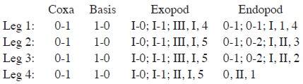
    Leg 1(Fig. 2C), leg 2 (Fig. 2D), and leg 3 with 3-segmented rami. Outer spines on exopods slender. Outer seta on basis of legs 1-4 naked. Leg 3 (Fig. 2E) similar to leg 2, except for having 3 spines and 2 setae on third endopodal segment. Leg 4 (Fig. 2F) with 3-segmented exopod and 1-segmented endopod; inner seta on coxa small and naked; endopod 1-segmented, with indistinct transverse line as articulation vestige; two distal spines 51 μm (outer one) and 88 μm (inner), respectively. Armature formula for legs 1-4 as follows:

    image

    Leg 5 represented by 1 dorsolateral seta on fifth genital somite and 1-segmented free exopod. Exopod (Fig. 2G) 73×35 μm, 2.09 times as long as wide, with fine spinules on outer margin, produced inner margin bearing pointed apex in midlength of exopod, and armed with 2 distal setae; outer seta 66 μm long, and inner seta 34 μm long. Leg 6 (Fig. 2H) represented by 1 small seta and 1 tooth-like process in genital aperture and 1 larger seta on lateral margin of double-somite.

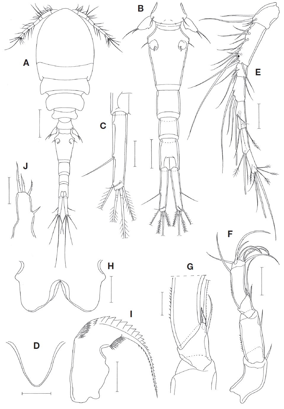
    Male. Body (Fig. 3A) resembling that of female, 1.24 mm long. Prosome 625 μm long. Cephalothorax 385×313 μm. Urosome (Fig. 3B) 6-segmented. Fifth pedigerous somite 80 μm wide. Genital somite 140×116 μm, longer than wide. Four abdominal somites 80×77, 72×66, 59×55, and 80×52 μm, respectively. Caudal ramus 113×23 μm, 4.91 times as long as wide.

    Rostrum as in female. Antennule as in female, but with 1 additional aesthetasc at distal region of second segment (at place indicated by dot in Fig. 1E). Antenna as in female.

    Labrum, mandible, maxillule, and maxilla as in female. Maxilliped (Fig. 3C) 4-segmented. First segment unarmed. Second segment with 2 naked seta at proximal 0.4 region and with 2 rows of spinules along inner margin. Small third segment unarmed. Fourth segment forming long, slender claw, with 1 unilaterally spinulose seta and 1 minute, needle-like seta at proximal region.

    Leg 1 endopod (Fig. 3D) with 2 spines and 4 setae on third segment; 2 distal spines 34 μm (outer) and 52 μm (inner) long, respectively. Third endopodal segment of leg 2 (Fig. 3E) with mid-distal process (indicated by arrowhead in Fig. 3E) more pronounced than that of female. Otherwise, leg 1-4 as in female.

    Leg 5 exopod (Fig. 3F) small, 25×15 μm, 1.67 times as long as wide, with few spinules on outer margin and 2 distal setae; inner distal seta spiniform, 30 μm long, with fine spinules; outer distal seta 42 μm long. Leg 6 represented by 2 unequal, naked setae on genital operculum (Fig. 3B).

    Etymology. The specific name turgipes is the combination of Latin words turg (=swollen) and pes (=the foot), alluding to the medially swollen exopod of leg 5 of the new species.

    DISCUSSION

    While describing Telestacicola xenophiothricis Doignon, Deheyn and Fiers, 2004, these authors dully transferred Doridicola claudus Humes and Stock, 1973 to the genus Telestacicola. They recognized the endopod of leg 4 of D. claudus as a one-segmented condition. The endopod is in fact one-segmented in our point of view, although Humes and Stock (1973) originally recognized it as two-segmented condition, with the expression “functionally 1-segmented, the line of articulation between the two segments being incomplete”. However, Doignon et al. (2004) did not notice that there were other two examples of the same condition in Doridicola: D. micropus Humes, 1973 and D. connexus Humes, 1986, both associated with the basket star Astroboa nuda (Lyman, 1874). In the original description of D. connexus, Humes (1986) described its endopod of leg 4 as “functionally 1-segmented, as in D. claudus and D. micropus”. Doridicola connexus and D. micropus have no reason to be maintained in the genus Doridicola, thus they are herewith transferred to Telestacicola.

    While comparing of congeners of Telestacicola one another, we have recognized one of them is very disharmonious with the remainings. It is T. sertus Humes, 1978 associated with a hydroid in the Moluccas (Humes, 1978), which has the following features unusual for Telestacicola: (1) the distal lash of the maxilla is perpendicular to the main axis of the maxilla; (2) the third exopodal segment of leg 4 carries 3 outer spines (formula III, I, 5); (3) the second endopodal segment of the antenna has a spine; and (4) the second antennular antennular segment in the male has two aesthetascs (1 proximal and 1 distal). These features are of the family Macrochironidae Humes and Boxshall, 1996. Because it has two terminal claw on the antenna it should be placed in the genus Pseudomacrochiron Reddiah, 1969 (see the key to genera of the Macrochironidae given by Humes and Boxshall, 1996), with a new name Pseudomacrochiron sertus (Humes, 1978). Major hosts of the Macrochironidae are hydroids (Boxshall and Halsey, 2004).

    With the removal of T. sertus and the inclusion of D. connexus and D. micropus, the genus Telestacicola now comprises seven valid species. Within the genus, T. xenophiothricis is unusual in having three large setae on the second endopodal segment of the antenna, compared to the size reduction of the distalmost seta which is spiniform and markedly shorter than other two in other six species. The remaining six species may be divided into two groups, based on the forms of the exopod of leg 5 in the female: one group (comprising T. connexus, T. lobophyti and T. micropus) having small exopod with two very unequal distal setae (short inner seta and much longer outer seta) and the other group (comprising T. angoti, T. claudus and T. turgipes n. sp.) having relatively large exopod with the inner seta being at least half as long as the outer one.

    The new species T. turgipes is very closely similar to T. angoti associated with alcyonacean corals in Madagascar (Humes and Stock, 1973; Humes, 1990). In particular, both species have in common an angular anterolateral apex of the genital double-somite. However, T. turgipes n. sp. is easily distinguishable from T. angoti and other congeners by its characteristic exopod of leg 5 having an inner swelling in the middle. Telestacicola angoti and T. xenophiothricis also have an inner swelling on the exopod of leg 5, but in these two species the swelling is rounded and position proximally.

    The seven species of Telestacicola may be differentiated using the following key.

참고문헌
  • 1. Boxshall GA, Halsey SH 2004 An introduction to copepod diversity. P.1-966 google
  • 2. Doignon G, Deheyn D, Fiers F 2004 Telestacicola xenophiothricis n. sp. (Copepoda, Poecilostomatoida) a remarkably well adapted commensal of the brittle star Ophiothrix purpurea (Echinodermata). [Belgian Journal of Zoology] Vol.134 P.67-73 google
  • 3. Humes AG 1973 Cyclopoid copepods associated with the ophiuroid Astroboa nuda in Madagascar. [Beaufortia] Vol.21 P.25-35 google
  • 4. Humes AG 1978 Cyclopoid copepods (Lichomolgidae) associated with hydroids in the tropical western Pacific Ocean. [Pacific Science] Vol.31 P.335-353 google
  • 5. Humes AG 1986 Two new species of Copepoda associated with the basket star Astroboa nuda (Ophiuroidea) in the Moluccas. [Zoologica Scripta] Vol.15 P.323-332 google cross ref
  • 6. Humes AG 1990 Synopsis of lichomologid copepods (Poecilostomatoida) associated with soft corals (Alcyonacea) in the tropical Indo-Pacific. [Zoologische Verhandelingen] Vol.266 P.1-201 google
  • 7. Humes AG, Boxshall GA 1996 A revision of the lichomologid complex (Copepoda: Poecilostomatoida), with the recognition of six new families. [Journal of Natural History] Vol.30 P.175-227 google cross ref
  • 8. Humes AG, Gooding RU 1964 A method for studying the external anatomy of copepods. [Crustaceana] Vol.6 P.238-240 google cross ref
  • 9. Humes AG, Stock JH 1973 A revision of the family Lichomolgidae Kossmann, 1877, cyclopoid copepods mainly associated with marine invertebrates. [Smithsonian Contributions to Zoology] Vol.127 P.1-368 google cross ref
OAK XML 통계
이미지 / 테이블
  • [ Fig. 1. ]  Telestacicola turgipes n. sp., female. A, Habitus, dorsal; B, Urosome, dorsal; C, Left caudal ramus, dorsal; D, Rostrum; E, Antennule (dot indicate the place of addition of an aesthetasc in male); F, Antenna; G, Third segment of antenna; H, Labrum; I, Mandible; J, Maxillule. Scale bars: A=0.2 mm, B=0.1 mm, C-F=0.05 mm, G-J=0.02 mm.
    Telestacicola turgipes n. sp., female. A, Habitus, dorsal; B, Urosome, dorsal; C, Left caudal ramus, dorsal; D, Rostrum; E, Antennule (dot indicate the place of addition of an aesthetasc in male); F, Antenna; G, Third segment of antenna; H, Labrum; I, Mandible; J, Maxillule. Scale bars: A=0.2 mm, B=0.1 mm, C-F=0.05 mm, G-J=0.02 mm.
  • [ Fig. 2. ]  Telestacicola turgipes n. sp., female. A, Maxilla; B, Maxilliped; C, Leg 1; D, Leg 2; E, Leg 3 endopod; F, Leg 4; G, Leg 5 exopod; H, Left genital aperture. Scale bars: A, B, G, H=0.02 mm, C-F=0.05 mm.
    Telestacicola turgipes n. sp., female. A, Maxilla; B, Maxilliped; C, Leg 1; D, Leg 2; E, Leg 3 endopod; F, Leg 4; G, Leg 5 exopod; H, Left genital aperture. Scale bars: A, B, G, H=0.02 mm, C-F=0.05 mm.
  • [ Fig. 3. ]  Telestacicola turgipes n. sp., male. A, Habitus, dorsal; B, Urosome, ventral; C, Maxilliped; D, Leg 1; E, Third endopodal segment of leg 2; F, Leg 5 exopod. Scale bars: A, B=0.1 mm, C-E=0.05 mm, F=0.02 mm.
    Telestacicola turgipes n. sp., male. A, Habitus, dorsal; B, Urosome, ventral; C, Maxilliped; D, Leg 1; E, Third endopodal segment of leg 2; F, Leg 5 exopod. Scale bars: A, B=0.1 mm, C-E=0.05 mm, F=0.02 mm.
  • [ ] 
(우)06579 서울시 서초구 반포대로 201(반포동)
Tel. 02-537-6389 | Fax. 02-590-0571 | 문의 : oak2014@korea.kr
Copyright(c) National Library of Korea. All rights reserved.