Six species of springtails (Collembola) are reported for the first time from North Korea: Pachyotoma takeshitai
Although many works have already been published about the springtails of the Korean Peninsula, the Collembola fauna of the region is still far from being exhaustively studied. This is also indicated by the fact that the recorded species’ number has increased significantly even recently (e.g., Kang et al., 2005; Lim and Park, 2011, 2013; Kang and Park, 2012; Park and Park, 2013; Dányi et al., 2014).
We report here further six species new to the Korean fauna:
For light microscopy, specimens were cleared in a mixture of lactic acid and glycerol (3 : 1), and examined under a Leica DM 1000 microscope (Leica, Wetzlar, Germany) with phase contrast optics. For using 1,000× magnification with oil immersion, specimens were mounted in Hoyer’s medium on permanent slides. Dark specimens were depigmented with Hüther’s fluid. Line drawings were prepared with a drawing tube. Photographs were taken with a Nikon CoolPix900 digital camera (Nikon, Tokyo, Japan) mounted on a Leica MZ75 stereo microscope (Leica, Wetzlar, Germany).
Dorsal chaetotaxy in Entomobryidae followed Jordana (2012). In Symphypleona, the chaetotaxic notation of abdominal VI segment followed Betsch (1997).
The specimens examined are deposited in the Soil Zoology Collection of Hungarian Natural History Museum (Budapest, Hungary) (HNHM) and in the National Institute of Biological Resources, Incheon, Republic of Korea (NIBR).
A list of abbreviations follows: Ant I-IV, antennal segments I-IV; Abd I-VI, abdominal segments I-VI; Tita I-III, tibiotarsus I-III; Emp, empodium; PAO, postantennal organ; Th I-III, thoracal segments I-III.
>
Material examined. North Korea: 1 male, Kangwon Province [Gangwon-do], Mt. Kumgang [Geumgangsan], moss sample, 18 Sep 1980, leg. Forró L, Topál Gy (As-430) (Korea 1980/701); 2 females, 3 males, 3 subadult females, 1 juvenile, Mt. Kumgang [Geumgangsan], moss sample collected along the foot-path to Kuryong Falls, extracted in Berlese-funnel, 7-800 m a.s.l., 19 Sep 1980, leg. Forró L, Topál Gy (As-433) (Korea 1980/713); 1 juvenile, Kangwon Province [Gangwon-do], Wonsan, soil sample collected in the park Songdown, 20 Sep 1980, leg. Forró L, Topál Gy (As-436) (Korea 1980/731).
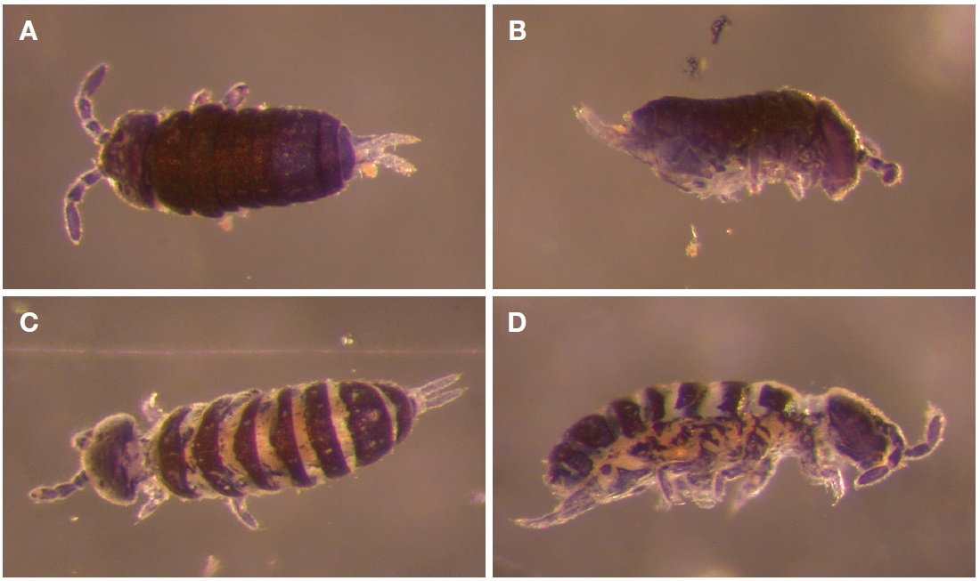
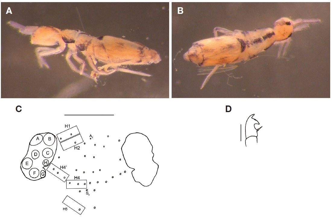
Description of the Korean specimens. Body size up to 1 mm. Head up to 0.25 mm long, as broad or slightly broader than long. Body of typical habitus for the genus (Fig. 1A-D).
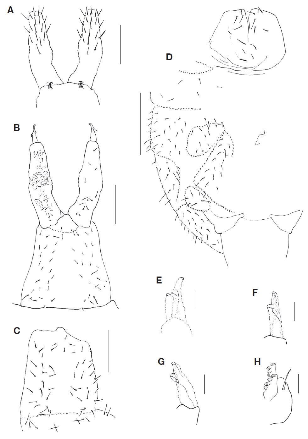
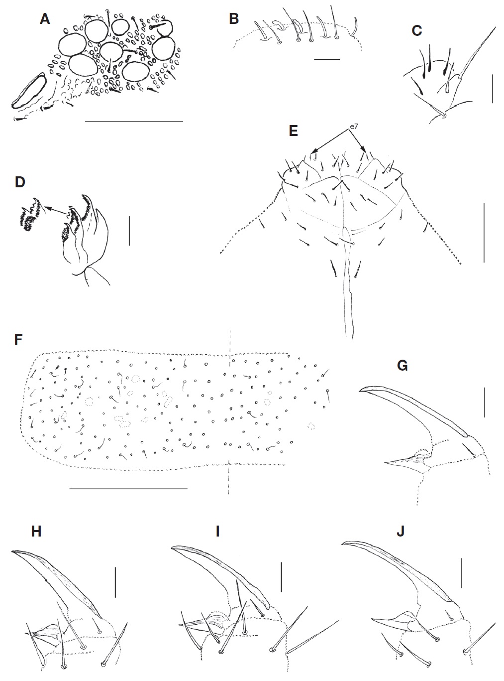
Colour bluish black, with paler appendages. In specimens swollen strongly in preservative, the pigmentation is broken into groups presenting characteristical striped pattern, which is however only an artifact (Fig. 1C, D). Integument with secondary granulation all over the body, without reticulation. 8+8 ocelli, G and H smaller; PAO broad elliptical without or with very weak constriction, 1.33-1.82 as long as ocellus diameter (Fig. 2A). Maxillary outer lobe with 4 sublobal hairs, maxillary palp bifurcate (Fig. 2C). Labral formula as 4/5,5,4. Clypeus with 17-21 chaetae. Labial palpus with 5 usual papillae (А-Е) and full set of guard chaetae, e7 moved forward, in some specimens up to in line with or in front of proximal chaetae (Fig. 2E). Labium with 3 proximal, 5 basolateral and 4 basomedian chaetae (Fig. 2E). Ventral side of head with 2+2 postlabial chaetae (in one specimen 3+4, in another 2+4). Maxilla with 6 lamellae as in Fig. 2D. Ant I with 2 small basal microsensilla (bms), dorsal and ventral, and 2 ventral sensilla (s), Ant II with 2 bms and without s, Ant III with 5 distal s (2 inner, 2 outer, and 1 lateral) (Fig. 2B) and without bms. No additional sensilla on first three antennal segments. Sensilla on Ant IV hardly differentiated, subapical organite small.
Body chaetotaxy. With numerous short chaetae on body (Fig. 2F). Normal dorsal axial chaetom of Th II-Abd IV as 5-7,5-6/4-5,4-5,4-5,8-10, but asymmetry often occurs. Macrochaetae absent, only a few larger chaetae are on Abd IV-VI (Fig. 3D). Tergites of Th II-Abd V covered with numerous sensilla along p-row and on medial and lateral parts. Sensilla and common cheatae along p-rows of the segments mostly alternately arranged (Fig. 2F). Tergal sensilla as long as or slightly longer than common chaetae. Sensillary formula as 19-27,11-14/9-12,12-16,11-17,18-19,11-13 (s), 1,1/1,1,1 (ms). Thorax without ventral chaetae.
Subcoxa 1 of leg I-III with 1,5,6 chaetae respectively. Subcoxa 2 of leg I-III with 0,6,7-8 chaetae respectively. Coxa I-III with 3,9,11, trochanter I-III with 9,11-12,11-12 chaetae respectively. Femur I-III with about 19,26,33 chaetae respectively. Tita I-III with 23,24,31 chaetae respectively. B-row on Tita I-II with 7 chaetae. Tibiotarsal tenent chaetae poorly developed, pointed. Claws of normal shape but in some specimens more elongated, sometimes with one weak inner tooth, empodium lamellate, without teeth (Fig. 2G-J). VT with 4-6+4-6 latero-distal and 3-4+3-4 posterior chaetae (Fig. 3D). Retinaculum with 4+4 teeth and 1 chaeta (Fig. 3I). Anterior (=anteriomedial) furcal subcoxae with 18-21 chaetae, anteriolateral furcal subcoxae (according to the terminology of Potapov et al., 2005) with 8-12 chaetae (Fig. 3D). Posterior furcal subcoxae with 7 chaetae. Anterior side of manubrium smooth, without chaetae. Posterior side of manubrium with about 48-52 chaetae on the main part and about 10 chaetae on its laterobasal lobes (Fig. 3B, C). Medial part of manubrial thickening simple, sometimes with irregular edge. Dens with 18-22 anterior setae, arranged as in Fig. 3A. Posterior side of dens granulated, with 11-13 chaetae arranged as in Fig. 3B. Mucro tridentate, lateral subapical tooth very strong, connected with the base by two lamella (Fig. 3E-G).
Remarks. Although some characters used in modern taxonomy of Isotomidae have not been described in
Distribution.
>
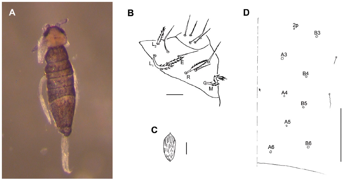
Material examined. Four specimens, North Korea: Hwanghaebuk-do, Mt. Bagyon [Bagyeonsan], San-chon tong [Sancheon-dong], about 10 km from Kaesong [Gaeseong], sweet chestnut woods, sod of grass (turf) beyond margin of woods, 8 Jun 1970, leg. Mahunka S, Steinmann H (Korea 119/3) (As-229).
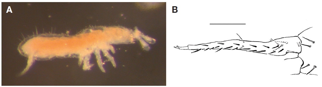
Diagnosis. Body size 0.8-1 mm without antennae. White, habitus as in Fig. 4A. Without Omma, PAO not constricted. Labrum with 4/5,5,4 chaetae. Cl without inner tooth, Emp without apical filament. Ret. with one chaeta and four teeth. Manubrium with 2+2 anterior chaetae, dens with 17-19 anterior and 5 posterior chaetae (Fig. 4B). Mucro with two teeth, as in Fig. 4B. Dorsomedial Mac on Th II-III absent, Th I-III without ventral chaetae, Abd without broadened sensilla, ms 1,0/1,0,0, sens on Abd I-III at the level of p-row.
Distribution.
>
Material examined. 1 specimen, North Korea: Kangwon Province [Gangwon-do], Mt. Kumgang [Geumgangsan], Hotel Kumgang, pitfall traps, 20 Sep 1980, leg. Forró L, Topál Gy (Korea 1980/724).
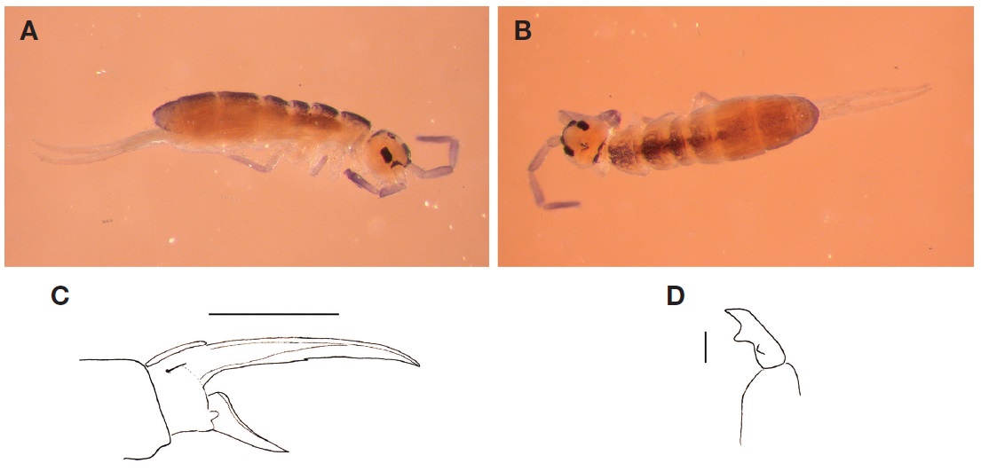
Diagnosis. Body size 1.6-3.1 mm without antennae. Coloration as in Fig. 5A and B. Ground colour greyish-green with dorsal parts of thorax and abdomen darkened. Extremities of same ground colour as body, antennae uniformly dark. Larger specimens (above 2 mm) almost entirely greyish-black (Fjellberg, 1986). Antennae long, 2.4-2.7 times longer than head diagonal. Head “prognathous” (with prominent mouth cone as in Fig. 5A). Body hairs fine and uniform, without differentiated macrochaetae. Ventral tube with about 11-22+11-22 frontal chaetae (12+12 in the Korean specimen). Claws long and slender, claw index about 2.8-3.2 (Fig. 5C). Furca strong, about 2.3-2.7 as long as head diagonal. Dens 2.1-2.4 as long as manubrium. Mucro without lateral chaeta, apical tooth larger than subapical (Fig. 5D).
Distribution. The species has been known only from the Pacific northwest region of North America till now (Fjellberg and Bernard, 2009). According to its presence in Korea, also
>
Material examined. 3 specimens, North Korea: Kangwon Province [Gangwon-do], Mt. Kumgang [Geumgangsan], Hotel Kumgang, soil traps baited with beer, in the forest behind the hotel, 20 Sep 1980, leg. Forró L, Topál Gy (As-434) (Korea 1980/724).
Diagnosis. Body colour pattern as in Fig. 6A and B. Length about 1.5-1.7 mm excluding antennae. 8 Omma, GH smaller than EF (Fig. 6C). Labrum with 4 smooth papillae. Length ratio of Abd IV/III>4. Claw with 3 teeth on internal edge, Emp with smooth external edge on leg III, spike-like. Furca length about 800-870 μm. Mucro with 2 teeth (subapical larger than apical one), mucronal spine present (Fig. 6D). Simplified chaetotaxy formula: 2-1-0-3-1/2-3/2-3/0-1-1/0-2-3-2-3. Head chaetotaxy as in Fig. 6C.
Remarks. The colour pattern found also in the Korean specimens is very characteristic for the species. The Korean specimens fit well the redescription given by Jordana (2012).
Distribution.
>
Material examined. 1 subadult, North Korea: Kangwon Province [Gangwon-do], Mt. Kumgang [Geumgangsan], Hotel Kumgang, soil traps baited with beer, in the forest behind the hotel, 20 Sep 1980, leg. Forró L, Topál Gy (As-434) (Korea 1980/724).
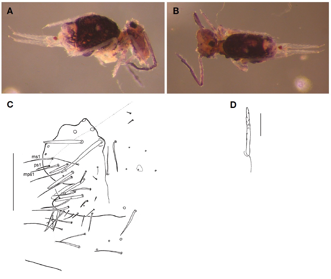
Diagnosis. Body size 0.9 mm without antennae. Scattered dark pigment on body, head a bit lighter (Fig. 7A). Ant IV with unilobed apical bulb. Labial base chaetae as MREL1L2 (Fig. 7B). Scales of the “long basal rib type” according to the nomenclature of Zhang et al. (2011), ordinary scales of tergites at most 4 times longer than wide (Fig. 7C). No scales on dens. The dorsal chaetotaxy of Abd IV (Fig. 7D) shows in the specimen an intermediate stage between those illustrated for the first instar by Szeptycki (1979) and for the adult by Zhang et al. (2011). Unguiculus lanceolate. Mucro with teeth subequal and basal spine short, reaching the apex of subapical tooth.
Remarks. Among all the species of the genus
Distribution. Subcosmopolitan from arctic to subtropical regions (Zhang et al., 2011). New to the fauna of the Korean Peninsula.
>
Material examined. 3 females, North Korea: Kangwon Province [Gangwon-do], Mt. Kumgang [Geumgangsan], Hotel Kumgang, soil traps baited with beer, in the forest behind the hotel, 20 Sep 1980, leg. Forró L, Topál Gy (As-434) (Korea 1980/724).
Diagnosis. Body size 1.8-2.0 mm without antennae. Habitus and colour as in Fig. 8A and B. Differentiated tibiotarsal chaetae serrate as in Fig. 8D. Tenent hairs acuminate. Dens with 3,2,1,1...1 chaetae anteriorly, posteriorly with 9 chaetae in row E (E3 : E2 ratio about 1.5), 5 in row P and 9 in row J. Both edges of mucro finely serrate. Claw with 2 lateral, 2 outer and 2 inner teeth. Female anal appendage smooth, weakly curved and acuminate; anal chaeta ps1 (sa) not spinelike, smaller than ms1 (A0) (Fig. 8C).
Distribution. The species have been reported from most European countries and also from the Lake Baykal in Russia (Bretfeld, 1999). New to the fauna of the Korean Peninsula.
Korean name:1*다께시다뚱보마디톡토기 Korean name:1*쌍털풀솜마디톡토기 Korean name:1*아길리스배마디톡토기, 2*시베리아털보톡토기 Korean name:1*검둥이비닐털보톡토기 Korean name:1*검은가시톱니둥근톡토기
Two species (