Copepods of the family Clausidiidae are external associates of various marine invertebrates. Their hosts include sponges, cnidarians, molluscs, polychaetes, crustaceans, vestimentiferans, and echiurans (Boxshall and Halsey, 2004). Some of them live in burrows of invertebrates. Copepods of the genus
Copepods of the genera
Specimens of the new speices of
>
Clausidium maximus n. sp. (
Material examined. 10 ♀♀, 13 ♂♂ (including 8 pairs in amplexus) from washings of
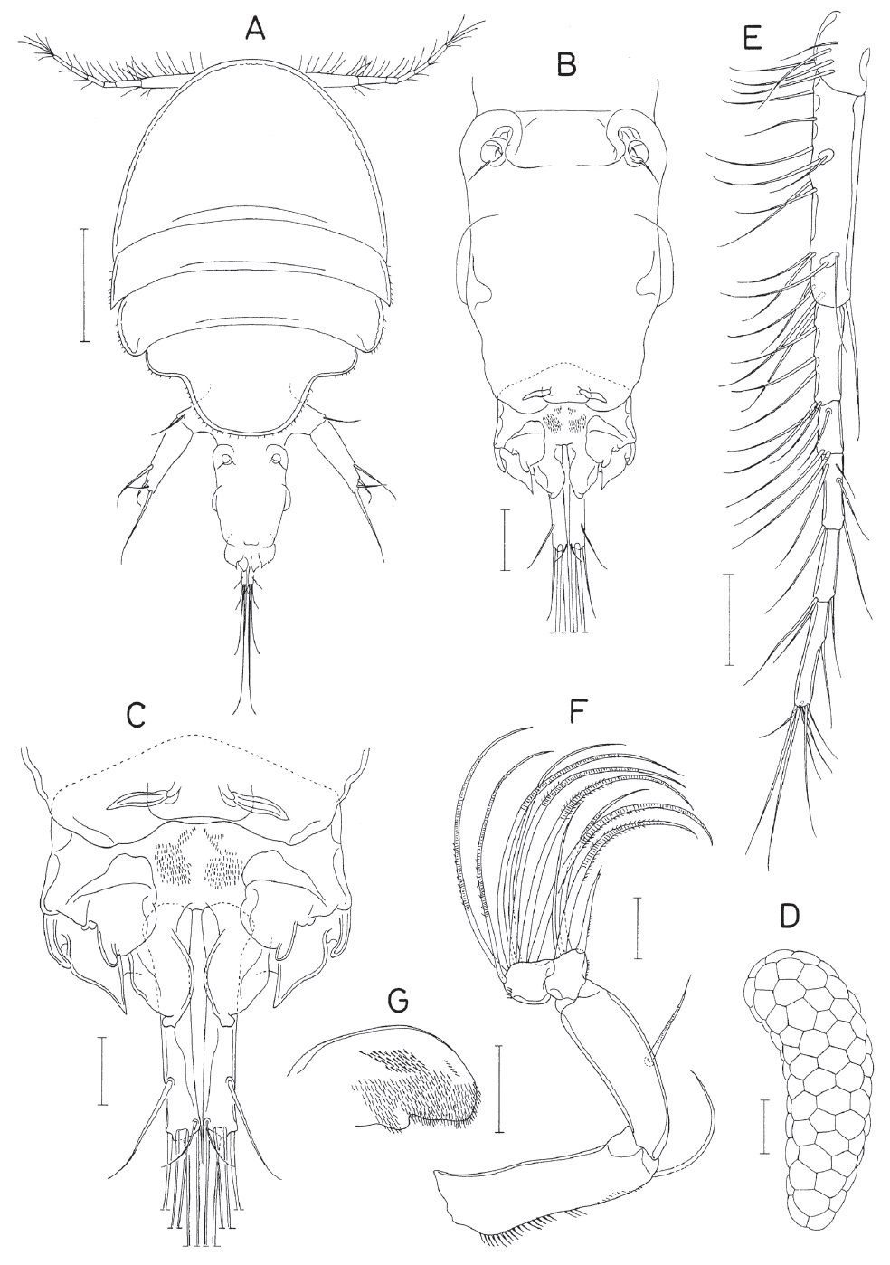
Female. Body (Fig. 1A) with flattened prosome and relatively small urosome. Length of dissected and figured specimen 2.35 mm. Prosome 1,692×1,238 μm, consisting of cephalothorax and 3 metasomites. Cephalothorax hemi-circular, 712×1,223 μm, much wider than long. Second pedigerous somite (first metasomite) widest among somites, with pointed posterolateral corners. Second and third pedigerous somites with fine setules on lateral margins. Third pedigerous somite slightly longer than preceding somite, with rounded posterolateral corners. Fourth pedigerous somite (third metasomite) consisting of broader anterior 1/3 and narrower posterior 2/3, the latter forming large posterior protuberance of somite, with fine setules along lateral and posterior margins. Urosome ambiguously 3-segmented, shorter than prosome, consisting of fifth pedigerous somite, genito-abdomen, and anal somite. Fifth pedigerous somite 431 μm wide and incompletely defined from next somite. Genito-abdomen (Fig. 1B) slightly tapering posteriorly, 465×338 μm, with transparent membrane near middle of lateral margins and 1 small posterodorsal flap bearing paired ribbon-like dorsal elements (Fig. 1C); genital apertures positioned dorsal proximal region (Fig. 1B). Anal somite (Fig. 1C) complicated, with 4 pairs of posterior processes (2 broad, 1 pointed, and 1 digitiform); dorsal surface with numerous minute spinules in middle; anal region uncertain. Caudal ramus (Fig. 1C) narrow, 178 ×37 μm (length : width ratio 4.81 : 1), directed straightly backwards, with 6 setae; all of setae naked; outer lateral seta (seta II) located at 77% region of ramus length. Egg sac (Fig. 1D) 1,077×315 μm, slightly tapering distally, and slightly curved near proximal third.
Rostrum absent. Antennule (Fig. 1E) elongate, 775 μm long, slender, and 7-segmented; armature formula 5, 15, 7, 4, 5, 3, and 8; all of setae naked and thin; terminal segment longer than each of its 3 proximal segments. Antenna (Fig. 1F) 4-segmented, consisting of basis and 3-segmented endopod. Basis with 1 seta distally and setules on inner margin; first endopodal segment with 1 inner seta at halfway of segment length; second and third endopodal segments similar in length, each wider than long and armed with 4 and 7 setae, respectively; inner margin of second endopodal segment produced.
Labrum (Fig. 2A) much wider than long, with spinules on posterior margin and setules on lateral margins. Mandible (Fig. 2B) armed distally with 2 massive, denticulate or spinulose elements and 1 small, spiniform seta. Paragnath (Fig. 1G) represented by spinulose lobe. Maxillule (Figs. 1, 2C) distally bilobed and armed with 8 setae (1 proximal, 4 on one lobe, and 3 on the other lobe) and several setules proximally. Maxilla (Fig. 2D) 2-segmented (syncoxa+allobasis); syncoxa with 3 inner distal setae (one of them inserted on proximal region of larger seta); allobasis terminated by large, spiniform process, with 2 spinulose setae, and 1 large, crenulate spine. Maxilliped (Fig. 2E) 4-segmented; first segment with 2 inner setae; second segment with produced inner margin and 2 setae at apical region of inner margin; small third segment with 1 distal seta; terminal segment also small and armed with 1 spiniform process (similar to nearby spines but lacking basal articulation), 2 spinulose spines, and 2 naked setae.
Legs 1-4 with 3-segmented exopod and endopod. Leg 1 (Fig. 2F) strongly modified. Coxa unarmed, but with spinules on outer distal region. Basis with 1 thin seta and several setules on outer margin, and with 1 large, sword-like element inner distally, the latter 192 μm long, 32 μm in greatest width, acutely pointed distally, with wavy transverse striations on ventral surface. Exopod 3-segmented, but incompletely segmented between first and second segments, with oblique rows of granules on outer ventral side (1 row each on first and second segment, and 3 rows on third segment); armature formula of exopod I-0; 1-0; 3, 2, 2; outer and distal setae on third exopodal segment spiniform. Endopod distinctly tapering; first segment with 1 large spiniform ventral process distally and 1 small inner seta. Second segment unarmed. Third segment with 2 sucking discs (1 proximal and 1 smaller distal ones) on inner side and 1 small distal seta (Fig. 2G); distal sucking disc accompanied by bifurcate element (Fig. 2G).
Leg 2 (Fig. 2H) and leg 3 with identical armature formula: coxa 0-1; basis 1-0; exopod I-0; I-1; III, I, 4; endopod 0-1; 0-2; I, II, 3. Inner coxal seta of these legs with spinule-like setules. Basis with spinules on outer margin and setules on inner margin. Endopod with 3 small sucking discs on outer margin: 1 distally on first segment and 2 (proximal and distal) on third segment. Distal spines on third endopodal segment of second leg 57 μm (outer spine) and 193 μm long (inner spine).
Leg 4 (Fig. 3A) similar to leg 2 or leg 3, except for the followings: inner seta on coxa weakly pinnate; third exopodal segment armed with 4 spines and 5 setae (armature formula III, I, 5); third endopodal segment of endopod armed with 3 spines and 2 setae (armature formula I, II, 2).
Leg 5 (Fig. 3B) 2-segmented, consisting of protopod and exopod. Protopod with 1 distal seta. Exopod slightly tapering distally; 365×112 μm (length : width ratio 3.26 : 1), armed with 4 setae (3 of them stiff); longest distalmost seta 350 μm long, slightly shorter than exopod; all of setae on leg 5 naked. Leg 6 (Fig. 3C) represented by 2 setae (one of them minute) in genital aperture.
Male. Body (Fig. 3D) different in form from that of female. Length 1.39 mm in dissected specimen. Prosome 870×670 μm and segmented as in female. Second and third pedigerous somites with pointed posterolateral corners. Fourth pedigerous somite with concave posterior margin, with posterior protuberance. Urosome (Fig. 3E) 6-segmented; all somites wider than long. Fifth pedigerous somite 255 μm wide. Genital somite 105×175 μm; genital opercula located ventrolaterally. First to third abdominal somites 87×129, 63×109, and 50×110 μm, respectively. Anal somite distinctly defined from caudal rami on ventral surface but obscurely defined from caudal rami on dorsal surface (Fig. 3F). Anal region uncertain. Caudal ramus 149×40 μm (ratio 3.73 : 1, measured ventrally) and evenly tapering.
Rostrum absent as in female. Antennule same as that of female. Antenna (Fig. 3G) similar to that of female, except for the followings: first endopodal segment with 1 blade-like ridge at proximal region and seta on this segment thicker than that of female; second endopodal segment with 3 setae and 1 bluntly tipped spine (indicated by arrow in Fig. 3G).
Labrum (Fig. 4A) more expanded posteriorly than that of female, with round lateral protrusion and small, digitiform ventrolateral process. Mandible and maxillule as in female. Maxilla (Fig. 4B) with row of large setules on ventral surface of syncoxa. Maxilliped (Fig. 4C, D) probably 3-segmented; first segment with 2 small distal setae; second segment distally complicated, with 2 setae, 1 bifurcate processes, 1 large, curved and spatulate process, and 2 digitiform processes (Fig. 4D); third segment armed with 2 claws, smaller one of them with 1 small spine proximally, and larger one with dentiform subsidiary process subdistally.
Leg 1 (Fig. 4E) with stiff, spiniform seta and large spinules on inner distal region of basis. Leg 2 (Fig. 4F) with only 1 inner seta on second endopodal segment; inner seta on coxa large, proximally plumose and distally spinulose. Legs 3-5 as in female. Leg 6 represented by 1 seta tipped distally on genital operculum (Fig. 3E).
Etymology. The specific name
Remarks. While describing
Of those 11 known species of
The hitherto known greatest body length in the genus
>
Hippomolgus limiticus n. sp. (
Material examined. One ♀ (holotype) from the muddy bottom sediments in the depth of 48 m, in the East China Sea (approximately 32°00ʹN, 125°00ʹE), 2 Jun 2015, collected by Lee J. Holotype ♀ (NIBRIV0000362225, dissected and mounted on a glass slide) has been deposited in the NIBR, Incheon, Korea.
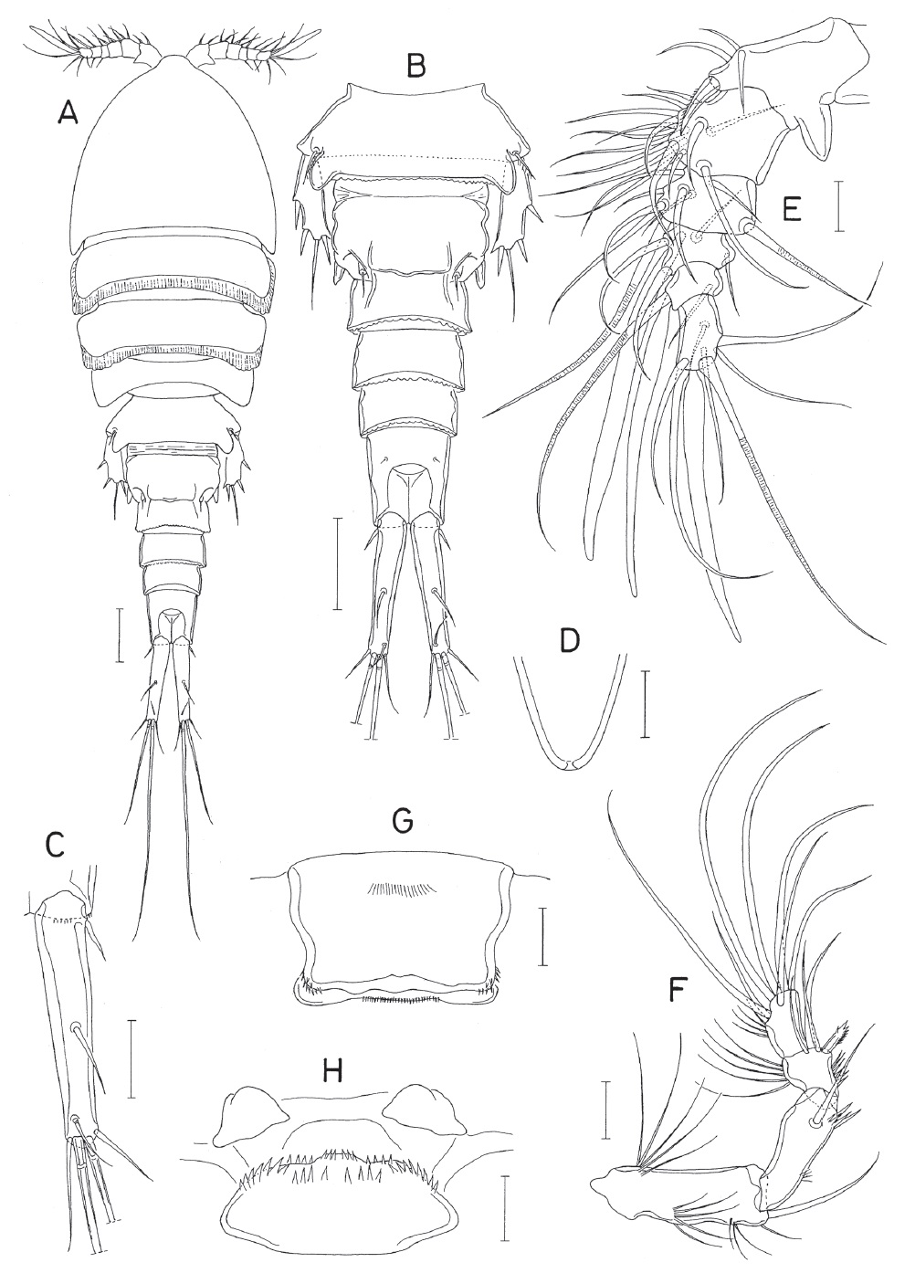
Female. Body (Fig. 5A) narrow, 1.26 mm long, with rather thick exoskeleton. Prosome 4-segmented, fusiform, and 650 μm long. Cephalothorax 342×385 μm, with rostral region slightly produced anteriorly. Second and third pedigerous somites with broad membranous rim along posterodorsal margin. Urosome (Fig. 5B) 5-segmented, gradually tapering. Fifth pedigerous somite 91×255 μm, distinctly wider than posterior somites, with lobately projected posterolateral corners dorsally. Genital double-somite 168×175 μm, consisting of broader anterior 3/5 and narrower posterior 2/5 (136 μm wide in this region); broader anterior part with stout spine at posterolateral corners; genital aperture positioned dorsally at posterior region of broader anterior part of double-somite. Three abdominal somites 68×125, 50×109, and 100×91 μm, respectively. Anal somite longer than wide, with large anal region. First to fourth urosomal somites with crenate membranous rim along their posterodorsal margins. Caudal rami (Fig. 5B, C) slightly divergent, about 1.6 times as long as anal somite, 164×33 μm (length : width ratio 4.97 : 1), weakly tapering distally, and armed with 7 seta; seta I (proximal seta) shortest among 7 setae but distinct; seta II located on dorsal surface of ramus at 54% region of ramus length; seta V longest, 418 μm long; seta IV second longest, 218 μm long; all of 7 setae naked.
Rostrum (Fig. 5D) evenly tapering, longer than wide, with blunt posterior apex. Antennule (Fig. 5E) stout, 159 μm long, less than half as long as cephalothorax, and 6-segmented. Armature formula: 3+spine, 15, 9, 4+aesthetasc, 2+aesthetasc, and 7+aesthetasc. All of setae naked. Aesthetascs on distal segments large, tapering, and basally narrowed. First segment with robust process posterodistally; spine on this segment located anterodistally, with fine spinules along anterior margin. Antenna (Fig. 5F) 4-segmented (basis+3-segmented endopod). Basis the longest, armed with 1 distal seta and several patches of long setules. First endopodal segment with 1 seta and 1 patch of spinules subdistally and 1 patch of minute spinules subproximally; second endopodal segment with 1 short spinulose spine and 3 setae at inner distal corner, spinules on inner margin and several long setules on outer margin; terminal segment longer than wide and armed with 7 setae (4 of them distinctly longer than other 3) and several outer setules. All of setae on segments naked.
Labrum (Fig. 5G) nearly rectangular, broader than long, ornamented with spinules at distolateral corners and on middle of distal margin. Labium (Fig. 5H) ornamented with spinules along anterior side. Mandible (Fig. 6A) distally armed with 2 thick, spinulose spines and 2 setae; ventral one of 2 spines distinctly larger than dorsal one; setae longer than spines, with minute spinules. Paragnath (Fig. 5H) lobate, smooth, and weakly bilobed distally. Maxillule (Fig. 6B) bilobed distally and armed with 7 thick setae (2 on narrower lobe and 5 on broader lobe). Maxilla (Fig. 6C) consisting of syncoxa and allobasis. Syncoxa unarmed, without ornamentation. Allobasis with 1 distal and 1 subdistal robust processes and 1 seta; distal process evenly tapering, with longitudinal row of spinules; subdistal process spiniform, not articulated at base, with spinules all over the surface. Maxilliped (Fig. 6D) 4-segmented; first segment with 1 distal seta; second segment with 2 subdistal setae of unequal lengths; third segment short and unarmed; terminal segment slender and tapering, with 1 seta and 1 spiniform process near middle and several spinules at distal region.
Legs 1-4 with 3-segmented exopod and endopod. Outer margin of these legs ornamented with spinules or setules. Intercoxal plate of legs 1-3 with long setules on both sides of posterior margin; that of leg 4 with thick setules. Posterior margin of basis ornamented with thick setules between rami. Inner margin of basis naked in leg 1, but with setules in legs 2-4. Outer margin of first exopodal segment of legs 1-4 spiniferous. Outer margin of endopodal segments setulose, except for spinulose in second endopodal segment of leg 1 and naked in third endopodal segment of leg 4. Inner spine on basis of leg 1 36 μm long, smooth, extending slightly beyond distal margin of first endopodal segment and accompanied by several setules near its base. Two inner spines on third endopodal segment of leg 1 setulose. Inner seta on coxa of leg 4 stiff and naked. Armature formula of legs 1-4 as follows:

Leg 5 (Fig. 5B) represented by 1 dorsolateral seta on fifth pedigerous somite and 1-segmented exopod; exopod (Fig. 6H) 99×37 μm (length : width ratio 2.68 : 1), armed with 3 spines and 1 seta, and with pointed mediodistal corner and ridge-like longitudinal elevation on dorsal surface; lengths of armature elements: 24, 28, and 35 μm for spines from outer to distal, respectively, and 80 μm for seta. Leg 6 represented by 1 small seta in genital aperture (Fig. 5B).
Male. Unknown.
Etymology. The specific name
Remarks. Three species of the genus
Although
Although Humes and Ho (1967) recorded the alcyonacean coral