Low temperature plasma (LTP) ionization mass spectrometry (MS) is one of the widely used ambient analysis methods which allows soft-ionization and rapid analysis of samples in ambient condition with minimal or no sample preparation. One of the major advantages of LTP MS is selective analysis of low-molecular weight, volatile and low- to medium-polarity analytes in a sample. On the contrary, the selectivity for particular class of compound also implies its limitation in general analysis. One of the critical factors limiting LTP ionization efficiency is poor desorption of analytes with low volatility. In this study, a home-built LTP ionization source with Peltier heating sample stage was constructed to enhance desorption and ionization efficiencies of analytes in a sample and its performance was evaluated using standard mixture containing fatty acid ethyl esters (FAEEs). It was also used to reproduce the previous bacterial identification experiment using pattern-recognition for FAEEs. Our result indicates, however, that the bacterial differentiation from FAEE pattern recognition using LTP ionization MS still has many limitations.
Ambient mass spectrometry (MS) allows rapid analysis of samples with minimal sample preparation and has attracted a lot of attention recently due to its potential application for portable in-situ analysis. Since the initial development of ambient MS based on desorption electrospray ionization (DESI)1 and direct analysis in real time (DART),2 a large variety of ambient ionization methods were reported.3 Ambient ionization methods are classified into two groups, a spray-based ionization and a plasma-based ionization. The latter has several advantages for portable MS application, including more direct analysis from intact sample surface, minimum use of solvents and generation of singly charged ions. The low temperature plasma (LTP) ionization4 is one of the plasma-based ionization methods sharing the same advantages. In addition, it is potentially the most favorable method for in-situ application of MS due to very simple and low-cost instrumentation.
LTP ionization source generates non-continuous micro plasmas by dielectric barrier discharge between a ground electrode and an AC-powered electrode separated by a dielectric material assisted by discharge gas flow. A simple high voltage (several kV) alternating (several kHz) current source is used to maintain fast plasma bullets. The LTP is a soft-ionization technique with minimal sample heating around 30 ℃, and thus, it is difficult to analyze molecules with low volatility.5 Several attempts were tried to overcome this limitation, including a heat gun,6 non-contact halogen lamp,7 and resistive heating plate.5 In the present study, we made a home-built LTP ionization source with Peliter heating sample stage to enhance desorption/ionization efficiency of the ionization source. The performance of the source was investigated and demonstrated by analysing fatty acid ethyl esters (FAEEs) in a standard mixture and a bacterial sample.7
Standard FAEE mixture (C4 - C24 even carbon saturated FAEEs, containing 1000 mg/mL each in hexane) used in this study was purchased from Sigma-Aldrich (St. Louis, MO, USA). For LTP ionization MS experiment, it was diluted with ethanol to prepare 100 mM working solution. About 100 mL of the working standard solution was loaded on a slide glass and dried before LTP ionization MS analysis.
>
Low temperature plasma ionization source
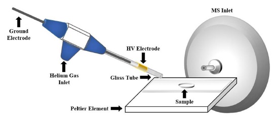
A LTP probe is assembled using a glass tube (O.D. 1/8 inch, I.D. 1/16 inch) with an central ground electrode (stainless steel, O.D. 1.0 mm) inserted inside the tube using a tee connector (Omnifit, 3-way connector), which is also used to allow discharge gas flow (Helium) through the glass tube. A high AC voltage was supplied to an outer electrode surrounding the glass tube, which was made by wrapping Cu tape (1 cm width) as shown in Figure 1. The gap between the outer HV electrode and the inner ground electrode was optimzed at about 10 mm. An alternating voltage of 0-5 kV with upto 10 kHz frequency was supplied using a AC power supply (KSC 300, Korea Switching Co., Seoul, Korea) regulated by a function generator (DG4062, RIGOL Tecnologies Inc., Beaverton, OR, USA). The He LTP was opimizaed at AC supply of 1.2-1.5 kV and 10 kHz.
>
Peltier heating of sample surface and temperature monitoring
A Peltier heating element in a square shape (50 mm × 50 mm) was installed very close to the MS inlet. A glass silde for sample deposition was attached on the Peltier surface a double sided adhesive tape for heat transfer. An adjustable DC supply upto 4 V was used to heat the Peltier element.
All of the experiments were performed using a linear ion trap mass spectrometer (Thermo LTQ, San Jose, CA, USA) with an electrospray ionization (ESI) source. For the LTP MS experiments, the ESI source was removed and the LTP source and sample stage were installed. A full MS scan over the m/z range from 100 to 500 was used for MS analysis of FAEEs. Capillary temperature was set at 250 ℃.
>
Heating characteristic of Peltier heated sample stage
The temperature of slide glasss surface heated by a Peltier heater was measured with an infrared thermometer. At 4 V DC supply, the temperature of sample surface increased upto 60 ℃ after 1 min. Due to the limited heating capability of the epoxy-sealed Peltier element (about 80 ℃), heating was limited to that temperature. In spite of this limitation, very cheap price of the element, small size and easy setup are clear advantages in instrumentation. Use of silicon-seal is expected to increase the operational temperature of Peltier upto 200 ℃.
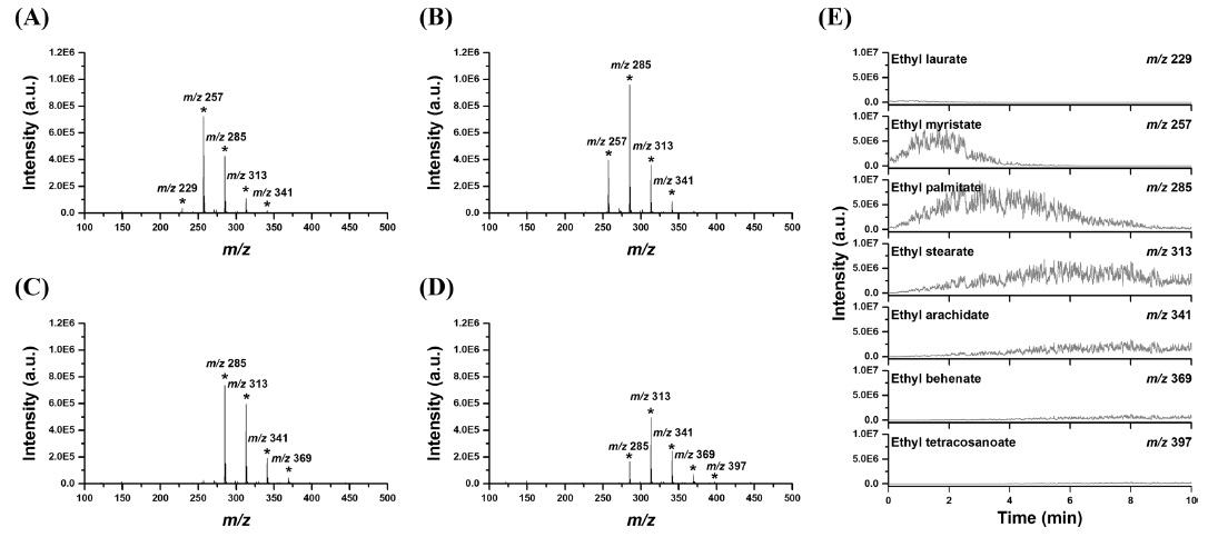
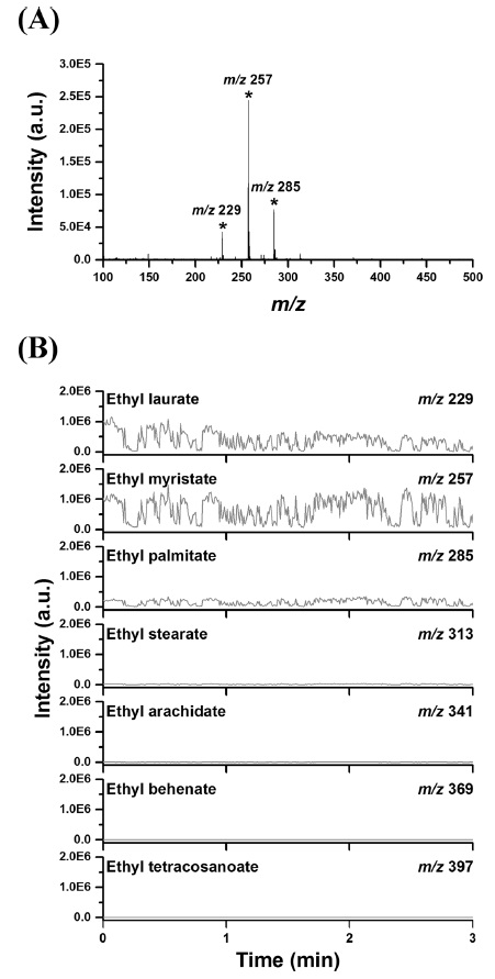
FAEE mixture sample, loaded on a slide glass about 10 nmol each, showed stable profile in the mass spectrum as shown in Figure 2. Figure 2B shows time-dependent ion intensities of [M+H]+ ions for various FAEEs in the standard mixture sample. Stable ion signals of FAEEs were obtained by the LTP ionization MS for several minutes, although there were some fluctuations of signals due to non-uniform deposition of FAEEs as shown in Figure 2B. A time-averaged MS spectrum is also displayed in Figure 2(A). Very volatile FAEEs smaller than ethyl decanoate (m/z 201, vapor pressure 3.1×10⁻² mmHg at 25 ℃) are most likely evaporated during sample deposition and drying or desorbed in the early stage of LTP ionization. In constrast, FAEEs larger than ethyl strearate (m/z 313, vapor pressure 3.01×10⁻⁵ mmHg at 25 ℃) didn’t show signal in the mass spectrum and illustrated desorption limitation of larger FAEEs with lower vapor pressure. In the non-heating condition, LTP showed maximum sensitivity for ethyl myristate (m/z 257, vapor pressure 1.57×10⁻³ mmHg at 25 ℃), followed by ethyl palmitate (m/z 285, vapor pressure 7×10⁻⁴ mmHg at 25 ℃) and ethyl laurate (m/z 229, vapor pressure 7.44×10⁻³ mmHg at 25 ℃). This narrow MS profile of FAEEs alone can impose a significant challenge to bacterial identification using FAEE profiling.
When we applied sample heating using the Peltier elements to 60 ℃, different profile was obtained for the FAEE standard mixture sample. As shown in Figure 3, non-volatile FAEEs upto ethyl tetracosanoate (m/z 397, no vapour pressure data available) could be observed time-dependently. FAEEs were desorbed in the order of their volatility. Still, we couldn’t observe very small FAEEs with relatively high vapour pressure due to their loss during early stage of analysis or sample preparation. The present experiment shows that it is quite challenging to obtain reproducible FAEEs profiles in LTP ionization MS even from a standard mixture without careful control of desorption and ionization conditions.
>
Direct analysis of FAEEs from an extract of E. coli
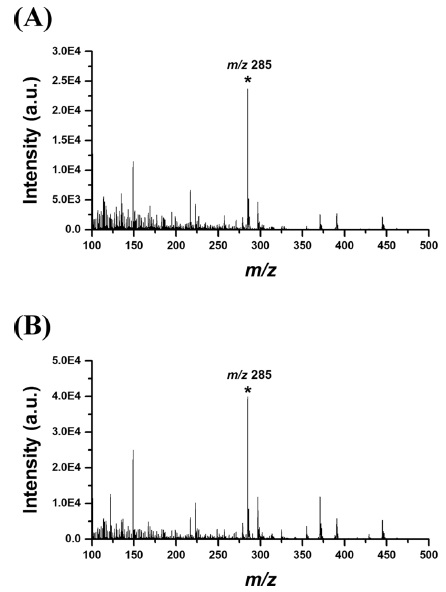
Zhang et al. reported recently that it was possible to distinguish several different bacterial strains by comparing the bacterial LTP mass spectral patterns from FAEEs in the m/z range of 200-300.8 To reproduce their experiment using our home-made LTP system, we cultured and analyzed a gram-negative bacteria,
A home-built LTP ionization MS system was developed to investigate its potential application to baterial strain identification using FAEE profiles. To overcome the volatility limitation of the LTP ionization, a low-cost Peltier heating sample stage was made with very simple instrumentation and it could increase the temperature of sample surface to 60 ℃. The increase in sample surface temperature allowed analysis of wider variety of FAEEs with lower volatilities using the LTP ionization MS. However, we failed to reproduce bacterial identification experiment of Zhang et al. for real