검색 전체 메뉴
PDF
맨 위로
OA 학술지
Streptococcus mutans로부터 분리한 Mutan이 흰쥐의 골결손부 치유에 미치는 영향 Effect of Mutan Isolated from Streptococcus mutans on the Healing of Bone Defect in Rat
ABSTRACT
Streptococcus mutans로부터 분리한 Mutan이 흰쥐의 골결손부 치유에 미치는 영향

We investigated the effects of a mutan (water-insoluble α-glucans) isolated from Streptococcus mutans on the healing of bone defect in rat. Sprague-Dawley rats were divided into control (saline-treated), lipopolysaccharide (LPS)-treated, and mutan-treated groups (n=6 per group). Experimental bone defects were surgically created with round fissure bur at the buccal surface of the left mandibular. The control groups was administered with saline solution (0.1 ml/100 g), while the LPS and mutan group was given LPS and mutan (1 mg/kg body weight) three times weekly. After 4 weeks the rats were sacrificed, the healing of bone defect was assessed by bone mineral density (BMD) and micro-computed tomography (μCT) examination. Percent bone volume (bone volume/tissue volume [BV/TV]), trabecular thickness (Tb.Th), and trabecular number (Tb.N) parameters of μCT showed higher values in control group than LPS and mutan group. Bone surface/volume ratio (BS/BV), trabecular bone pattern factor (Tb.Pf), and structure model index parameters of μCT showed higher values in LPS group than mutan group. BMD values of mutan treated-alveolar bones were significantly lower for than that of the LPS group. Therefore, we suggest that mutan, water-insoluble α-glucans from S. mutans may be induce the induction of periodontal diseases.

KEYWORD
Bone defect , Micro-computed tomography , Mutan , Streptococcus mutans , α-1,3-glucan
  • 서 론

    치주질환은 치주조직의 소실을 동반하는 염증 및 면역반응으로 치조골 흡수 및 치주낭 형성을 특징으로 하는 염증성 질환이다. 치주질환의 주요 원인은 치근면에 존재하는 병원균의 효소와 독소이며, 이는 조직으로 침투하여 질환의 상태를 악화시킨다1). 특히 독소와 효소들은 염증과 면역반응에 영향을 주어 급성 또는 만성적인 치주조직 파괴를 유도한다2). 또한 구강 내 감염성 질환을 일으키는 세균의 주요인자 가운데 하나는 부착인자이다. 부착인자는 세균이 숙주에 부착하는 세균의 표면 구조물로서, 부착능이 없는 치주병원균이 부착능이 강한 세균과 결합함으로써 치주국소에 정착이 가능하도록 한다. 치아표면에 부착하는 최초의 세균의 예는 그람 양성균인 Streptococcus mutansActinomyces viscosus이다. 이들 세균은 치아표면과 치주조직에 부착 및 증식하여 치면세균막(dental plaque)을 형성하며, 그 주변 부위는 화학적, 물리적인 변화가 일어나 유사한 세균 또는 다른 세균들의 응집 및 증식이 일어난다3). 이 같은 현상은 구강 내에서 관찰되는 대표적인 바이오필름이며, 이에 관한 연구는 치아우식증의 주원인균인 S. mutans에서 가장 많은 연구가 이루어졌다.

    S. mutans에서 생성되는 부착성 다당류는 글루칸(glucan)으로4,5) α-1,6-glucose linkage로 구성된 덱스트란(dextran), α-1,6-glucose linkage가 주된 결합인 수용성 글루칸(water-soluble α-glucan), 그리고 α-1,3-glucose linkage가 주된 결합인 비수용성 글루칸(water-insoluble α-glucan), 즉 mutan (뮤탄)으로 구분된다6). Mutan은 점착성이 높은 비수용성 다당류로서 치면에 S. mutans의 부착을 용이하게하여, 각종 세균을 응집시켜 치면세균막을 형성하고7,8), 탄수화물 대사과정에서 다량의 유산을 생성하여 치아 표면을 탈회시킴으로써 치아우식증을 유발한다. 따라서 이러한 세포외 다당류는 세균과 구강표면 간의 특이적인 상호작용을 할 수 있는 부착체계를 가지고 있어 많은 연구자들의 관심 대상이 되어왔다9). 이 같은 mutan은 치아 표면 또는 구강 연조직에 쉽게 노출되는 다당류이므로 치주조직에 미치는 염증 및 면역 반응에 관여할 가능성이 있음에도 불구하고 현재까지 연구가 거의 이루어지지 않았다. 구강 캔디다증을 일으키는 Candida albicans와 같은 모든 진균류의 세포벽 성분은 β-glucan 다당체로 구성된 특징을 갖고 있다. 이러한 β-glucan은 인체의 면역세포인 대식세포에 존재하는 dectin-1 수용체(receptor)에 인식되어 제거된다. Dectin-1 수용체의 존재가 규명됨에 따라 진균류에 대한 인체의 내재 면역반응(innate immune response)의 생화학적 경로가 밝혀졌으나, 충치균에서 생성하는 α-glucan에 관한 연구는 많지 않다10). 지금까지 구강세균의 다당류에 관한 주요 연구로는 비수용성 글루칸이 대식세포 활성화에 관여하여 염증반응을 유도11,12), Streptococcus sobrinus의 비수용성 글루칸은 단핵구를 자극하여 tumor necrosis factor (TNF)-α와 interleukin (IL)-8을 생성하고 다형핵 백혈구에서 hydrogen peroxide 생성을 유도, 또한 비수용성 글루칸에 의해 유도된 면역활성 반응은 구강 내 염증성 질환의 진행에도 관여하는 것으로 보고된 바 있다13). Chia 등14)S. mutans의 glucosyltransferases는 단핵구로부터 전구 염증성 사이토카인(cytokine)인 TNF-α, IL-1β, 그리고 IL-6의 형성을 촉진하여 연쇄상 구균성 쇽(shock) 증후군과 감염성 심내막염을 유도하는 것으로 보고하였다. Mutan에 대한 연구는 대부분 비수용성 글루칸의 구조적 특징 및 분해 효소인 dextranase 및 mutanase를 이용한 plaque 분해에 대한 연구가 주를 이루어 왔으며6,15,16), 면역세포의 활성에 따른 골흡수를 동반하는 치주조직에 대한 작용기전은 보고된 바 없다.

    본 연구는 S. mutans로부터 mutan을 분리 정제하여 얻은 후, 외과적으로 형성된 흰쥐 치조골 결손부에 주입하여 mutan이 치조골의 치유에 미치는 영향을 도립현미경을 통해 골 변화양상을 관찰하고, micro-computed tomography(μCT) 촬영을 시행하여 골밀도(bone mineral density, BMD)와 3차원적 골형태(bone morphometry)를 비교 분석하고자 하였다.

    연구대상 및 방법

       1. 실험재료

    1) 균주 및 배지

    실험에 사용한 S. mutans ATCC 25175 균주는 미생물자원센터에서 분양 받아 사용하였으며, 균주 보관 및 배양은 brain heart infusion (Difco Lab., Detroit, MI, USA) 배지를 이용하였다.

    2) Mutan의 분리 및 정제

    Mutan을 분리하기 위해, 먼저 균체 외 조 다당류를 정제하고 불용성 mutan을 분리하였다. S. mutans를 brain heart infusion 배지에 접종하여 2×10⁷ cells/ml가 될 때까지 전 배양하였다. 전 배양된 균을 본 배양용 배지에 접종하고 5% CO2 incubator에서 배양시킨 후 8,000g으로 원심분리하였다. 수획된 상층액에 trichloroacetic acid (TCA)를 최종 농도 2.5%가 되도록 첨가하였다. TCA가 첨가된 배지에 70%농도가 되도록 99% ethanol을 첨가한 후 24시간 동안 4°C에서 냉침하였고, 다시 9,600g에서 10분간 원심분리하여 조다당류를 침전시켰다. 침전된 조다당류 분획은 다시 멸균수에 희석시킨 후, dialysis membrane bag (molecular weight cut off: 10,000)에 넣고 흐르는 물에서 5일 동안 투석시켜 저분자물질을 제거시켰다. 최종적으로 투석된 분획물들은 원심분리한 후 동결건조하여 조다당류를 얻었다. 조 다당류의 순도를 높이기 위하여 gel column chromatography를 실행하였다. 먼저 시료 10 mg을 1 ml의 0.3 N NaOH에 용해시켜 Sepharose CL-4B (Sigma-Aldrich, St. Louis, MO, USA) column에서 용출시켰다. Column chromatography에서 얻은 용출물은 phenol- sulfuric acid로 발색시킨 후 490 nm에서 흡광도를 측정하여 당의 위치를 확인하고, 투석 후 동결 건조하여 mutan을 분리하였다.

    3) 실험 동물

    실험동물은 180∼200 g의 Sprague-Dawley계 암컷 흰쥐를 대한바이오링크(Eumseong, Korea)에서 구입하여 실험실 환경적응을 위하여 1주간 예비 사육 후 실험을 실시하였다. 본 실험은 순천향대학교 동물실험윤리위원회의 승인(SCH12_05_01) 하에 표준작업 지침서에 따라 수행되었으며 사육실 조건은 23±2°C, 50±5%의 상대습도와 12시간의 명암주기를 유지하고 물과 사료(Dhbiolink)는 자유롭게 섭취하도록 하였다. 실험목적에 따라 saline을 처리한 대조군, lipopolysaccharide (LPS) 처리군, mutan 처리군으로 하여 각 군에 6마리씩을 배정하였다. 실험동물의 체중변화는 Table 1과 같다.

    [Table 1.] Bone Histomorphometry of the Alveolar Bone Defects in All the Study Groups

    label

    Bone Histomorphometry of the Alveolar Bone Defects in All the Study Groups

       2. 치조골 결손부 형성 및 외과적 처치

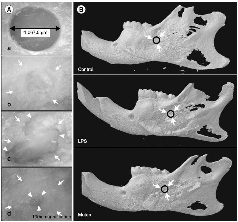
    치조골 결손부를 형성하기 위하여 실험 대상 흰쥐를 ethyl ether로 흡입 마취시킨 후, 하악골 좌측 피부털을 제거하여 표피를 노출시켰다. 외과용 blade를 이용하여 구외측으로 하악골 좌측 협면에 구각부위에서 1 cm 정도 떨어진 부위를 시작점으로 하여 약 2 cm로 표층 피부를 절개하였다. 표피를 하방의 근육들로부터 하악각 쪽으로 넓게 박리하여 분리하다. 교근을 주변 인접근육들로부터 박리한 뒤, 하악골 부착부에서 절단하고 하방의 골막에 절개를 하여 하악골을 노출시켰다. 하악의 협측을 덮고 있는 하악골의 중앙부위인 제3대구치 치근 첨단부의 후하방에 저속엔진 하에서 치과용 절삭 bur를 이용하여 골 결손부를 형성하였다. 하악골을 천공하지 않도록 주의하면서 골 결손부를 폭 1.5 mm×길이 3 mm의 크기로 형성한 후, 봉합사를 층별로 골막과 표피의 봉합을 수행하였다. 24시간 후, 골 결손부에 각 군당 양성대조군인 LPS (1 mg/kg body weight; serotype O111:B4; Sigma-Aldrich) 그리고 mutan (1 mg/kg body weight)을 총 4주 동안(주 3회, 총 12회) 매회 당 20 μl씩 골결손부 피하에 주사하였다. 각 시료는 20°C 냉동고에 보관한 후 실험에 사용하였다.

       3. Three-dimensional (3D) μCT 촬영

    하악골의 골치유 정도를 알아보기 위해 관찰부위는 미세 단층촬영기 μCT (SkyScan 1172; Bruker, Antwerp, Belgium)에 의해 촬영되었다. 시료가 미세단층촬영기 내에서 특정 방향을 향하도록 한 후 촬영하였다. 화소 크기는 26.5 μm이며 60 kA의 전압과 167 μA의 전류의 X-선을 방사하여 0.5 mm 알루미늄 필터를 거쳐 시료를 투과하는 방식으로 미세영상이미지를 획득하였다. 획득된 미세영상이미지는 Nercon Ver 1.3 (SkyScan)을 이용하여 gray scale level로 재건하였으며, 재건된 2차원 영상은 CTAn (SkyScan) 소프트웨어를 이용하여 3차원 모델을 재구성하였다.

       4. 통계분석

    정량적인 결과는 평균값과 표준편차로 표시하였다. 통계적인 차이는 PASW Statistics ver. 18.0 (IBM Co., Armonk, NY, USA)을 이용하여 Student’s t-test로 분석하였고 p값이 0.05 이하인 경우 통계적으로 유의한 것으로 판단하였다.

    결 과

       1. 실험동물의 체중변화

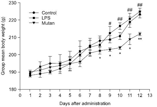
    실험기간 동안의 실험동물의 체중변화는 Fig. 1에 나타냈다. 각 시험군의 체중은 4주 동안 총 12 회 투여 중 1∼7회차까지는 처리군 간에 유의한 차이가 없었으나, 8회차부터 mutan 처리군에서 saline을 처리한 대조군에 비해 유의한 체중 증가를 보였다(p<0.05). 9회차에는 mutan 처리군이 대조군과 LPS 처리군에 비해 유의한 체중 증가를 보였으며(p<0.05), 10∼12회차에서도 LPS 처리군과 비교하여 유의한 체중 증가를 나타냈다(p<0.01). 이는 실험 기간 중 시료 투여 및 외과적 시술이 정상적인 식이 투여에 영향을 미치지 않고, 성장했음을 시사한다.

       2. 도립현미경 및 μCT를 통한 골치유 관찰 소견

    하악골에 인위적인 골 결손부를 형성한 후 4주가 경과한 후에 mutan에 의한 골치유 여부를 도립 현미경과 3차원 micro CT (3D μCT)를 통해 관찰해 본 결과는 Fig. 2와 같다. 동일한 양의 saline을 처리한 대조군은 자연적인 골치유 반응으로 인해 정상적인 골형성 소견이 관찰되었다. Mutan 처리군은 골 결손부 변연으로부터 심부로 진행된 골형성 소견이 관찰되었으나, LPS 처리군에 비해 완전한 골 결손부의 회복이 이루어지지 않았다. 반면에 LPS 처리군은 전체적인 골 결손부의 회복은 보이지만, 골형성뿐만 아니라 골흡수도 진행되어 기존의 인공적인 골 결손부 외에 주변 변연부로의 진행된 골흡수 소견이 관찰되었다.

       3. 3차원 μCT 미세구조의 형태계측학적 지표값 분석
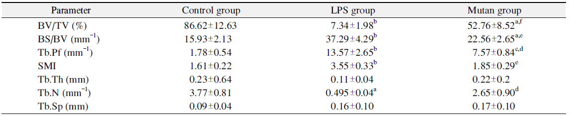
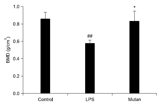
    Table 1Fig. 3에서 보는 바와 같이, 시각화된 μCT 상의 데이터를 수치화하기 위해 3차원 미세구조의 형태계측학적 지표(mophometric parameter)인 tissue volume (TV), bone volume (BV), percent bone volume (BV/TV), bone surface (BS), interception sulface, bone surface density (bone surface/bone volume [BS/BV]), trabecular bone pattern factor (Tb.Pf), structure model index (SMI), trabecular thickness (Tb.Th), trabecular number (Tb.N), trabecular separation (Tb.Sp), BMD 총 12개 항목을 분석하였다. 전체 조직에서 골소주의 부피를 나타내는 BV/TV는 전체 TV에서 BV의 비율을 %로 나타낸 것으로 값이 높을수록 골강도가 강한 것으로 판단하며, LPS 처리군(7.34±1.98)은 각각 대조군(86.62± 12.63)과 mutan 처리군(52.76±8.52)에 비해 골강도가 유의하게 낮은 것을 확인할 수 있었다. BS/BV는 관심 용적내의 골용적에 대한 골표면 면적의 비율이며, 구조의 복잡성(complexity)을 특징짓는 영상지표로서, parameter 값이 낮을수록 골강도가 강한 것으로 판단하며, mutan 처리군(22.56±2.65)은 대조군(15.93±2.13)보다 다소 낮은 골강도를 보였으나, LPS 처리군(37.29±4.29)에 비해 유의하게 높은 골강도를 나타냈다(p<0.01). 골소주 사이의 연결성을 나타내는 Tb.Pf는 낮은 값이 좋은 연결성을 나타내며, 전체 골소주 면적에서 오목함(concavity)과 볼록함(convexity)의 상대적인 비에 기초한 연결성 지표이다. 오목함은 둘러싸인 골수강의 존재를 의미하고 볼록함은 끊어져 있는 골소주 지주들(trabecular struts)을 의미한다. LPS 처리군, mutan 처리군, 대조군 순으로 연결성이 좋은 것으로 판단되며 특히 mutan 처리군에서 대조군에 비해 유의하게 좋은 연결성을 보였다(p<0.001). SMI는 골소주의 형태가 막대모양(rod-like structure) 혹은 판모양(plate-like structure) 중 어느 형태가 우세한지 알려주는 지표로 대개는 0∼3까지의 지수로 표현되며 0에 가까울수록 판모양, 그리고 3에 가까울수록 막대 모양으로 표현된다. 측정 결과, LPS 처리군은 막대형, 나머지 처리군은 판형에 가깝게 나타났다. Tb.Th는 골소주의 평균 두께를 나타내며, 골소주 내에 수많은 구(sphere)를 위치시켜 이 구들의 직경의 평균을 내어 얻어진다(parallel plate-like model). 골소주의 두께가 두꺼울수록 골강도가 크므로 LPS 처리군, mutan 처리군, 대조군으로 갈수록 골강도가 증가함을 확인하였다. 골소주의 평균 개수를 나타내는 Tb.N는 mutan 처리군(2.65±0.90)에서 LPS 처리군(0.50±0.04)보다 골소주의 평균 개수가 높게 나타났다. Tb.Sp (mm)는 골소주 간극, 즉 골소주 사이의 평균길이를 의미하며 mutan 처리군에서 가장 높은 수치를 보였다. BMD는 hydroxyapatite (CaHA)에 대한 체적당 질량의 단위로서 g/cm3로 나타내며 0.25 g/cm3와 0.75 g/cm3인 팬텀(phantom)을 기준으로 골밀도를 측정해주는 지표로서, 측정 결과는 Fig. 3과 같다. 대조군의 골밀도치가 LPS 처리군과 mutan 처리군의 골밀도치보다 높았으며 mutan 처리군의 골밀도치는 LPS 처리군과 비교하여 유의한 차이를 나타내었다(p<0.01).

    고 찰

    감염성 미생물에 존재하는 수많은 종류의 탄수화물은 숙주 면역세포 표면에 존재하는 다양한 수용체에 인식되어 면역 활성과 염증 등을 유도한다17). 본 연구는 S. mutans에서 생성되는 부착성 다당류인 비수용성 글루칸인 mutan이 치주조직의 면역학적 활성인자로 작용하여 치주질환을 유발하는지 확인하였다. 지금까지 mutan은 치아우식증의 부착인자로써 많은 연구가 되었으나, 골흡수 및 골재생에 관여하는 경로에는 거의 연구가 이루어지지 않았다. 우선 하악골 부위를 외과적으로 절개한 후 치조골부분을 절삭하고, 결손부위에 mutan을 주사할 경우, 생체 내 골치유 효과는 생리식염수를 주사한 대조군(control group)과 비교하여 LPS와 mutan 처리군에서 골재생이 지연되는 것이 확인되었다. μCT의 측정 결과에서도 대조군은 자연적인 골치유 반응으로 인한 우수한 골형성이 관찰되었으나 mutan 처리군은 초기 골결손부의 외형이 대부분이 회복되는 소견을 보였으나 대조군과 비교하여 치유가 완전하게 이루어지지 않았다. Mutan의 양성대조군으로 사용된 LPS 처리군은 mutan 처리군보다 뚜렷한 골흡수 소견이 관찰되어 치주염과 같은 염증성 골흡수를 유도하는 것으로 생각된다. 이 같은 LPS는 그람음성세균의 외막 성분으로 치주질환 발생에 중요한 독성인자로 여겨왔고, 염증성 사이토카인의 분비를 자극하여 골흡수를 유도하거나 골치유를 지연하는 물질로 보고되어 왔다18). Rogers 등19)과 Kim 등20)Actinobacillus actinomycetemcomitans에서 분리한 LPS를 흰쥐의 치주조직에 주입하였을 때, LPS를 주사하지 않은 대조군보다 심한 골흡수와 전구염증성 사이토카인을 유도한다고 하였으며, Yoshinaga 등21), Lee 등22), Gürkan 등23)은 치주질환 동물 모델을 이용한 연구에서 LPS는 치주염을 유도하여 골흡수에의 영향을 주는 인자로 보고하였다. 따라서 본 연구에서는 실험군으로 사용된 세포외 다당류인 mutan이 골흡수 유도물질인 LPS와 비교하여 치주조직의 염증을 유도하여 골흡수 촉진 및 골치유 지연에의 작용을 규명하고자 하였다. 이와 관련된 최근의 연구에 따르면 S. sobrinus로부터 분리된 수용성과 비수용성 글루칸이 마우스와 사람에서 대식세포와 다형핵 백혈구를 활성화함으로써 염증성 면역반응을 유도한다고 하였으며, 치아우식증의 병인뿐만 아니라 구강 내의 치주질환과 같은 염증성 질환에도 기여하는 것으로 보고된 바 있다13).

    본 연구결과 그람 양성균의 다당체인 mutan은 그람음성균의 LPS보다 비교할 만큼 골재생억제에 영향을 미치진 않았으나, 어느 정도 골치유를 지연시키는 것으로 관찰되었다. μCT 촬영 결과, 형태계측학적 지표 값을 분석하여 3차원 골 미세구조 지표를 알아보기 위해 골 결손 주변부에 관심부위를 설정하여 3차원 모델을 재건하여 분석하였다. μCT 미세구조 지표의 분석에 있어 골밀도와 μCT 지표들 Tb.Th와 BV/TV가 골밀도와 밀접한 상관관계가 있다고 보고된 바 있으며, μCT 지표를 연구 변수로 사용하는 것이 타당하다고 보고된 바 있다24-26). 본 연구에서 하악의 치조골을 μCT로 촬영하여 생성된 이미지를 관찰한 결과, 대조군에 비해 mutan 및 LPS 처리군에서 골치유 능력이 상대적으로 저하된 것을 확인하였다. BV/TV는 LPS 처리군이 다른 처리군보다 골강도가 유의하게 낮게 나타났으며, 따라서 mutan 처리군은 골소주의 부피에 직접적인 영향을 주지 않음을 알 수 있었다. BS/BV는 mutan 처리군이 LPS 처리군에 비해 유의하게 높은 골강도를 나타냈으며, Tb.Pf의 경우 mutan 처리군이 LPS 처리군보다 유의하게 골소주 사이의 우수한 연결성을 보였다. SMI는 LPS 처리군은 막대형인 반면, 대조군 및 mutan 처리군은 판형에 가깝게 나타났다. 골소주 모양이 막대보다는 판모양일 때 골강도가 높기 때문에 대조군과 mutan 처리군이 LPS 처리군보다 단단하며 판모양을 보인다고 생각된다. Tb.Th는 LPS 처리군, mutan 처리군, 대조군으로 갈수록 골강도가 증가함을 확인하였다. Tb.N는 mutan 처리군에서 LPS 처리군보다 골소주의 평균 개수가 높게 나타나 mutan 처리군이 LPS 처리군 대비 골강도가 유의하게 높은 것으로 생각된다. Tb.Sp는 mutan 처리군에서 가장 높은 수치를 보여 골소주들 간의 공간이 증가되어 골강도가 낮은 것으로 생각된다. BMD의 경우, mutan의 골밀도치는 대조군과 뚜렷한 차이를 보이지는 않았으나, LPS 처리군에 비해 유의하게 골밀도치가 높은 것이 확인하였다. 이러한 결과는 LPS 처리군이 대조군과 mutan 처리군에 비해 관심용적 내의 골소주의 양과 골소주의 두께가 감소하였으며, 골 용적에 대한 골 표면적에 대한 비율도 감소하는 경향을 보인다고 생각된다.

    본 연구의 한계점은 주요 실험이 in vivo에서 μCT 관점에서 이루어졌으므로 학술적 가치를 주장하기 위해서는 조직학적 및 면역학적 연구가 보완되어야 한다. 또한 S. mutan에 존재하는 mutan이 염증을 유발하고 골치유를 지연시키는 영향을 규명하기 위해서는 진균류의 베타글루칸 수용체인 dectin-1의 발견처럼 in vitro에서 mutan에 대한 innate immune을 담당하는 면역세포의 mutan 수용체 발견과 그들의 신호경로(signal pathway)에 관한 연구가 요구된다.

    결론적으로 LPS를 처리한 실험군은 염증유도에 의한 골 흡수를 증가시킴으로써, 실험동물에서 골조직의 치유를 억제하고 골 소실을 현저히 증가시켰으며, S. mutans에서 분리한 mutan 다당류는 LPS보다는 작지만 골치유 과정에서 골조직 형성이나 흡수에 영향을 주는 것으로 판단된다.

참고문헌
  • 1. Kunnen A, van Doormaal JJ, Abbas F, Aarnoudse JG, van Pampus MG, Faas MM 2010 Periodontal disease and pre-eclampsia:a systematic review [J Clin Periodontol] Vol.37 P.1075-1087 google cross ref
  • 2. Page RC 1991 The role of inflammatory mediators in the pathogenesis of periodontal disease [J Periodontal Res] Vol.26 P.230-242 google cross ref
  • 3. Lemos JA, Quivey RG Jr, Koo H, Abranches J 2013 Streptococcus mutans: a new Gram-positive paradigm? [Microbiology] Vol.159 P.436-445 google cross ref
  • 4. Loesche WJ 1986 Role of Streptococcus mutans in human dental decay [Microbiol Rev] Vol.50 P.353-380 google
  • 5. Lee HW, Lee P, Kwon HJ, Han KI, Han MD 2013 Antimicrobial activity of extracts from some traditional oriental medicinal plants against dental caries bacteria [J Dent Hyg Sci] Vol.13 P.45-52 google
  • 6. Pleszczynska M, Wiater A, Szczodrak J 2010 Mutanase from Paenibacillus sp. MP-1 produced inductively by fungal a-1,3-glucan and its potential for the degradation of mutan and Streptococcus mutans biofilm [Biotechnol Lett] Vol.32 P.1699-1704 google cross ref
  • 7. Rolla G, Scheie AA, Ciardi JE 1985 Role of sucrose in plaque formation [Scand J Dent Res] Vol.93 P.105-111 google
  • 8. Kwon HJ, Han MD 2012 Effect of brazilin from Caesalpinia sappan L. on the growth of Streptococcus mutans ATCC 25175 [J Dent Hyg Sci] Vol.12 P.209-215 google
  • 9. Han MD 2001 Immunological activities of Streptococcus mutans KCTC 3056 polysaccharide [J Dent Hyg Sci] Vol.1 P.1-6 google
  • 10. Brown GD, Gordon S 2001 Immune recognition: a new receptor for β-glucans [Nature] Vol.413 P.36-37 google
  • 11. Shibata Y, Abiko Y, Ohishi T, Tamaya H, Takiguchi H 1989 Free arachidonic acid source for PGE2 and TXB2 production in guinea pig peritoneal macrophages exposed to insoluble glucan from Streptococcus mutans [Int J Biochem] Vol.21 P.1043-1045 google cross ref
  • 12. Abiko Y, Shibata Y, Fukushima K, Murai S, Takiguchi H 1983 The stimulation of macrophage prostaglandin E2 and thromboxane B2 secretion by Streptococcus mutans insoluble glucans [FEBS Lett] Vol.154 P.297-300 google cross ref
  • 13. Okamoto S, Terao Y, Kaminishi H, Hamada S, Kawabata S 2007 Inflammatory immune responses by water-insoluble α-glucans [J Dent Res] Vol.86 P.242-248 google cross ref
  • 14. Chia JS, Lien HT, Hsueh PR, Chen PM, Sun A, Chen JY 2002 Induction of cytokines by glucosyltransferases of Streptococcus mutans [Clin Diagn Lab Immunol] Vol.9 P.892-897 google
  • 15. Wiater A, Szczodrak J, Pleszczynska M 2008 Mutanase induction in Trichoderma harzianum by cell wall of Laetiporus sulphureus and its application for mutan removal from oral biofilms [J Microbiol Biotechnol] Vol.18 P.1335-1341 google
  • 16. Shimotsuura I, Kigawa H, Ohdera M, Kuramitsu HK, Nakashima S 2008 Biochemical and molecular characterization of a novel type of mutanase from Paenibacillus sp. Strain RM1: identification of its mutan-binding domain, essential for degradation of Streptococcus mutans biofilms [Appl Environ Microbiol] Vol.74 P.2759-2765 google cross ref
  • 17. Kim MJ, Rye HW, Cho GH, Kim HW 2008 Expression of inflammatory cytokines by beta-glucan in macrophage cell line [J Pharm Soc Korea] Vol.52 P.73-78 google
  • 18. Wilson M 1995 Biological activities of lipopolysaccharides from oral bacteria and their relevance to the pathogenesis of chronic periodontitis [Sci Prog] Vol.78 P.19-34 google
  • 19. Rogers JE, Li F, Coatney DD 2007 Actinobacillus actinomycetemcomitans lipopolysaccharide-mediated experimental bone loss model for aggressive periodontitis [J Periodontol] Vol.78 P.550-558 google cross ref
  • 20. Kim CC, Cui DZ, Kim YJ 2007 The effect of Actinobacillus actinomycetemcomitans lipopolysaccharide on rat periodontal tissues [J Korean Acad Periodontol] Vol.37 P.297-310 google cross ref
  • 21. Yoshinaga Y, Ukai T, Nakatsu S 2014 Green tea extract inhibits the onset of periodontal destruction in rat experimental periodontitis [J Periodontal Res] Vol.49 P.652-659 google cross ref
  • 22. Lee BA, Lee HS, Jung YS 2013 The effects of a novel botanical agent on lipopolysaccharide-induced alveolar bone loss in rats [J Periodontol] Vol.84 P.1221-1229 google cross ref
  • 23. Gurkan A, Emingil G, Nizam N 2009 Therapeutic efficacy of vasoactive intestinal peptide in Escherichia coli lipopolysaccharide-induced experimental periodontitis in rats [J Periodontol] Vol.80 P.1655-1664 google cross ref
  • 24. Parfitt AM, , Mathews CH, Villanueva AR, Kleerekoper M, Frame B, Rao DS 1983 Relationship between surface, volume, and thickness of iliac trabecular bone in aging and in osteoporosis. Implications for the microanatomic and cellular mechanisms of bone loss [J Clin Invest] Vol.72 P.1396-1409 google cross ref
  • 25. Huh KH, Yi WJ, Jeon IS 2006 Relationship between two-dimensional and three-dimensional bone architecture in predicting the mechnical strength of the pig mandible [Oral Surg Oral Med Oral Pathol Oral Radiol Endod] Vol.101 P.363-373 google cross ref
  • 26. Iwata H, Yana S, Nasu M, Yosue T 2005 Effect of chitosan oligosaccharides on the femur trabecular structure in ovariectomized rats [Oral Radiol] Vol.21 P.19-22 google cross ref
OAK XML 통계
이미지 / 테이블
  • [ Table 1. ]  Bone Histomorphometry of the Alveolar Bone Defects in All the Study Groups
    Bone Histomorphometry of the Alveolar Bone Defects in All the Study Groups
  • [ Fig. 1. ]  Mean body weight (g) of each experimental group. Control groups: saline treated, experimental groups: lipopolysaccharide (LPS)-treated group and mutan (water insoluble α-1,3-glucan)-treated group. Data are expressed as mean±standard error (n=6 in each group). *p<0.05, **p<0.01 versus control group, and #p<0.05, ##p<0.01 versus LPS group.
    Mean body weight (g) of each experimental group. Control groups: saline treated, experimental groups: lipopolysaccharide (LPS)-treated group and mutan (water insoluble α-1,3-glucan)-treated group. Data are expressed as mean±standard error (n=6 in each group). *p<0.05, **p<0.01 versus control group, and #p<0.05, ##p<0.01 versus LPS group.
  • [ Fig. 2. ]  Effect of mutan on bone healing in Sprague-Dawley rats. (A) Photomicrographs of mandibles from rats treated with mutan for 4 weeks after healing. Arrowheads point to the original defect margins, and the arrows show bone healing margin. a: after 1 day defect formation, b: saline administration after defect formation, c: lipopolysaccharide (LPS) (1 mg/kg of body weight) administration after defect formation, d: mutan (1 mg/kg of body weight) administration after defect formation. (B) Micro-computed tomography images of rats after healing on alveolar bone defect (arrows and black circles: bone defect and healing area). Bone was visualized using an upright microscope (CKX41; Olympus Corporation, Tokyo, Japan).
    Effect of mutan on bone healing in Sprague-Dawley rats. (A) Photomicrographs of mandibles from rats treated with mutan for 4 weeks after healing. Arrowheads point to the original defect margins, and the arrows show bone healing margin. a: after 1 day defect formation, b: saline administration after defect formation, c: lipopolysaccharide (LPS) (1 mg/kg of body weight) administration after defect formation, d: mutan (1 mg/kg of body weight) administration after defect formation. (B) Micro-computed tomography images of rats after healing on alveolar bone defect (arrows and black circles: bone defect and healing area). Bone was visualized using an upright microscope (CKX41; Olympus Corporation, Tokyo, Japan).
  • [ Fig. 3. ]  Bone mineral density of the alveolar bone defects in all the study groups. Analysis of micro-computed tomography (μCT) bone mineral density (BMD) were assessed using SkyScan (Bruker, Antwerp, Belgium) CT-analysys software. Values are presented as mean±standard error (n=6 in each group). *p<0.05 versus lipopolysaccharide (LPS) group, and ##p<0.01 versus control group.
    Bone mineral density of the alveolar bone defects in all the study groups. Analysis of micro-computed tomography (μCT) bone mineral density (BMD) were assessed using SkyScan (Bruker, Antwerp, Belgium) CT-analysys software. Values are presented as mean±standard error (n=6 in each group). *p<0.05 versus lipopolysaccharide (LPS) group, and ##p<0.01 versus control group.
(우)06579 서울시 서초구 반포대로 201(반포동)
Tel. 02-537-6389 | Fax. 02-590-0571 | 문의 : oak2014@korea.kr
Copyright(c) National Library of Korea. All rights reserved.