The present study summarizes the taxonomic notes and korean distribution of 29 taxa of the desmids (Chlorophyta) collected from several swamps, reservoirs, rivers, and high land wetlands in South Korea from 2009 to 2013. All of these consisting of 9 genera (Tetmemorus 1 taxon, Pleurotaenium 5 taxa, Triploceras 1 taxon, Euastrum 7 taxa, Cosmarium 6 taxa, Staurastrum 5 taxa, Xanthidium 1 taxon, Hyalotheca 2 taxa, and Desmidium 1 taxon) are newly described in Korean freshwater algal flora. In this study, light microscopy of all of these are presented and briefly discussed with regard to their taxonomy, distribution and ecology within South Korea.
Many investigations on fresh-water algal flora have been carried out in Korea since Kawamura (1918) reported a species of
Although many diverse species of desmids are found within freshwater algal flora, only a few studies on this flora have been conducted in Korea (Skvortzov 1932, Choi 1976, Chung and Lee 1986, Kim 1996). Also, all of the most floristic or taxonomic studies on the desmids from Korea have investigated from lowland artificial lakes, swamps, reservoirs and ponds, but the investigation of freshwater algae on characteristic habitats such highland moorlands, mountainous
More than 500 samples were collected from various habitats throughout the country and were investigated for establishing fresh-water algal flora in Korea. This study reports 29 taxa of desmids newly recorded in Korea.
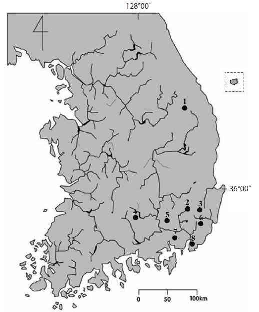
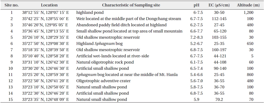
The samples were collected at 15 different water bodies such as lowland swamps, reservoirs, mountainous wetlands, sphagnum bogs, orum (very small and shallow caldera lakes) from 2009 to 2013 (Fig. 1 and Table 1). The samples were obtained by means of spoid, plankton net (mesh size, 25 μm) or by squeezing submerged macrophytes. Living materials was immediately examined. After first examination, materials were fixed with 5% formalin for permanent preservation and detail identification. Microscopic examinations were made at ×200-1000 magnification under an Axio Imager A2 microsope (Carl Zeiss, Jena, Germany) and photomicrographs were taken with an AxioCam HRC camera (Carl Zeiss). Water temperature, pH, and conductivity were measured in the field by means of a HI8314 membrane pH meter (HANNA instruments, Smithfield, RI, USA) and a HI9835 EC meter (HANNA instruments). All the taxa recorded are illustrated with photomicrographs. The materials were deposited in the National Institute of Biological Resources (NIBR) and Department of Biology Kyungpook National University. The following abbreviations are used: Dist., distribution; rr, very rare; r, rare; c, common; cc, abundant.
[Table 1.] Informations of sampling sites
Informations of sampling sites
References: Krieger 1937, p. 419, pl. 45, fig. 1; Prescott et al. 1975, p. 113, pl. 47, fig. 15.
Dist.: 5 (r), 7 (r), 9 (r); numbers indicate sampling sites in Fig. 1 and Table 1.
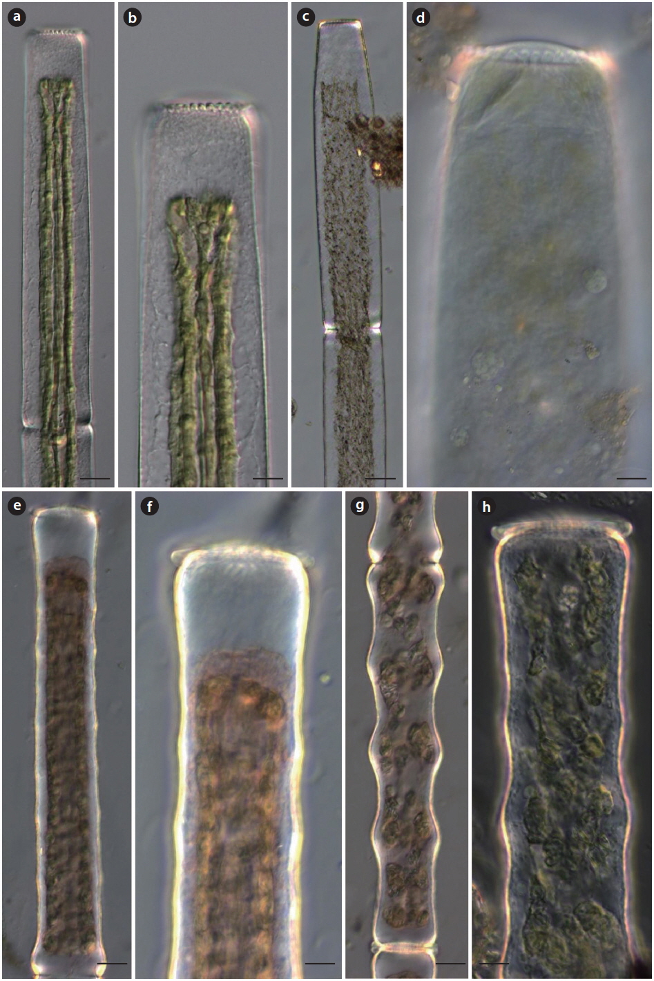
Description: Cells cylindrical, 7-13 times longer than broad, slightly constricted in the middle, lateral margins parallel; semicell with evident basal inflation, apices slightly broadened, with 10 to 20 tubercles; cells length 400-420 μm, breadth 25-27 μm.
In the present study, this species was rarely occurred from several lowland mesotrophic to eutrophic reservoirs.
References: Krieger 1937, p. 420, pl. 45, fig. 3; Prescott et al. 1975, p. 114, pl. 47, figs. 16-17; Förster 1982, p. 117, pl. 14, fig. 8; Yamagishi and Akiyama 1998, p. 55.
Dist.: 9 (rr).
Description: Cells large, about 11-18 times longer than broad, very slightly constricted in the middle, lateral margins straight; semicells cylindrical throughout, apices rounded or truncate with 12-17 tubercles in front view; cells length 560-950 μm, breadth 46-75 μm. A variety differ from nominate by its larger size, greater length and in not being broader at the apex. This species very rarely occurred only at oligotrophic, shallow pond in Jeju Island.
References: Krieger 1937, p. 425, pl. 46, fig. 3; Hirose and Yamagishi 1977, p. 517. pl. 175, fig. 3; Yamagishi and Akiyama 1987a, p. 59.
Dist.: 3(r).
Description: Cells large, about 11-19 times longer than broad, slightly constricted in the middle, lateral margins with 7-10 undulations beyond the basal inflation to the apices; semicells cylindrical, conspicuously inflated at the base, not attenuated from base to apex, apex slightly dilated, with 13 to 15 rounded tubercles; cells length 520-900 μm, breadth 40-69 μm. It rarely occurred in mountainous oligotrophic, slightly acidic wetlands in this study.
References: Krieger 1937, p. 421, pl. 45, fig. 7; Yamagishi and Akiyama 1996, p. 62.
Dist.: 3(r).
Description: Cells large, cylindrical, about 10-14 times longer than broad, slightly constricted in the middle; semicells typically with 4 large, equal prominent undulation, sometimes with small inflation between the large undulations, tapering slightly to the apex; apices rounded-truncate, with a crown of 11-15 conical tubercles; cells length 540-590 μm, breadth 56-62 μm. It rarely occurred in mountainous oligotrophic, slightly acidic wetlands in this study.
References: Prescott et al. 1977, p. 126, pl. 47, figs. 6, 8-11; Yamagishi and Akiyama 1997, p. 81; Coesel and Meesters 2007, p. 68, pl. 34, figs. 1-3; John et al. 2011, p. 696, pl. 170, fig. E.
Dist.: 5(r), 15(c).
Description: Cells medium, about 8-14 times longer than broad, slightly constricted in the middle; semicells with 5-9 undulated margins, apex smooth, rounded or truncately rounded; wall punctuate or scrobiculate; cells length 510-1,115 μm, breadth 59-85 μm.
References: West and West 1904, p. 222, pl. 32, figs. 11-16; Prescott et al. 1975, p. 149, pl. 56, figs. 1-3, 6-8; Rŭžička 1981, p. 366, pl. 56, figs. 9-15; Yamagishi and Akiyama 1986, p. 91; Coesel and Meesters 2007, p. 70, pl. 39, figs. 1-2.
Dist.: 11 (r).
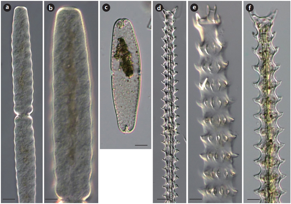
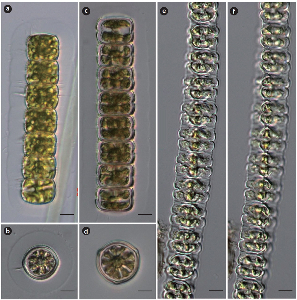
Description: Cells cylindrical or subcylindrical, 4-6 times longer than broad, slightly constricted in the middle, lateral margins straight or slightly concave, towards the ends slightly attenuated; apices broadly rounded with a deep, conspicuous median incision; cell wall with clearly defined longitudinal rows of punctures; chloroplast starshaped with 3-5 longitudinal ridges and 2-5 pyrenoids in each semicell; cells length 107-134 μm, breadth 22-26 μm. This species probably is acidophilic; mainly occurred in acidic peaty pools and
References: Krieger 1937, p. 442, pl. 52, figs. 1-7; Prescott et al. 1975, p. 143, pl. 51, figs. 7-14; Hirose and Yamagishi 1977, p. 519, pl. 176, fig. 5; Rŭžička 1977, p. 288, pl. 44, figs. 9-13; Förster 1982, p. 131, pl. 16, figs. 1-3; Yamagishi and Akiyama 1984, p. 96.
Dist.: 9(c).
Description: Cells medium size, subcylindrical, about 10-19 times longer than broad, slightly constricted in the middle; semicells slightly attenuated from base to apex, with 9-15 whorls of 10-14 low, each nodulation with a series of short spine; spines in lower whorls horizontal but spines in upper whorls directed upward; apex dilated and divided into two parts tipped with a pair of spines; cells length 190-230 μm, breadth 20-23 μm. It commonly occurred only at oligotrophic, shallow pond in Jeju Island.
References: Krieger 1937, p. 627, pl. 90, figs. 16-17; Prescott et al. 1977, p. 30, pl. 80, figs. 17-18; Hirose and Yamagishi 1977, p. 623, pl. 194, fig. 13; Yamagishi and Akiyama 1985a, p. 33.
Dist.: 11(r).
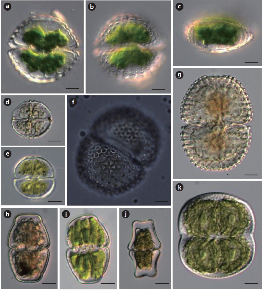
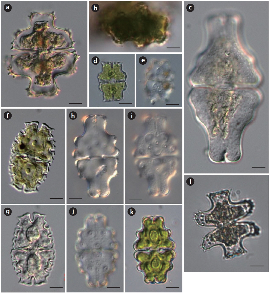
Description: Cells quadrate in outline, about 1.3 times longer than broader, deeply constricted in the middle, sinus acutely open outward; semicells 3-lobed, lateral lobes broadly rounded and transversely extended; polar lobe rectangular, the margins slightly diverging or nearly parallel, apex slightly retuse or flat, apical angles broadly rounded or slightly swollen, the margins of the apical angles and the basal lobes with sharp short spines; the center above the isthmus with a prominent protuberance, provided with concentric series of granules, and with a small inflation disposed by the imperfectly concentric series of granules in both sides of the central protuberance; in vertical view narrowly elliptic, poles broadly rounded, with a group of spines; lateral view elongate rectangular with a prominent median granulated protuberances, apical angles rounded with a group of short spines; cells length 46-78 μm, breadth 39-63 μm, isthmus 11-16 μm.
References: Krieger 1937, p. 519, pl. 67, figs. 4-6; Yamagishi and Akiyama 1988, p. 42.
Dist.: 10(r).
Description: Cells medium size, narrowly oval in outline, about two times longer than broad, deeply constricted in the middle, sinus linearly closed, slightly open at the extremity; semicells pyramidal with 5 protuberances; cell wall punctuate, with two mucilage pores vertically arranged at the center; lateral view narrowly pyramidal, basal margins with 2 inflations at the each side; cells length 116-122 μm, breadth 56-61 μm, isthmus 15-16 μm.
References: Krieger 1937, p. 587, pl. 81, figs. 8-11; Prescott et al. 1977, p. 98, pl. 75, figs. 14-14b; Hirose and Yamagishi 1977, p. 667, pl. 205, fig. 6; Rŭžička 1981, p. 490, pl. 82, figs. 1-6. Yamagishi and Akiyama 1994, p 33; Coesel and Meesters 2007, p. 81, pl. 47, figs. 4-5; John et al. 2011, p. 683, pl. 166, fig. F.
Dist.: 12(r).
Description: Cells small size, rectangular in outline, about 1.3-1.5 times longer than broad, deeply constricted in the middle, sinus linearly closed, slightly open at the extremity; semicells rectangular or trapeziform, basal angles broadly rounded with a minute spine; lateral margins retuse, apical angles with a short spine, apical margin elevated with a median incision; central protuberance with 3 large granules and a pair of mucilage pores, cell wall with granules within the margins of all lobes; vertical view elliptic; lateral view narrowly oval; cells length 26-41 μm, breadth 20-30 μm, isthmus 6.5-8 μm.
References: Krieger 1937, p. 623, pl. 88, figs. 24-26; Hirose and Yamagishi 1977, p. 64, pl. 199, fig. 18; Yamagishi and Akiyama 1989a, p. 35.
Dist.: 12(c).
Description: Cells medium size, broadly oval in outline, about 1.6-1.7 times longer than broad, deeply constricted in the middle, sinus linearly closed, rounded at the extremity; semicells 5-lobed; lateral lobes divided two lobules, the margins deeply retuse; polar lobe with deeply opened median incision and convex apex, apical angles with a short spine, incision between polar and lateral lobes deep and open; semicell with protuberance just above the isthmus and a pair of scrobiculations in the central region; in vertical view subrectangular, sides with a protrusion in the middle; lateral view ovate, apex narrowly rounded, sides inflated at the lower part, each side of the upper part with a large granule; cells length 40-50 μm, breadth 28-38 μm, isthmus 6.5-8 μm.
References: Krieger 1937, p. 502, pl. 63, fig. 1; Prescott et al. 1977, p. 104, pl. 61, figs. 1-2; Hirose and Yamagishi 1977, p. 627, pl. 195, fig. 2; Rŭžička 1981, p. 425, pl. 69, fig. 8; Yamagishi and Akiyama 1985a, p. 38.
Dist.: 3(c), 6(c), 11(c).
Description: Cells medium size, broadly oval in outline, about 2 times longer than broad, deeply constricted in the middle, sinus linearly closed; semicells truncate-pyramidate, 3-lobed; polar lobe elongate rectangular with concave lateral margins and apex truncate, apex truncate and convex with deep closed incision, angles rounded; basal part of semicell rectangular, lateral margin slightly retuse; incision between polar and upper lateral lobes concave; lateral view of semicell elongate truncate-pyramidate with two inflation at the lower part, apex convex; vertical view elliptic; cells length 60-67 μm, breadth 30-34 μm, isthmus 8.5-10 μm.
References: Krieger 1937, p. 503, pl. 63, figs. 2-3; Prescott et al. 1977, p. 105, pl. 60, fig. 15; Hirose and Yamagishi 1977, p. 627, pl. 195, fig. 4; Rŭžička 1981, p. 426, pl. 69, figs. 11-12; Förster 1982, p. 339, pl. 44, fig. 2.
Dist.: 3(c), 6(c), 11(c).
Description: Cells medium size, broadly oval in outline, about 1.5 times longer than broad, deeply constricted in the middle, sinus linearly closed; semicells truncate-pyramidate, 3-lobed; polar lobe elongate rectangular with concave lateral margins and apex truncate, apex truncate and convex with deep closed incision, angles rounded; basal part of semicell rectangular, lateral margin slightly retuse; incision between polar and upper lateral lobes concave; lateral view of semicell elongate truncate-pyramidate with two inflation at the lower part, apex convex; vertical view elliptic; cells length 60-67 μm, breadth 30-34 μm, isthmus 8.5-10 μm.
References: Krieger 1937, p. 629, pl. 90, figs. 21-24; Hirose and Yamagishi 1977, p. 622, pl. 194, fig. 8.
Dist.: 13(r).
Description: Cells medium size, slightly longer than broad, deeply constricted at the middle, sinus acutely opened; semicells 3-lobed; polar lobe rectangular with retuse apex and parallel sides, angles with a few small spines and within margin with 2-3 small spines; lateral lobes horizontally extended, lateral margins convex and slightly divergent and furnished with a number of spines; central part above the isthmus with a concentric series of wart-like granules, in both sides of the central granules-group with a few small spines inside the lateral lobes; lateral view ovate, with a granular protuberance on each side; cells length 47-60 μm, breadth 41-53 μm, isthmus 11-14 μm.
References: West and West 1905, p. 145, pl. 63, figs. 8-9; Prescott et al. 1981, p. 116, pl. 166, figs. 2-4; Coesel and Meesters 2007, p. 113, pl. 68, fig. 27.
Dist.: 1(r).
Description: Cells medium size, circular in outline, as long as broad or slightly broader than long, deeply constricted in the middle, sinus narrow linear and slightly open at the extremity; semicells semicircular, with about a dozen crenations, and 2 or 3 concentric series of undulations within the margin, basal angles slightly rounded; in vertical view of semicell fusiform, undulate toward the sharp poles; cells length 46-56 μm, breadth 50-58 μm, isthmus 16-28 μm.
References: Prescott et al. 1981, p. 120, pl. 224, fig. 8; Yamagishi and Akiyama 1985a, p. 19.
Dist.: 10(r).
Description: Cells medium size, slightly broader than long, deeply constricted in the middle, sinus linear but open, and slightly widening outwards; semicells semicircular, each basal angles rectangular with a small papilla, lateral margins undulate, undulations 4 to 5 on either side, apical margin slightly convex, with intramarginal of 4 small teeth; in vertical view of semicell elliptic, poles truncate, with 3 slight prominences in the mid-region of both side and with 10 minute teeth arranged in an oval around the center; cells length 25-32 μm, breadth 26-32 μm, isthmus 6-9 μm.
References: Prescott et al. 1977, p. 121, pl. 267, fig. 11; Hirose and Yamagishi 1977, p. 601, pl. 190, fig. 2; Förster 1982, p. 192, pl. 32, fig. 9; Yamagishi and Akiyama 1984, p. 11.
Dist.: 12(r), 14(r).
Description: Cells large, about 1.3 times longer than broad, deeply constricted in the middle, sinus narrowly linear, a slightly dilated extremity; semicells truncate-prymidal, basal angles rounded, lateral margins convex and upwardly convergent, upper angles obtusely rounded, apex nearly straight; cell wall uniformly granules disposed in oblique decussating series as well as in vertical and horizontal rows, each granules surrounded by 6 triangular pits throughout the surface of the semicell, the pits diminishing in size toward the margins of the cell; in vertical view of semicell elliptic, with a slight but conspicuous inflation in the mid-region on both side, and with a punctuate area destitute of granules in the center; cells length 64-86 μm, breadth 47-63 μm, isthmus 22-33 μm.
References: West and West 1908, p. 4, pl. 65, fig. 6.
Dist.: 11(c).
Description: Cells moderate, about 1.6-1.8 times longer than broad, slightly constricted in the middle, sinus narrowly linear, a slightly dilated extremity; semicells broadly pyramidal, apical and basal angles rounded, apex slightly convex, upper lateral margins just below the apex concave; in lateral and vertical view of semicell broadly elliptic; cell wall finely punctuate; cells length 47-55 μm, breadth 31-33 μm, isthmus 15-17 μm.
References: Prescott et al. 1981, p. 237, pl. 203, figs. 8, 12-13; Yamagishi and Akiyama 1987b, p. 24.
Dist.: 9(r).
Description: Cells large, about 1.1-1.2 times longer than broad, median constriction very shallow; semicells semielliptic; chloroplast with 4-7 longitudinal ridges in semicell, each with 2-3 pyrenoids; in vertical view, semicell circular or nearly so; cells length 65-70 μm, breadth 59-65 μm, isthmus 50-53 μm.
References: West and West 1908, p. 46, pl. 64, figs. 11-12; Yamagishi and Akiyama 1989b, p. 20.
Dist.: 6(c), 11(c).
Description: Cells medium in size, about 1.7-1.8 times longer than broad, median constriction deep, sinus narrowly linear, a slightly dilated extremity; semicells trapeziform, lateral margins and apex concave, basal angles slightly protrude; cell wall punctuate; in vertical and lateral view, semicell elliptic; cells length 40-50 μm, breadth 24-28 μm, isthmus 14-15 μm.
References: West and West 1923, p. 193, pl. 157, fig. 5; Hirose and Yamagishi 1977, p. 749, pl. 221, fig. 1; Prescott et al. 1982, p. 129, pl. 410, fig. 6; Yamagishi and Akiyama 1986, p. 78; Coesel and Meesters 2013, p. 174, pl. 101, fig. 2.
Dist.: 5(r), 12(r).
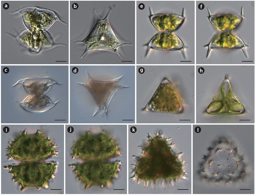
Description: Cells large, about 1.5 times longer than broad without processes, deeply constricted in the middle, sinus acutely open; semicells subelliptic to subspherical, and with two superimposed whorls of processes, lower whorl consisted of 9 processes horizontally disposed, upper whorl consisted of 6 upwardly divergent processes, all the processes nodulated and tipped with 3 sharp teeth; in vertical view showing 2 whorls of processes, with a marginal series of 9 processes and intramarginal series of 6 processes; cells length 50-96 μm without processes, 86-155 μm long with processes, broad 36-68 μm without processes, 77-160 μm breadth with processes, isthmus 24-33 μm.
References: West and West 1923, p. 32, pl. 134, fig. 4; Hirose and Yamagishi 1977, p. 686, pl. 208, fig. 12; Prescott et al. 1982, p. 142, pl. 363, fig. 13; Yamagishi and Akiyama 1987b, p. 69; Coesel and Meesters 2013, p. 72, pl. 39, figs. 1-2.
Dist.: 4(c), 9(c).
Description: Cells medium size, about as broad as long, deeply constricted in the middle, sinus obtuse at the extremity, widely open; semicells subelliptic or subcuneate, lateral margins convex, apical angles slightly produced, with a pair of long, sharp superimposed spines, spines long and strongly downturned and sometimes recurved, apical margins flat, truncate or broadly convex; cell wall smooth; in vertical view of semicell triangular, the margins flat or slightly concave, the poles bifurcate and furnished with two stout spines; cells length 26-33 μm, breadth 29-33 μm without spines, 48-56 μm with spines, isthmus 9-14 μm.
References: Prescott et al. 1982, p. 243, pl. 363, fig. 15; Yamagishi and Akiyama 1987a, p. 76.
Dist.: 3(r).
Description: Cells medium size, about as long as broad with spines, deeply constricted in the middle, sinus obtuse at the extremity and widely open; semicells suboviform or trapeziform, lateral and apical margins convex, angles with 2-3 spines; in vertical view of semicell triangular, the margins flat or slightly convex, the poles with 2-3 spines; cell wall smooth; cells length 32-35 μm, breadth 29-34 μm without spines, isthmus 11-14 μm.
References: Prescott et al. 1982, p. 316, pl. 345, figs. 10, 13; Yamagishi and Akiyama 1998, p. 72.
Dist.: 3(c), 6(c), 11(c)
Description: Cells medium size, about as broad as long, deeply constricted in the middle, sinus acutely open outwardly; semicells subtrapeziform, apical and basal angles with bispinate verruca, with a transverse series of 6 verrucae across the midregion of semicell; in vertical view of semicell triangular, the margins slightly convex, with a stout bi- or trispinate verrucae at the angles, and with 4-6 bi-spinate verrucae at the margins; within the margin 2 concentric circles of verrucae; cells length 45-53 μm, breadth 42-50 μm, isthmus 12-13 μm.
References: Yamagishi and Akiyama 1985b, p. 85.
Dist.: 12(r).
Description: Cells large, broadly elliptic, slightly longer than broad, deeply constricted in the middle, sinus slightly open like narrow fusiform but acuminate at the extremity; semicells semicircular, basal angles slightly produced toward obliquely downward and slightly thickened; in vertical view of semicell triangular, the margins straight, angles truncate and forked; cell wall minutely granulated; cells length 105-125 μm, breadth 88-100 μm, isthmus 22-36 μm.
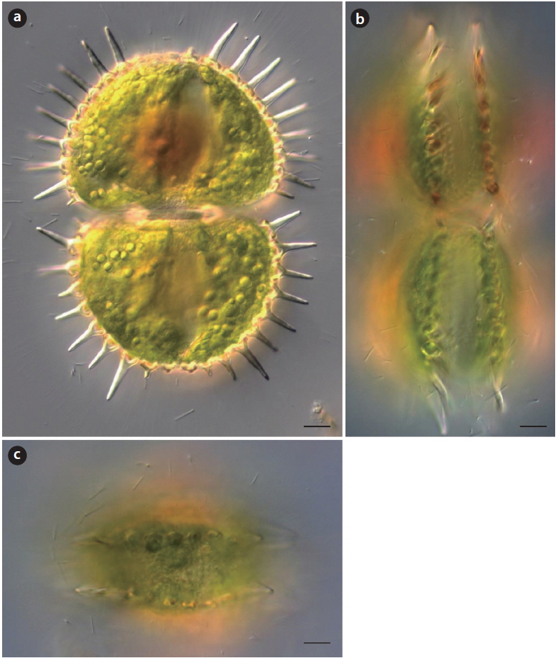
References: Hu and Wei 2006, p. 894, pl. XIV-85, figs. 1-3; Scott and Prescott 1961, p. 5, pl. 37, fig. 5.
Dist.: 6(r).
Description: Cells large, about 1.2-1.5 times longer than broad without spines, deeply constricted in the middle, sinus narrowly linear; semicells semicircular, apical margin rounded or slightly straight, basal angles with 1-2 papillae or short spine, margins of semicells with 15-16 equidistant pairs of long stout spines, apex with 3-4 pairs of conical short spines; vertical view of semicells elliptic, with two low of spines; cells length 110-130 μm without spines, breadth 75-80 μm without spines, spine 15-20 μm. It rarely occurred in mountainous oligotrophic, slightly acidic wetlands in this study.
References: West and West 1923, p. 232, pl. 161, figs. 20, 26; Croasdale et al. 1983, p. 28, pl. 460, fig. 13.
Dist.: 2(r), 3(r).
Description: Cells small, slightly broader than long, transversely rectangular, slightly constricted in the middle, sinus depressed and widely open; semicell subtrapeziform, later margins convex; vertical view of semicell circular with a slight papilla in opposite disposition; cells length 14-33 μm, breadth 17-36 μm, isthmus 14-30 μm. This variety differs from nominate variety by having vertical view of semicell with a slight papilla in opposite disposition. It rarely occurred in mountainous oligotrophic, slightly acidic wetlands in this study.
References: West and West 1923, p. 233, pl. 161, figs. 21; Croasdale et al. 1983, p. 28, pl. 461, fig. 1.
Dist.: 11(r).
Description: Cells small, slightly broader than long, transversely rectangular, slightly constricted in the middle, sinus depressed and widely open; This variety differs from nominate variety in that vertical view of semicell contains three tiny mamillates equidistant around the periphery of the circle; cells length 18-24 μm, breadth 26-37 μm, isthmus 23-33 μm. It rarely occurred in mountainous oligotrophic, slightly acidic wetlands in this study.
References: Croasdale et al. 1983, p. 39, pl. 463, fig. 9.
Dist.: 9(r).
Description: Cells medium size, about 1.5 times broader than long, moderately constricted in the middle, sinus acute and linear; semicells transversely narrowly oblong, lateral angles broadly rounded, later margins convergent to the apex, with a small basal inflation on either side of the isthmus; apex concave in the middle, with a cavity between the adjacent cells; vertical view of semicells triangular, sometimes quadrangular, angles broadly rounded, sides concave; This variety differs from the nominate variety in that the sinus is much deeper and linear, with a slightly dilated extremity; cells length 15-16 μm with processes, breadth 21-23 μm, isthmus 15-16 μm. It rarely occurred in mountainous oligotrophic, slightly acidic wetlands in this study.