나노물질(nanomaterials)은 입자의 외형적인 치수를 나타내는 가로, 세로, 직경 등의 규격 중 하나 이상이 100nm 이하인 물질을 총칭하며(Bae et al., 2009) 다양한 분야에서 가장 널리 이용되는 나노입자(nanoparticle) 이외에도 그 용도에 따라 나노선(nanowire), 나노튜브(nanotube), 나노막대(nanorod), 나노판(nanoplate) 등의 다양한 형태의 나노물질이 전자, 통신, 재료, 의료, 환경, 에너지 및 항공우주 등의 분야에서 널리 이용되고 있다(Guo et al., 2006; Meng et al., 2011, Sund et al., 2011). 나노물질은 같은 성질의 물질을 기반으로 하는 벌크(bulk)물질과는 전혀 다른 성격을 가지게 되는데(Saafan et al., 2010), 이는 나노물질의 경우 크기가 매우 작을 뿐 아니라 표면적이 극대화되기 때문으로 촉매로 사용될 경우에는 보다 높은 전환성능과 화학적 반응성을 나타내게 되고 산화물의 경우에는 큰 표면 전도도를 띠게 되는 등의 특징을 가진다(Zhao et al., 2011; Zhu et al., 2012).
나노물질의 종류별로는 탄소계 나노물질, 금속계 나노물질, 덴드리머(Dendrimers) 및 복합체 등으로 구분할 수가 있는데(U.S.EPA, 2007) 가장 많은 종류의 나노물질이 속하는 금속계 나노물질의 경우에는 의류 및 살균, 향균 등에 이용되는 은(Ag)나노물질과(Marambio-Jones and Hoek, 2010) 자외선차단, 반도체, 광촉매 등에 사용되는 이산화티타늄(TiO2) 나노물질(Shephard et al., 2002; Villalobos-Hernandez and Muller-Goymann, 2006), 메모리, MRI 조영제, 합금 등에 이용되는 산화철(Fe2O3)나노물질(Ali et al., 2014) 등이 포함된다. 탄소계 나노물질은 풀러렌(Fullerene), 카본블랙(Carbon black) 등의 나노물질이 포함되는데 풀러렌의 경우 연료전지, 스포츠 용품 및 IT 기기에 사용되며(Arie et al., 2009; Withers et al., 1997) 카본블랙은 타이어, 고무, 프린터 토너 등에 다양하게 사용되고 있다(Relsetter et al., 2011).
이처럼 나노물질이 산업과 실생활 및 다양한 분야 전반에서 널리 사용됨에 따라 인체 및 환경에 대한 나노물질의 위해성과 안전성이 대두되고 있는 실정이다. 나노물질이 제조, 사용 및 폐기되는 과정에서 인체와 환경에 노출됨으로 인한 위해성에 대한 많은 연구가 진행되고 있으나 정확한 독성 기작에 관해서는 논쟁의 여지가 있다(Cordeiro et al., 2014). 특히 생태계에 대한 나노위해성 연구는 2000년대 이후로 꾸준히 증가하고 있으며(Lee and An, 2010) 다양한 나노물질에 대한 생태독성 연구가 진행되었다. 또한 나노물질이 생태계로 유입될 경우에 발생할 수 있는 생물들 간의 먹이사슬(food chain) 및 영양단계(trophic level)를 통한 나노물질의 전이(transfer) 및 생물농축(biomagnification)에 관한 연구들이 초기단계를 지나 다양한 생물종과 나노물질에 대한 다수의 연구들이 발표되고 있다.
본 연구에서는 Lee and An (2011)의 논문 이후 활발하게 진행된 수생태계에서 나노물질 영양단계 전이 연구동향을 포괄적으로 조사하였으며, 연구에 이용된 나노물질들과 생물종, 연구기법들을 심층적으로 분석하였다. 또한 이를 바탕으로 앞으로의 나노물질 영양단계 전이의 연구방향에 관하여 논하였다.
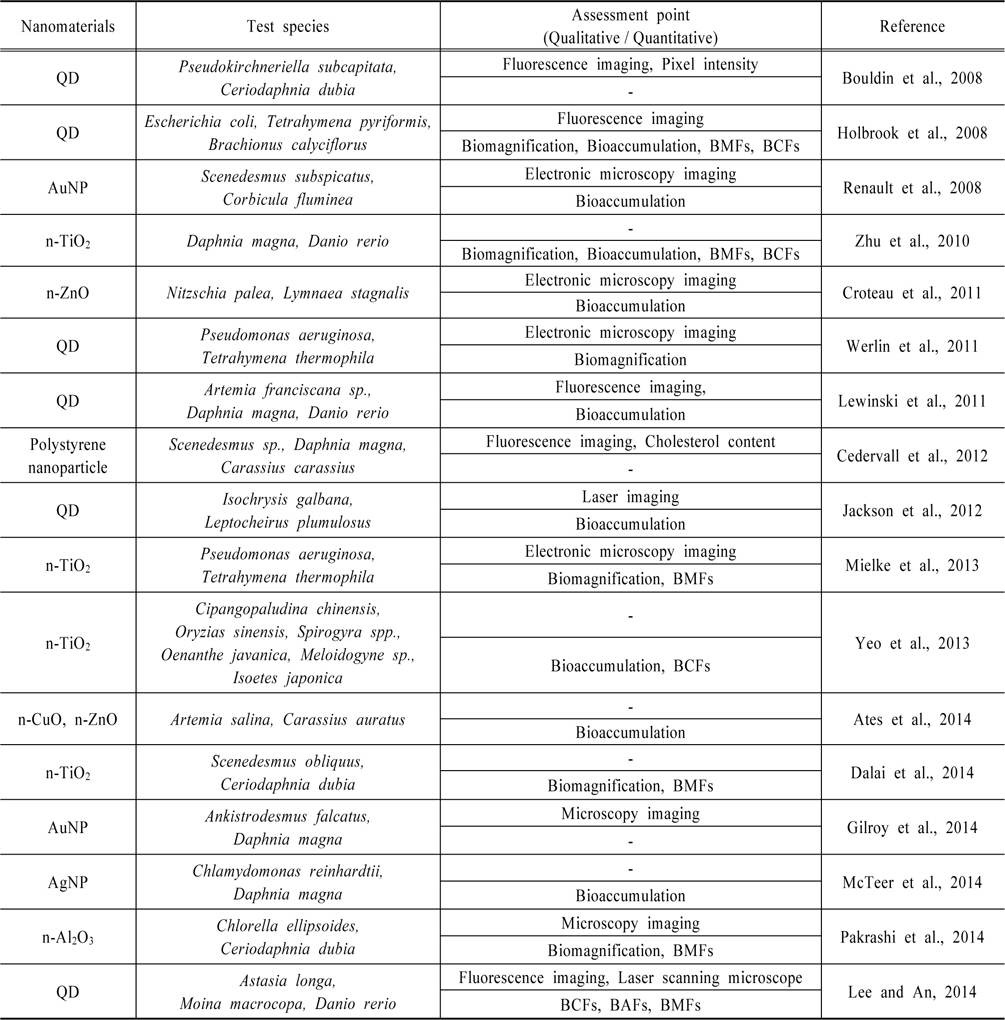
담수생태계에서의 나노물질 영양단계전이 연구는 지금까지 약 17편 정도가 발표된 것으로 조사되었다(Table 1). 그 중 가장 많은 연구가 진행된 나노물질은 Quantum dot(QD)으로, 총 6편의 연구가 진행되었는데, Bouldin et al. (2008)은 QD를 이용하여 나노물질의 담수생태계에서의 영양단계 전이를 연구한 최초의 논문을 발표하였다. Bouldin et al.은 carboxyl QD를 이용하여 조류인 Pseudokirchneriella subcapitata와 물벼룩인 Ceriodaphnia dubia 간의 나노물질 영양단계전이 실험을 진행하였는데, 나노물질의 전이는 QD의 형광을 이용하여 먹이노출로 물벼룩 내에 QD가 축적된 것을 형광현미경을 통해 관찰하였으며 이를 Pixel intensity를 통해 정량화하였다. Holbrook et al. (2008) 또한 carboxylated QD와 biotinylated QD를 이용한 나노물질의 영양단계 전이를 연구하였으며 박테리아(Escherichia coli), 섬모충 (Tetrahymena pyriformis), 담륜충(Brachionus calyciflorus)의 세 단계 먹이사슬을 이용하였다. Holbrook et al. 은 confocal laser scanning microscopy를 이용하여 QD의 형광을 관찰하였는데, QD 에 노출된 E. coli 를 섭취한 T. pyriformis 에서는 나노물질의 영양단계 전이가 관찰되지 않았다. 이는 E. coil 내에 QD가 축적되지 않았기 때문으로 생각되며 T. pyriformis 가 E. coli cell만을 선택적으로 섭취하였기 때문으로 보여진다. 반면, T. pyriformis의 QD 직접노출에서는 QD의 세포 내 축적을 공초점 레이져 현미경(CLSM)으로 확인할 수 있었으며 T. pyriformis 에서 B. calyciflorus 로의 먹이노출에서는 B. calyciflorus 내 QD가 영양단계 전이를 통해 축적된 것을 확인할 수 있었다. 또한 동위원소희석 유도결합플라스마 질량분석법(Isotope Dilution Inductively Coupled Plasma Mass Spectrometric, ID-ICP-MS)을 이용한 정량화를 통하여 수중 생물체 내 물질농도와 수중 물질농도의 농도비를 뜻하는 생물농축계수(Bioconcentration Factors, BCFs)와 먹이 연쇄를 통하여 물질농도가 상위단계로 갈수록 증가하는 비율을 의미하는 생물농축확대계수(Biomagnification Factors, BMFs)를 도출하였다. Werlin et al. (2011)의 경우, 박테리아(Pseudomonas aeruginosa)와 섬모충(Tetrahymena thermophile)을 이용하여 Cd/Se QD의 영양단계 전이를 연구하였는데, 주사투과전자현미경(STEM)을 이용하여 P. aeruginosa를 섭취한 T. thermophile 내의 QD의 축적을 확인하였으며, 이 과정에서 의미있는 BMFs인 4.82(cell volume base)와 5.37(dry biomass base)을 산출할 수 있었다. 이는 박테리아가 생태계 내의 먹이사슬에서 가장 기초적인 먹이원이기 때문이라고 보고하였다(Werlin et al., 2011). Lewinski et al. (2011)은 동물성플랑크톤(Artemia franciscana sp.)과 물벼룩(Daphnia magna), 어류(Danio rerio) 3단계의 영양단계에 걸친 전이 연구를 진행하였으며 이 과정에서 QD의 영양단계전이를 확인하였으나 유의한 BMFs는 산출하지 못하였다. Jackson et al. (2012)은 조류(Isochrysis galbana)와 단각류(Leptocheirus plumulosus)를 이용한 QD의 영양단계 전이 연구에서 레이저 절삭 유도 결합 플라즈마-질량 분석 측정법(LA-ICP-MS)을 이용하여 L. plumulosus 에 대한 QD의 먹이노출과 직접노출을 시각적으로 비교하였다. 그 결과 먹이노출로 QD에 노출된 L. plumulosus 는 체내에 QD가 분산되어 축적된 반면, 직접 QD에 노출된 L. plumulosus에서는 QD가 주로 소화관 부분에 집중적으로 축적되어있는 것으로 나타났다. 마지막으로 가장 최근에 발표된 Lee and An (2014)의 연구에서는 원생동물(Astasia longa), 물벼룩(Moina macrocopa), 어류(Danio rerio)의 3 trophic chain을 이용한 QD의 전이를 확인하였으며 Bioimaging을 통하여 A. longa 부터 D. rerio 까지의 나노물질의 전이를 시각적으로 관찰하였다. 또한 각 영양단계전이 별로 BCFs 값을 산출하였으나 생물농축확대(Biomagnification) 현상은 발생하지 않았음을 확인하였다.
QD 다음으로 많은 영양단계전이 연구가 진행된 나노물질은 이산화티타늄(Titanium dioxide, TiO2) 나노물질로서 총 4편의 연구가 존재했다. 2010년에 발표된 Zhu et al. (2010)의 연구에서는 물벼룩(Dphnia magna)와 어류(Danio rerio)를 이용한 n-TiO2의 전이를 확인하였는데, Zhu et al. (2010)은 직접노출보다 먹이노출에 의한 n-TiO2의 체내 축적량이 더 높게 측정되는 것은 수생태계의 고등생물에게 있어 먹이를 통한 나노물질의 섭취가 나노물질에 노출되는 가장 주요한 경로이기 때문이라고 설명하였다. 박테리아(Pseudomonas aeruginosa)와 섬모충(Tetrahymena thermophile)을 이용하여 n-TiO2의 영양단계 전이 연구를 진행한 Mielke et al. (2013) 의 경우, n-TiO2의 직접노출에서는 T. thermophile의 개체군의 수율이 낮아지는 경향을 보였으나, P. aeruginosa를 통한 먹이노출의 경우에는 개체군 수율과 성장률이 모두 저하되는 경향이 나타났다고 보고하였다. Yeo et al. (2013) 은 TiO2 nanoparticle (TiO2NP)과 TiO2 nanotube (TiO2NT)를 이용한 마이크로코즘 연구를 진행하였으며, 실험에는 달팽이(Cipangopaludina chinensis), 어류(Oryzias sinensis), 조류(Spirogyra sp.), 수생식물(Oenanthe javanica, Isoetes japonica), 선충(Meloidogyne sp.) 등이 이용되었다. 실험결과, 마이크로코즘 연구에서는 나노물질의 노출 후에 즉시 농도의 감소가 일어나는 것으로 나타났으며 2종류의 나노물질 모두 바이오필름에서 농도가 가장 높았다. 또한 O. javanica 의 뿌리에서도 높은 Ti 농도가 검출되었는데 뿌리를 잠재적인 먹이원으로 삼는 Meloidogyne sp. 와 C. chinensis 로의 영양단계 전이가 높은 수준으로 나타났다. 또한 바이오필름을 먹이로 삼는 Spirogyra sp. 에서 O. sinensis 로의 전이도 비슷한 수준으로 나타났다. Dalai et al. (2014) 은 n-TiO2를 이용한 조류(Scenedesmus obliquus)와 물벼룩(Ceriodaphnia dubia)의 영양단계전이 연구에서 세가지 노출환경에 따른 나노물질의 체내 축적 정도를 비교평가하였다. 첫째로 조류와 물벼룩을 n-TiO2에 함께 노출시키는 경우, 두번째로 조류를 n-TiO2에 48시간 노출시킨 후 물벼룩을 추가로 노출시키는 경우, 셋째로 n-TiO2에 노출시킨 조류를 원심분리하여 워싱한 후 이를 먹이로 물벼룩에게 주는 경우로 나누어 실험을 진행하였다. 실험결과, 조류를 통한 먹이노출이 물벼룩의 나노물질 섭취의 약 70% 정도를 차지하는 것으로 나타났으며, 조류의 삼출물 등으로 코팅된 나노입자가 그렇지 않은 나노입자보다 물벼룩이 섭취하기 용이한 것으로 나타났다.
다음으로 금나노입자(AuNP)를 이용한 연구가 두 편 존재하였는데, Renault et al. (2008) 은 조류(Scenedesmus subspicatus)에서 조개(Corbicula fluminea)로 AuNP의 전이를 연구하였으며 세포 내 AuNP의 분산은 투과전자현미경(Transmission Electron Microscope, TEM)을 이용하여 확인하였으며 MS-ICP를 이용하여 생물 내 나노입자의 축적을 확인하였다. TEM 촬영을 통하여 AuNP가 조류의 세포벽에 강하게 흡착된 것을 확인할 수 있었으며 조류를 섭취한 C. fluminea 의 위와 아가미, 소화샘의 상피세포에서 AuNP를 확인하였다. Gilroy et al. (2014) 은 조류(Ankistrodesmus falcatus)와 물벼룩 (Daphnia magna)을 이용한 AuNP의 영양단계 전이를 평가하였으며 UV-Vis spectroscopy를 이용하여 AuNP의 거동을 관찰하였다. Gilroy et al. (2014)은 AuNP가 조류의 표면에 강하게 흡착하므로 이를 섭취하는 물벼룩의 경우 소화기관과 소화기관 내 섭취한 조류에 나노물질이 축적되어 물벼룩이나 조류를 섭취하는 어류를 인간이 섭취하게 될 경우 나노물질에 노출될 가능성이 있음을 제시하였다.
산화아연(ZnO) 나노물질을 이용한 연구 또한 두 편이 존재하였으며 Croteau et al. (2011) 의 경우 규조류(Nitzschia palea)와 달팽이(Lymnaea stagnalis)의 먹이사슬을 이용한 n-ZnO의 전이를 연구하였는데, 그 결과 높은 농도의 n-ZnO에 노출된 N. palea를 섭취한 L. stagnalis의 경우 대조군에 비해 더 적은 양을 섭취하고 배변의 양도 감소하는 등 소화력이 저하되는 경향이 나타났다. 실험에는 주사전자현미경(Scanning Electron Microscope, SEM)이 사용되었으며 N. palea의 표면에 n-ZnO가 다량으로 흡착되어있는 것과 고농도 n-ZnO 노출군의 N. palea 를 섭취한 L. stagnail의 배설물에서 소화되지 않은 N. palea가 저농도 노출군보다 다량으로 존재하는 것을 확인하였다. Ates et al. (2014) 은 n-ZnO와 산화구리(CuO) 나노물질을 이용한 조류(Artemia salina)에서 어류(Carassius auratus)로의 영양단계전이를 연구하였다. C. auratus 의 간과 아가미에서의 n-ZnO와 n-CuO의 산화스트레스로 인한 영향을 평가하기 위하여 유지의 산화로 생성되는 물질인 말론디알데히드(malondialdehyde, MDA)의 농도를 측정하였으며, 대조군과 비교했을 경우, n-ZnO 은 먹이노출 시 아가미에서 저농도와 고농도 모두 유의한 증가가 나타나지 않았고 n-CuO의 경우에는 저농도에서는 거의 증가하지 않았으나 고농도에서는 유의하게 증가한 것으로 나타났다. n-ZnO는 먹이노출과 직접노출 모두 간에서 미미한 MDA의 증가가 나타났으나 n-CuO의 경우에는 두 노출방법 모두에서 유의한 MDA의 증가가 나타났다.
이외의 나노물질들을 이용한 연구들로는 Cedervall et al. (2012) 의 금속계 나노물질이 아닌 나노플라스틱 polystyrene nanoparticle을 이용한 조류(Scenedesmus sp.), 물벼룩(Daphnia magna) 그리고 어류(Carassium carassius) 총 3단계의 영양단계 전이 연구가 존재하였다. 연구결과, 형광을 띠는 나노플라스틱이 조류에서 물벼룩으로 전이된 것을 확인하였으며, C. carassius 의 무게 감소, 혈청 내의 트리글리세라이드:콜레스테롤의 비율, 근육과 간 사이의 콜레스테롤의 분포가 모든 노출군에서 대조군과 다르게 나타났다. 이는 C. carassius 가 나노입자를 흡수함에 따라 체내에서 지질의 대사에 교란이 온 것으로 Cedervall et al. 은 설명하였다. McTeer et al. (2014) 은 조류인 Chlamydomonas reinhardtii 와 물벼룩 Daphnia magna 를 이용한 은나노입자(AgNP)의 영양단계전이를 평가하였으며 AgNP의 실험 결과와 비교하기 위하여 질산은(AgNO3)을 이용한 실험을 동일하게 진행하였다. 또한 Ag의 전이에 PO43−가 미치는 영향을 알아보기 위하여 low PO43− medium과 high PO43− medium을 각각 사용하여 실험하였다. 결과적으로 AgNP와 AgNO3 모두 D. magna 로 축적되는 Ag의 양은 유사하였으며 PO43− 의 가용성이 은이온의 축적과 D. magna 로의 전이에 영향을 미치는 것으로 나타났다. 이는 Ag+의 독성을 감소시키는 잠재적인 리간드(ligand) 역할을 하는 PO43−가 부족한 환경으로 AgNP가 유입될 경우, 환경 중에 서식하는 생물들이 Ag+에 의해 더 큰 피해를 입을 수 있음을 시사한다. 마지막으로 Pakrashi et al. (2014) 은 산화알루미늄(Al2O3)나노입자를 이용한 조류(Chlorella ellipsoides)와 물벼룩(Ceriodaphnia dubia)의 영양단계전이를 평가하였으며 n-Al2O3가 먹이노출을 통해 C. ellipsoids 에서 C. dubia 로 전이되는 것을 확인하였다. 또한 먹이노출이 D. magna 의 섭식활동에 미묘한 변화를 유발하는 것으로 관찰되었으며 생식행동에는 영향을 미치지 않는 것으로 나타났다.
담수생태계에서의 나노물질의 영양단계 전이 연구는 총 17건의 연구 중 2단계 전이 연구가 12건, 3단계 전이 연구가 4건 존재하였으며 미소생태계 연구는 단 한 건 만이 존재하였다. 그 중 가장 많은 연구가 진행된 물질은 형광 나노물질인 QD였으며 그 다음으로는 다양한 분야에서 널리 사용되고 있는 n-TiO2 순이었다. 연구대상 나노물질은 총 7개의 금속계 나노물질과 1개의 비금속계 나노물질이 사용되었다. 시험종의 경우, 가장 많이 사용된 시험종은 물벼룩(Water flea)으로 D. magna 가 5건, C. dubia 가 3건, Moina macrocopa 가 1건의 연구에서 시험종으로 이용되었다. 가장 많이 사용된 먹이사슬은 조류(Algae)-물벼룩의 조합으로 나타났다. 나노물질과 시험종이 연구마다 동일하지 않으므로 직접적인 나노물질의 영양단계 전이 및 독성은 비교할 수 없었으나 나노물질의 종류과 시험종에 관계없이 대부분의 연구에서 생물축적(Bioaccumulation)과 생물농축확대(Biomagnification) 현상을 확인할 수 있었다.
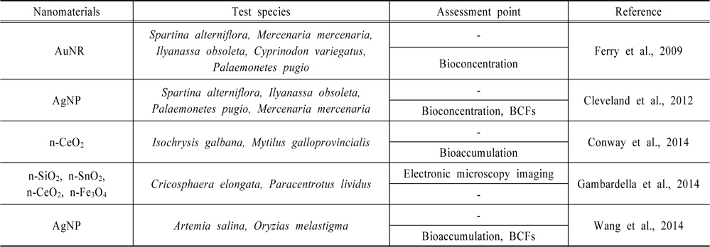
해양생태계에서의 나노물질 영양단계전이 연구는 지금까지 약 5편 정도가 발표되었으며(Table 2) 담수생태계와 비교해, 보다 다양한 나노물질들에 대한 연구가 진행된 것으로 조사되었다. 먼저 두 건의 AgNP에 대한 연구결과가 존재했는데, Cleveland et al. (2012) 은 AgNP를 이용한 메소코즘 연구를 진행하였으며 해수와 퇴적물, 바이오필름, 연안 초본(Spartina alterniflora), 진흙 달팽이(Ilyanassa obsoleta), 새우(Palaemonetes pugio), 대합(Mercenaria mercenaria) 유생을 실험 대상으로 하였다. 또한 나노물질 뿐 아니라 나노물질이 사용된 양말이나 약품, 장난감 등의 소비재를 노출물질로 이용하였다. ICP-MS 분석을 통하여 실험종 및 해수, 퇴적물, 바이오필름 간의 BCFs와 영양단계를 통해 물질이 피식자로부터 포식자로 전이되는 비율을 나타내는 영양전이계수(Trophic Transfer Factors, TTFs)를 산출하였으며 M. mercenaria와 퇴적물 간의 BCFs가 78에서 1,000으로 가장 높게 나타났다. Wang et al. (2014) 은 20nm와 80nm 두 종류의 AgNP를 이용하여 새우(Artemia salina)와 어류(Oryzias melastigma)에서의 영양단계 전이 연구를 진행하였다. 28일의 만성실험결과, 먹이노출을 통한 O. melastigma로의 AgNP의 흡수율은 6% 미만으로 낮았으며 0.01에서 0.04 정도의 낮은 영양단계 전이율을 나타냈다. 또한 고농도의 AgNP에 노출된 A. salina 를 섭취한 O. melastigma에서는 성장 저해와 수분 함량 감소가 관찰되었다.
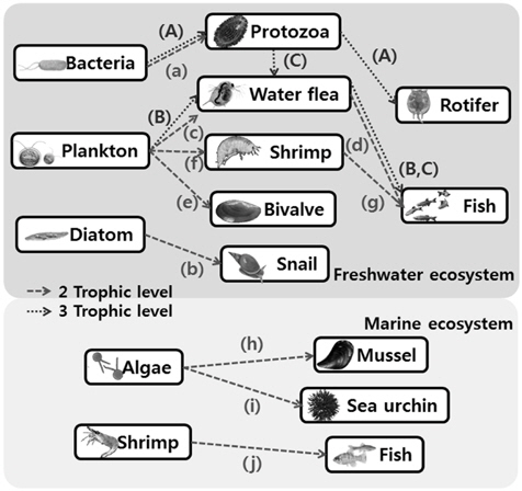
[Fig. 1.] Examples of nanoparticle trophic transfer shown in the previous studies. (A) Holbrook et al., 2008 (Bacteria-Protozoa-Rotifer) (B) Lewinski et al., 2011; Cedervall et al., 2012 (Plankton-Water flea-Fish) (C) Lee and An, 2014 (Protozoa-Water flea-Fish) (a) Werlin et al., 2011; Mielke et al., 2013 (Bacteria-Protozoa) (b) Croteau et al., 2011 (Diatom-Snail) (c) Bouldin et al., 2008; Dalai et al., 2014; Gilroy et al., 2014; McTeer et al., 2014; Pakrashi et al., 2014 (Plankton-Water flea) (d) Zhu et al., 2010 (e) Renault et al., 2008 (Plankton-Bivalve) (f) Jackson et al., 2012 (Plankton-Shrimp) (g) Ates et al., 2014 (Shrimp-Fish) (h) Conway et al., 2014 (Algae-Mussel) (i) Gambardella et al., 2014 (Algae-Sea urchin) (j) Wang et al., 2014 (Shrimp-Fish).
AgNP 이외에는 Ferry et al. (2009) 이 처음으로 나노물질의 메소코즘(Mesocosm)에서의 거동과 축적에 관한 연구를 발표하였는데, 실험물질은 금나노막대(Gold nanorod, AuNR)를 이용하였으며 해수, 퇴적물, 해중식물(Spartina alterniflora), 바이오필름, 달팽이(Ilyanassa obsoleta), 조개(Mercenaria mercenaria), 새우(Palaemonetes pugio), 그리고 어류(Cyprinodon variegatus)를 이용하여 메소코즘을 형성, 실험을 진행하였다. 실험결과, 가장 나노물질이 많이 축적된 곳은 여과섭식자인 M. mercenaria 였으며 그 다음으로 바이오필름인 것으로 나타났다. Conway et al. (2014) 은 제조 나노물질인 산화세륨(CeO2) 나노입자를 이용하여 식물성플랑크톤(Isochrysis galbana)과 해양생태계 내에서 여과섭식을 하는 홍합(Mytilus galloprovincialis) 사이의 영양단계 전이를 확인하였다. I. galbana 의 경우 n-CeO2를 1h 이내에 흡착하는 것으로 나타났으며 M. galloprovincialis 에 의해 약 99%의 n-CeO2가 흡착되어 위분(Pseudofaeces)으로 배출되는 것으로 확인되었다. 직접노출과 먹이노출 간 홍합의 조직이나 위분에서의 세륨의 농도는 크게 차이를 보이지 않았다. Gambardella et al. (2014) 은 조류 Cricosphaera elongata 와 이를 먹이원으로 하는 성게인 Paracentrotus lividus의 유생을 대상으로 다양한 금속계 나노물질인 이산화규소(SiO2), 산화주석(SnO2), 산화세륨(CeO2), 산화철(Fe3O4) 나노입자들을 이용하여 실험을 진행하였다. n-SiO2 와 n-CeO2에 노출된 C. elongata 를 섭취한 P. lividus 에서 급격한 생존률의 감소가 관찰되었으며, 이상발달, 골격퇴보와 초기 성장 변화가 네 가지 각각의 NPs에 노출된 C. elongata 를 섭취한 P. lividus 에서 관찰되었다. 결론적으로 n-SiO2 와 n-CeO2는 성게 유생의 생존능력을 감소시키며 모든 실험 나노물질들이 P. lividus 에 악영향을 미치는 것으로 확인되었다.
해양생태계에서 나노물질의 영양단계 전이 연구는 담수생태계에서의 연구결과보다 그 수가 절반 이하로 적었다. 두 건의 메소코즘 연구가 존재하였으나 아직까지 3단계 전이 연구결과는 발표되지 않은 것으로 조사되었다. 연구대상물질은 총 8개의 금속계 나노물질이 이용되었다. 해양생태계에서의 나노물질 영양단계 전이 연구는 그 수가 담수생태계에 비해 적으나 다양한 시험종을 이용한 메소코즘 연구가 두 건 존재하여 비교적 많은 종류의 시험종이 연구에 이용되었다. 한 건의 연구(Gambardella et al., 2014)만을 제외한 모든 연구에서 생물축적 및 생물농축(Bioconcentration)을 확인하였으며 생물농축계수를 산정한 연구는 그 중 두건이 존재하였다.
현재까지 발표된 수생태계에서의 나노물질의 영양단계 논문들은 담수생태계가 약 17편, 해양생태계가 약 5편으로 총 22편 정도가 발표되었다. 그 중 가장 많이 연구가 진행된 나노물질은 QD로, 금속계 나노물질이기 때문에 ICP, AAS 등을 이용한 정량화가 가능하고 다른 금속계 나노물질과 다르게 형광을 발현하므로 QD가 영양단계 전이를 통하여 생체나 세포 내에 축적된 것을 시각적으로 확인할 수 있어 가장 활발한 연구가 진행된 것으로 보인다. 다음으로 다수의 연구가 진행된 n-TiO2의 경우에는 자외선차단제나 광촉매, 백색도료 및 반도체 등 매우 광범위한 분야에서 다량으로 사용되기 때문에(Shephard et al., 2002; VillalobosHernandez and Muller-Goymann, 2006) 최근에 그 안전성에 대한 우려가 확산되며 다수의 연구가 진행되고 있다. 그 외 다양한 금속계 나노물질에 대한 연구가 보고된 바 있으며, 금속계 나노물질 이외에는 나노플라스틱인 polystyrene nanoparticle에 대한 연구가 한 건 보고되었다.
2008년부터 나노물질의 영양단계 전이 연구가 발표된 이후로 담수생태계에서는 12건의 2단계 전이 연구와 2건의 3단계 전이, 그리고 한 건의 미소생태계 연구가 발표되었는데, 3단계 전이 연구는 2단계 전이 연구 보다 비교적 실험이 용이하지 않으며, 미소생태계 연구 또한 실험과 데이터 분석이 쉽지 않으므로 다수의 연구가 발표되고 있지는 않다. 해양생태계의 경우에는 메소코즘 연구와 2단계 수준의 연구가 발표되었고 아직 3단계 수준의 연구는 발표된 바 없다. 영양단계 전이를 정성적 및 정량적으로 확인하는 방법론으로는 나노물질에 형광이 존재하는 경우에는 형광현미경이나 CLSM 등을 이용하는 방법들이 있으며, 대부분 전자현미경을 이용하여 나노물질의 전이를 확인하였다. 정량적 측정법으로는 ICP와 AAS를 이용한 방법만이 사용되었다.
본 연구에서는 영양단계와 먹이사슬을 통한 나노물질의 개체간 전이에 관한 연구들을 수집하여 연구에 사용된 나노물질과 생물종, 기법 등을 분석하였으며 연구 동향을 파악하였다. 2008년 처음 관련 연구들이 발표되기 시작하면서 현재는 많은 연구자들과 연구기관들이 나노물질의 생태독성과 환경 중에서의 거동, 영양단계를 통한 전이와 생물농축에 대한 연구를 진행하고 있어 이제 나노물질의 영양단계 전이에 대한 연구는 성숙기에 접어든 것으로 보인다. 그러나 아직까지 대부분의 연구들이 2단계 전이에만 치중되어있어 상대적으로 3단계 전이 및 미소생태계 연구가 부족한 점과 더욱 다양한 나노물질에 대한 연구가 부족한 점, 그리고 단순 나노물질의 전이만을 확인하는 것이 아닌 영양단계 전이 연구를 통한 매체와 생물, 생물과 생물 간 영향에 대한 연구가 부족하다는 한계점이 존재하는 것을 확인하였다. 미소생태계 연구를 제외한 대부분의 연구에서는 아직까지 매체를 통한 생물체로의 전이에 관한 연구가 추가적으로 이루어지지 않고 있어 향후 관련 연구가 필요할 것으로 사료된다. 또한 지금까지의 영양단계전이 연구에서 사용된 나노물질은 대부분 0차원(0-dimension) 입자성 물질들로, 이와는 다른 특성을 가진 1차원 및 2차원 나노물질에 대한 연구도 앞으로 진행되어야 할 것으로 판단된다.