검색 전체 메뉴
PDF
맨 위로
OA 학술지
First Records of Two Species of Genus Clavelina (Ascidiacea: Aplousobranchia: Clavelinidae) from Korea
  • 비영리 CC BY-NC
  • 비영리 CC BY-NC
ABSTRACT
First Records of Two Species of Genus Clavelina (Ascidiacea: Aplousobranchia: Clavelinidae) from Korea
KEYWORD
taxonomy , Ascidian , Clavelina elegans , Clavelina miniata , Korean waters
  • INTRODUCTION

    The genus Clavelina is defined as comprising solitary or colonial species of Clavelinidae, in which the zooids are connected by basal stolon or completely or partially embedded in common test, having no less than 4 rows of stigmata and no less than 20 stigmata per row, with posterior abdominal vascular stolon’s branch which do not form a network, with branchial and atrial opening without lobes, having neither folds nor papillae in the branchial sac, with languets on the dorsal lamina, having an abdomen containing the gut loop, and developing eggs in the distal part of the oviduct or in the anterior part of the abdomen (Savigny, 1816; Kott, 1990). Additionally, the colors of living zooid are important key to identify Clavelina (Tokioka and Nishikawa, 1976; Kott, 1990).

    Among the 47 recorded species of Genus Clavelina, C. fasciculate has been reported in Korean waters (Rho, 1966). Clavelina elegans has been reported only from Japan (Nishikawa and Tokioka, 1976; Tokioka and Nishikawa, 1976). Clavelina miniata has been reported only from Japan (Watanabe and Tokioka, 1973; Nishikawa and Tokioka, 1976). We expand the distribution range to Korea and provide detailed descriptions and illustrations of C. elegans and C. miniata as the first record of Korean fauna.

    MATERIALS AND METHODS

    Materials of Clavelina elegans examined in this study were collected at subtidal zone (19 m depth) of Beomseom in Jeju-do by SCUBA diving on 23 May 2013. Materials of C. miniata examined in this study were collected at Chagwido in Jeju-do by SCUBA diving on 8 June 2001.

    Collected specimens were anesthetized with menthol for 8 h, and then fixed in 4% formalin with seawater. Materials were kept in the solution which was composed of 1% chromic acid and 50% acetic acid in the ratio of 1 to 10 for a day and then they were replaced into the solution of 1% chromic acid for a few hours before dissecting.

    For identification, each specimen was examined for morphological characteristics such as colony organization, thoracic muscles formula, stigmata, stomach, gonad, larvae and coloration under stereomicroscopes (SZX7; Olympus, Tokyo, Japan). The color of each part was recorded with a color code based on the color chart (Pantone color formula guide 747XR). Images of the collected living Clavelina colonies prior to fixation were taken by a digital camera (WG2; Pentax, Tokyo, Japan). The images of zooids and larvae were taken by a stereomicroscope with camera (UHCCD05000KPA; Touptek Photonics, Zhejiang, China). The size of the zooid and larvae were then measured using an image analyzer (Toupview 3.7; Touptek Photonics).

    The systematic scheme of ascidians was adopted from Kott (1990), Monniot and Monniot (2001), and Sanamyan (2013a, 2013b). Specimens are deposited in Natural History Museum, Ewha Womans University, Seoul, Korea (EWNHMAS 1-3).

    SYSTEMATIC ACCOUNTS

    Order Aplousobranchia Lahille, 1886

    Family Clavelinidae Forbes and Hanley, 1848

    Genus Clavelina Savigny, 1816

    1*Clavelina elegans(Oka, 1927) (Figs. 1, 2)

    Dendroclavella elegans Oka, 1927: 555-557, figs. A, B.

    Clavelina elegans (Oka, 1927), Nishikawa and Tokioka, 1976: 68-73, figs. 3-5; Tokioka and Nishikawa, 1976: 341-345, figs. 1, 2.

    Material examined. Two colonies with 9 zooids and 5 zooids, Korea, Jeju-do, Beomseom, 30 May 2013, Seo SY (EWNHMAS 1-2), 17 m deep by SCUBA diving.

    Diagnosis. Zooids partially embedded in common stalk. Small zooid. Cobalt blue color with white patch around aperture. Longitudinal thoracic muscles.

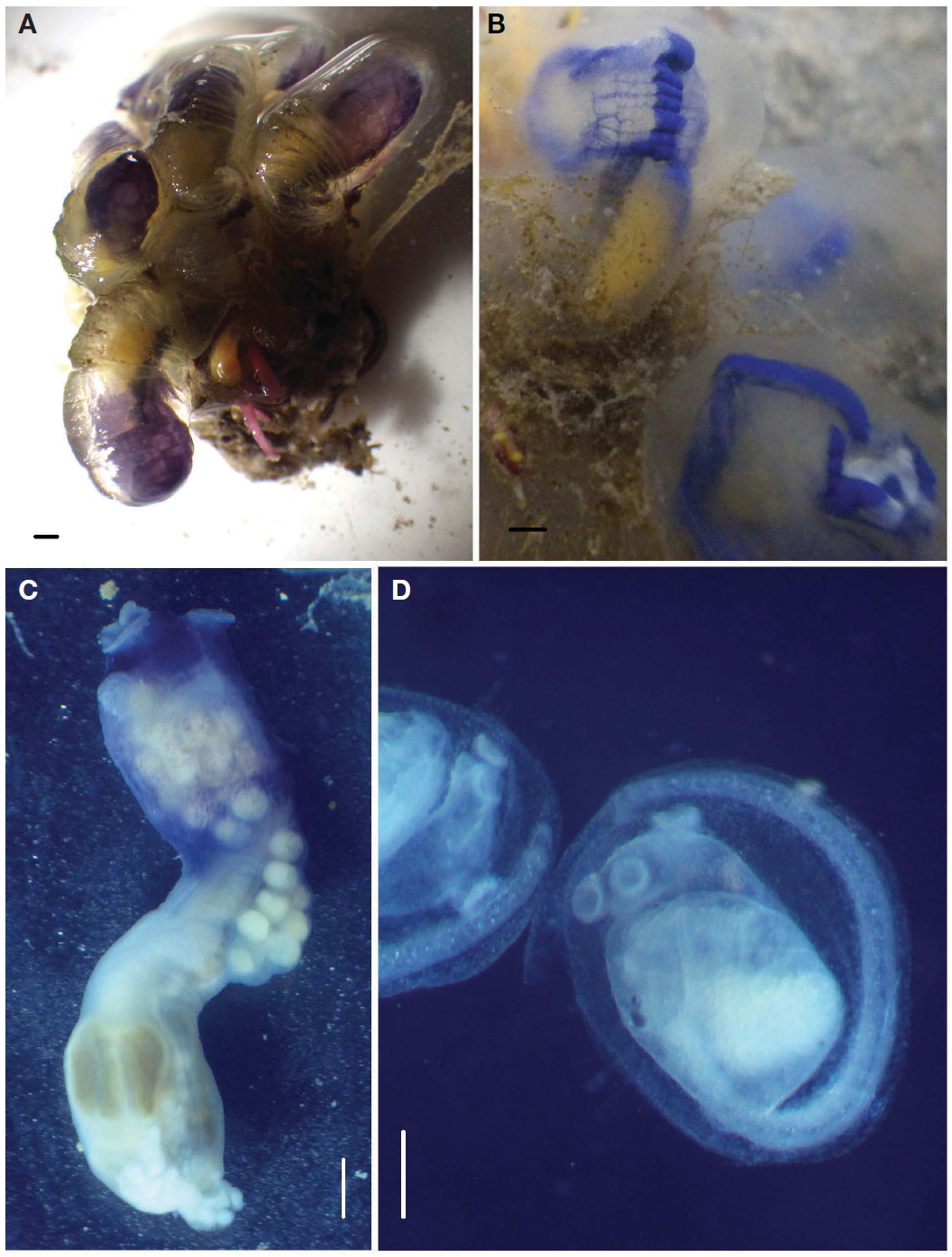
    Description. Two colonies consist respectively of 5 and 9 zooids crowded on a common basal mass up to 6.2 mm in thickness. Zooids fanning out from the upper surface of basal mass in which posterior ends of zooids embedded. Long portion (12.5-15.7 mm) of zooids separated, while the proximal part (1.2-2.2 mm) of zooids embedded in common basal mass (Figs. 1A, 2A).

    Zooidal test fragile and transparent, while test of the stalk somewhat tough and whitish translucent. Zooid transparent with dark cobalt blue pigments and white patch in living. Dark cobalt blue pigments (Pantone 286C) in band along each side of endostyle, on dorsal midline, and around branchial apertures in living. White patch U shaped and inside dark blue patch between both apertures in living (Fig. 1B). Zooids become purple blue (Pantone 2755C) in formalin solution (Fig. 1C).

    Zooids small 12.9 mm long on average, up to 14.1 mm long in contracted condition. Thorax 3.8-5.4 mm long and 2.0-3.8 mm wide, while abdomen 6.9-8.7 mm long and 1.4-1.7 mm wide. Thorax shorter than abdomen in both extracted zooids and contracted zooids (Figs. 1C, 2B).

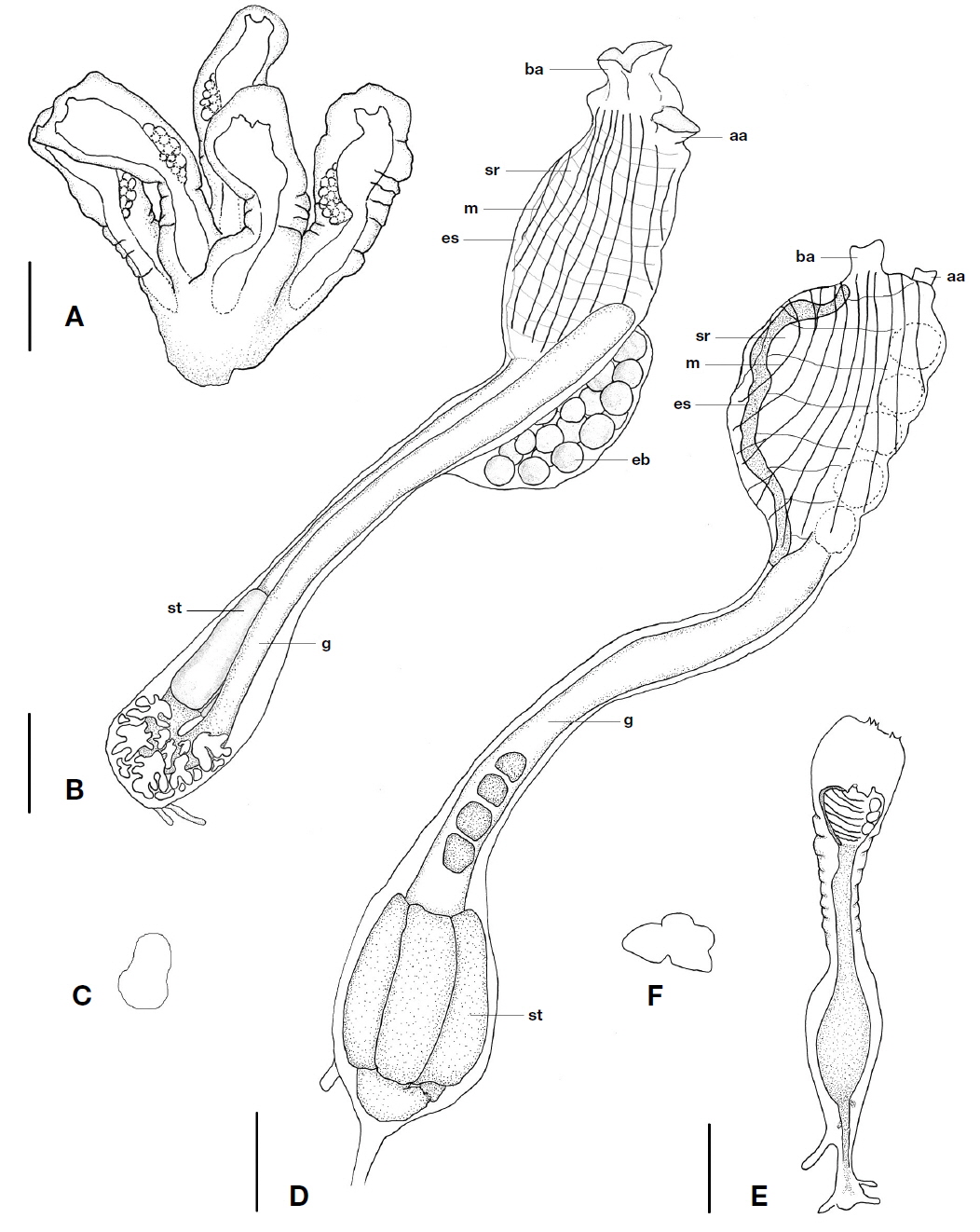
    Strong longitudinal muscles extend along length of thorax. Muscles formula 1-2E5-7B2D=8-11.

    Both apertures open at upper terminal of thorax. Branchial apertures have two lips. Number of tentacles 9-10 alternated large and small. Opening of neural gland simple and longitudinal slit. Dorsal languet fine pointed tongue shape. 14-16 stigmata rows of 46-52 stigmata. Transverse vessels provided each with a well-developed membrane.

    Oesophageal neck more or less long, narrow, and occupy about two thirds of total length of abdomen in zooid.

    No prestomach and large posterior stomach occurs at posterior end of descending limb of gut loop. Pear-shaped stomach have smooth surface with a few rather distinct plication (Figs. 1C, 2C). Anus opens at level as high as posterior three stigmata row. Atrial apertures have smooth border (Fig. 2B).

    Gonads usual form in gut loop posterior to stomach. Male follicles spill out over posterior end of gut loop obscuring it. Brood pouch extends across posterior end of right side of thorax from postero-dorsal corner and have 27-48 embryos. Embryos complete their development in peribranchial cavity. Larvae yellow in living (Pantone 114C) and small; trunk 0.63-0.69 mm long; tail three quarters of distance around trunk. Larvae have an ocellus and an otolith. Frontal plate well developed. Three adhesive organs lie on expanded lobes from frontal plate. Two on its left upwards while one on its right upwards (Fig. 1D).

    Distribution. Pacific Ocean: Korea (Beomseom, Jeju-do), Japan (Sagami Bay, Amakusa rooky reef, Minabe).

    Remarks. The present species, Clavelina elegans is the first record from Korean waters.

    This species very resembles Amakusa species of Tokioka and Nishikawa (1976) in colony organization, zooid size, thoracic muscles, ciliate groove, stomach surface, stomach location and brood pouch. They slightly differ in that this species has 14-16 stigmata rows of 46-52 stigmata but Amakusa species 17-18 stigmata rows of 50-60 stigmata. Tokioka and Nishikawa (1976) did not describe white patches in living zooid from Amakusa.

    The present species very resembles Clavelina fasciculate from Korea and Japan. But C. elegans have the stomach with a few rather distinct plications, whereas C. fasciculate has the stomach with 4 ridges (Van Name, 1945; Rho, 1977). The larval trunks of this species are 0.63-0.69 mm long and are larger than 0.42-0.46 mm of C. fasciculate, and the zooids of the one are up to 14.1 mm long and are smaller than 20 mm of the other.

    This species resembles Clavelina fecunda from Australia and Philippines in colony organization and zooid musculature. However, they differ in that C. fecunda has stumpy zooids up to 10 mm at length, and the bright yellow color in the living specimens (Kott, 1990).

    1*Clavelina miniata Watanabe & Tokioka, 1973 (Figs. 2, 3)

    Clavelina miniata Watanabe and Tokioka, 1973: 99-107, figs. 1, 2; Nishikawa and Tokioka, 1976: 78

    Material examined. One colony with 50 zooids, Korea, Jeju-do, Chagwido, 8 Jun 2001, Song JI (EWNHMAS 3), by SCUBA diving.

    Diagnosis. Zooids connected by basal stolon. Small zooid. Yellow color. Longitudinal thoracic muscles.

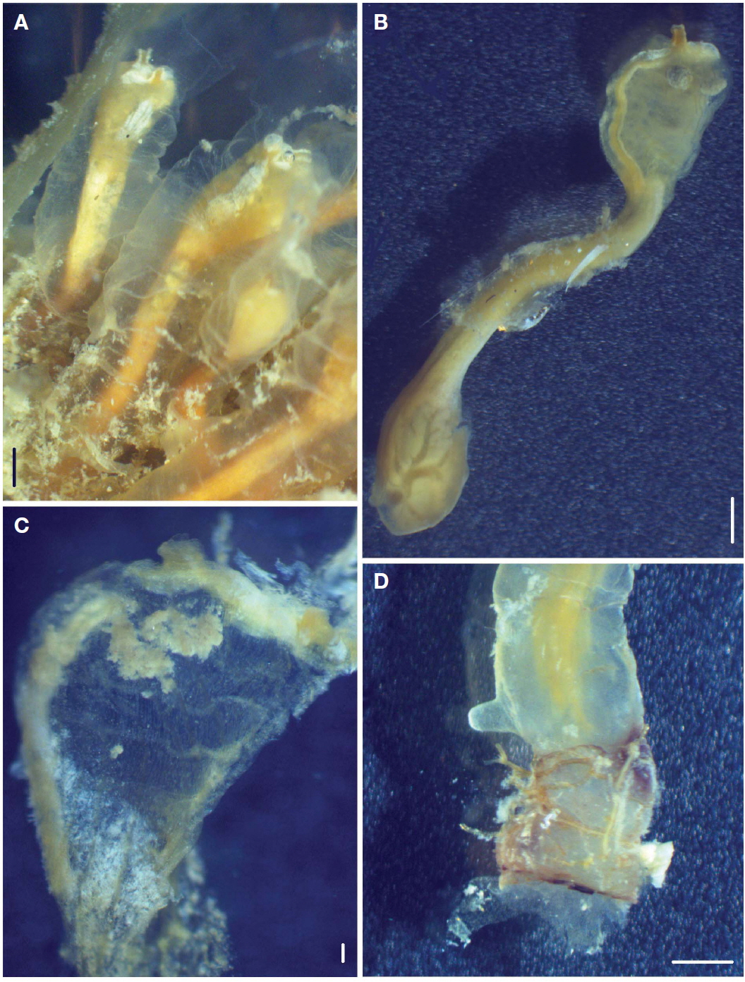
    Description. Fifty zooids independently attached to an oyster shell by stolon and form social colony of 30-40 mm in extent (Figs. 2E, 3D). Some hydrozoans grow along marginal attachment part of zooidal test. Zooidal test thin, gelatinous, colorless and very transparent so that mantle body wholly seen through it (Fig. 3A). Bright orange pigments (Pantone 128U) in band along each side of endostyle and on dorsal midline in formalin solution (Fig. 3B).

    Zooids small 14 mm long on average, up to 17 mm long in contracted condition. Thorax 4 mm long and 2-2.8 mm wide, while abdomen 10 mm long and 1.2-2.1 mm wide. Thorax shorter than abdomen in extracted zooids and contracted zooids. Thorax occupies one fourth of whole body, while abdomen occupies three-fourths of it in preserved specimen (Figs. 2D, 3B).

    Nearly longitudinal muscles extend along length of thorax. Muscles formula 4-5E3B3D=10-11.

    Branchial aperture opens at terminal of thorax and atrial aperture subterminal. Anus opens at level as high as posterior one stigmata row. Branchial aperture short moderately with plain margin. Atrial apertures have smooth border. Number of tentacles 12 alternated large and small. Opening of neural gland small, elliptical and slightly transverse. Dorsal languet ribs. 8-10 stigmata row of up to 40 stigmata. Stigmata row around middle of branchial sac have largest number of stigmata (Fig. 3C). Transverse vessels provided each with a well-developed membrane.

    Oesophageal neck very long, narrow, and occupy about one half of total length of abdomen in zooid.

    Large posterior stomach occupy from one fourth to one fifth of abdomen, occurs at posterior end of descending limb of gut loop. Stomach globular, has 4-5 short striations on the right side of surface, and distinctly represented by thickened wall on inner surface (Fig. 2F).

    Gonads occupy left side of intestinal loop posterior to the pyloric end of stomach. Ovarian eggs 0.75 mm in diameter at maximum.

    Distribution. Pacific Ocean: Korea (Chagwido, Jeju-do), Japan (West of Shimonda).

    Remarks. The present species, Clavelina miniata is the first record from Korean waters.

    This species most closely resembles Clavelina miniata (Watanabe & Tokioka, 1973) from Tatado, west of Shimoda in colony organization, zooid size, thoracic longitudinal muscles, ciliate groove, stomach surface, stomach location, gonad and brood pouch. However the present specimens are slightly different from Clavelina miniata in the many individual grow densely aggregated (not aggregated in former) and the number of longitudinal muscles.

    This species resembles Clavelina oliva from Australia and Philippines in colony organization. But C. oliva has more oblique thoracic muscles than longitudinal muscles of this species (Kott, 1990).

참고문헌
  • 1. Kott P 1990 The Australian Ascidiacea, part 1, Aplousobranhia (1) [Memories of the Queensland Museum] Vol.29 P.33-67 google
  • 2. Monniot F, Monniot C 2001 Ascidians from the tropical western Pacific [Zoosystema] Vol.23 P.201-383 google
  • 3. Nishikawa T, Tokioka T 1976 Contributions to the Japanese ascidian fauna 29. Notes on some clavelinids from the Japanese waters [Publication of the Seto Marine Biological Laboratory] Vol.23 P.63-82 google
  • 4. Oka A 1927 Ueber Dendroclavella, eine gattung von socialen ascidian [Proceedings of the Imperial Academy] Vol.3 P.555-557 google
  • 5. Rho BJ 1966 Taxonomy study on the Prochordates from Korea [Korean Culture Research Institute Transactions] Vol.8 P.209-216 google
  • 6. Rho BJ 1977 Ascidiacea. In: Illustrated encyclopedia of fauna and flora of Korea (Porifera, Hydrozoa and Ascidiacea) P.101-136, 289-374 google
  • 7. Sanamyan K, Shenkar N, Gittenberger A, Lambert G, Rius M, Moreira Da, Rocha R, Swalla BJ, Turron X 2013 Clavelina elegans (Oka, 1927), 2013. Ascidiacea World Database [Internet]. World Register of Marine Species google
  • 8. Sanamyan K, Shenkar N, Gittenberger A, Lambert G, Rius M, Moreira Da, Rocha R, Swalla BJ, Turron X 2013 Clavelina miniata Watanabe & Tokioka, 19732013. Ascidiacea World Database [Internet], World Register of Marine Species google
  • 9. Savigny JC 1816 Memoires sur les animaux sans vertebres, Part II P.1-239 google
  • 10. Tokioka T, Nishikawa T 1976 Contribution to the Japanese ascidian fauna. 30. Further notes on Japanese clavelinids [Publication of the Seto Marine Biological Laboratory] Vol.23 P.341-350 google
  • 11. Van Name WG 1945 The North and South American ascidians [Bulletin of American Museum of Natural History] Vol.84 P.1-476 google
  • 12. Watanabe H, Tokioka T 1973 On a new species of Clavelina from Japan, with remarks on its mode of budding [Publication of the Seto Marine Biological Laboratory] Vol.21 P.99-107 google
OAK XML 통계
이미지 / 테이블
  • [ Fig. 1. ]  Clavelina elegans. A, Colony in preservative; B, Zooid in living; C, Contracted zooid; D, Larvae with three adhesive organs. Scale bars: A-C=1 mm, D=0.2 mm.
    Clavelina elegans. A, Colony in preservative; B, Zooid in living; C, Contracted zooid; D, Larvae with three adhesive organs. Scale bars: A-C=1 mm, D=0.2 mm.
  • [ Fig. 2. ]  A, Colony of Clavelina elegans; B, Zooid of C. elegans; C, Cross section of stomach in C. elegans; D, Zooid of Clavelina miniata; E, Colony of C. miniata; F, Cross section of stomach in C. miniata. aa, atrial apertures; ba, branchial apertures; eb, embryo; es, endostyle; g, gut; m, muscles; sr, stigmata row; st, stomach. Scale bars: A, D=5 mm, B, E=2 mm.
    A, Colony of Clavelina elegans; B, Zooid of C. elegans; C, Cross section of stomach in C. elegans; D, Zooid of Clavelina miniata; E, Colony of C. miniata; F, Cross section of stomach in C. miniata. aa, atrial apertures; ba, branchial apertures; eb, embryo; es, endostyle; g, gut; m, muscles; sr, stigmata row; st, stomach. Scale bars: A, D=5 mm, B, E=2 mm.
  • [ Fig. 3. ]  Clavelina miniata. A, Colony in preservative; B, Contracted zooid in preservative; C, Expanded thorax; D, Terminal of abdomen. Scale bars: A, B, D=1 mm, C=0.2 mm.
    Clavelina miniata. A, Colony in preservative; B, Contracted zooid in preservative; C, Expanded thorax; D, Terminal of abdomen. Scale bars: A, B, D=1 mm, C=0.2 mm.
(우)06579 서울시 서초구 반포대로 201(반포동)
Tel. 02-537-6389 | Fax. 02-590-0571 | 문의 : oak2014@korea.kr
Copyright(c) National Library of Korea. All rights reserved.