검색 전체 메뉴
PDF
맨 위로
OA 학술지
조피볼락(Sebastes schlegeli) 선충(Nematode: Philometridae)에 대한 분자생물학적 동정 및 PCR 검출법 개발 Molecular Identification and Development of a PCR Assay for the Detection of a Philometrid Nematode in Rockfish Sebastes schlegeli
  • 비영리 CC BY-NC
  • 비영리 CC BY-NC
ABSTRACT

Nematode infection in the epithelial tissue of cultured rockfish Sebastes schlegeli was first reported in 2012. Since then, nematode infections have caused serious economic losses in rockfish aquaculture on the west coast of Korea. Taxonomic and life cycle information for this parasite are currently unknown. In this study, 18S rRNA and cytochrome c oxidase subunit I (COI) genes were used for molecular identification and polymerase chain reaction (PCR) to detect the invisible stages of this parasite. Nucleotide sequences of the 18S rRNA of the rockfish nematode showed 98% identity with that of Philometra morii. Therefore, this rockfish nematode was classified to the Philometridae family. However, we could not identify it to genus level using 18S rRNA. Its COI nucleotide sequences shared 85% and 82% identities with those of Bursaphelenchus sinensis and Philometra overstreeti, respectively. In addition, two gene-specific primer sets were designed based on the 18S rRNA gene to detect the intermediate host and nematode larvae. These primers were specific to this rockfish nematode without cross-reacting to other pathogens. The detection limit of the PCR assay using these primers was 1,000 copies of nematoda plasmid DNA. Therefore, the PCR assay described here is suitable for the detection of nematode DNA within rockfish. In addition, this PCR assay could be used to detect nematode larvae and the intermediate host.


KEYWORD
Philometrid nematode , Rockfish , 18S rRNA gene , Cytochrome c oxidase subunit I gene , PCR
  • 서 론

    조피볼락(Sebastes schlegeli)은 한국 연안을 비롯한 북서태평양 지역에서 넓게 서식하며, 넙치와 함께 국내 해산어 양식을 대표하는 어종으로 2014년에는 24,598톤을 생산되어 국내 총 양식 생산량의 약 30%를 차지하고 있다(KOSIS, 2015). 2012년 서해안 천수만 지역의 양식 조피볼락에서 상피에 감염되는 선충이 최초로 확인되었으며, 2013년부터 약 1년간 조피볼락 선충의 감염현황을 조사한 결과 서해안 천수만지역의 조피볼락에서 50%이상 감염되어 있는 것으로 확인되었다. 2014년 조사 결과, 선충이 감염된 조피볼락은 대부분 폐사가 발생하지 않았지만, 여름철 고수온기에 선충이 어체를 뚫고 나간 상처를 통하여 병원세균의 2차 감염이 발생함으로써 높은 폐사가 확인되었다. 여름철 이후 가을철 조사에는 선충의 감염이 확인되지 않았으며 겨울철 조사 이후에는 다시 선충의 재감염이 확인되었다(Seo et al., 2014).

    Nematode (phylum) Secernentea (class) Camallanida (order)에 속하는 Philometridae (family) 선충은 어류를 최종숙주 혹은 중간숙주로 하여 기생을 하며, 현재까지 650종 이상이 어류에 감염되는 것으로 보고되어 있다. 어류에 감염되는 Philometridae선충으로는 Afrophilometra, Alinema, Buckleyella, Caranginema, Clavinema, Dentiphilometra, Margolisianum, Nilonema, Philometra, Philometroides, Philonema, Phlyctainophora, RumaiSpirophilometra 의 속(genus)이 보고되어 있다(Moravec and Harrison, 1989; Moravec and Wang, 2002; Wijová et al., 2006). 선충은 가늘고 긴 원통형 또는 실 모양의 형태를 하고 있으며 종숙주인 어류의 근육, 비늘 밑, 생식소, 부레 등에 기생하지만 소화관에 기생하는 것이 가장 많다. 선충은 암수가 따로 존재하는 자웅이체로서 교미를 통하여 번식하며 대부분은 난생으로서 수중에서 난이 부화하여 자충이 방출되지만, Philometridae 선충의 경우 태생으로서 암컷이 자충을 수중으로 방출한다. 선충은 생활사에서 중간숙주를 필요로 하며, 자충은 갑각류와 같은 중간숙주에 먹힌 뒤 어류와 같은 종숙주가 최종적으로 먹어 어류 내에서 성충으로 성장한다(Edward, 1996). Seo et al. (2014)이 보고한 조피볼락 선충은 성충의 경우 상피층에 위치하여 육안으로 쉽게 확인이 가능하지만 자충의 경우 육안으로 확인이 불가능하여 진단에 어려움이 따른다. 따라서 조직 내에 위치한 선충의 자충을 확인하기 위해서는 외부관찰 이외의 다른 진단법이 필요하다고 생각하였다. Stephanie et al. (2011)은 South Carolina 퇴적지에서 찾아낸 philometrids의 생활사를 밝혀내기 위하여 어류의 장에 기생하는 선충의 자충을 현미경으로 확인한 뒤 Polymerase Chain Reaction (PCR) 법을 통하여 양성체를 찾아내었다. Kang et al.(2008)도 고래회충 유충의 검출을 위하여 육안적 검사와 더불어 Polymerase Chain Reaction-Restriction Fragment Length Polmorphism (PCR-RFLP)법을 통하여 양성체를 찾아내었다. 본 연구에서도 마찬가지로 조피볼락 선충의 생활사를 밝히기 위하여 선충 자충의 탐색이 필요하다고 생각되었으며, 자충이 포함된 중간숙주 역시 육안적인 방법 이외의 다른 검출법이 필요하다고 생각되었다. 특정 유전물질을 대량으로 증폭하는 PCR법은 최근 어류의 질병진단에 많이 사용되고 있는 방법으로서 샘플의 처리, 병원체에 대한 특이성 및 재현성이 높아 각광 받고 있는 진단법이다.

    분자생물학적 종 분류 및 동정을 위한 유전자 마커로는 ribosomal RNA (rRNA), mitochondrial DNA (mtDNA) 및 microsatellite DNA등이 일반적으로 사용된다. 진핵생물의 단백질 합성에 있어 중추역할을 하는 ribosomal DNA는 유전자의 계통발생학적 분석에 사용되는 rRNA를 code하는 분자 마커로써 18S, 5.8S 및 28S 유전자와 internal 및 external transcribed spacers (ITS and ETS)로 구성되어 있다. 이중 18S rRNA는 유전자의 변이가 심하지 않기에 다양한 생물군의 분류에 사용되고 있다(Nyaku et al., 2013). mtDNA는 고변이성 유전자로서 종의 진화적 유연관계를 나타내고 있으며, 그 중 cytochrome c oxidase subunit I (COI)는 다양한 해양 무척추동물들의 유전학적 연구에 사용되는 유전자 마커이다(Edmands et al., 1996). Cytochrome 은 생체세포 내에 존재하는 헴단백질 색소 중 하나로서 고등동식물의 세포, 세균, 곰팡이 및 효모 등의 미토콘드리아에 존재한다. Cytochrome은 철 포르피린의 구조, 단백질과의 결합 상태에 따라 a, b, c 및 d의 4군으로 구별되는데, 이 중에서 cytochrome c는 산화환원반응에 관여하는 효소로서 미토콘드리아의 외막에 존재하며 호흡과 순환에 의해 세포내에 운반된 산소를 활성화하여 세포호흡을 원활하게 하는 데 필요하다. Cytochrome c oxidase는 cytochrome c 에서 전자를 직접 분자상 산소로 넘겨주는 효소활성을 하는 호흡효소를 뜻하는데 동물군은 미토콘드리아의 COI를 표준바코드 유전자로 이용하여 종 동정에 사용하기도 한다. Herbert (2003)는 DNA barcoding 기법이 유전자 서열을 이용하여 종을 동정하는 방법으로서 적당하며 동물의 동정으로 미토콘드리아의 COI 유전자 서열을 사용할 것을 제안하기도 하였으며, 최근의 다른 연구에서는 미토콘드리아 COI 유전자 염기서열의 다형성을 바탕으로 어류간 계통 유연관계를 확인하기도 하였다(Han et al., 2014).

    따라서 본 연구에서는 선충의 18S rRNA 및 COI 유전자를 증폭시킬 수 있는 primer를 사용하여 선충 유전자 분석을 실시하였으며, 획득한 염기서열을 바탕으로 조피볼락에서 분리된 선충의 계통발생학적 분석을 실시하였다. 또한 어류 조직 내에 위치한 선충 자충의 분석 및 중간숙주의 탐색에 사용하기 위하여 18S rRNA 유전자를 증폭시킬 수 있는 primer를 제작하고, 선충을 분자생물학적으로 검출할 수 있는 PCR법을 개발하였다.

    재료 및 방법

      >  선충 시료 채취 및 DNA 추출

    2014년도 1월, 서해안 천수만 일대에 양식중인 조피볼락에서 선충을 분리 후 100% ET-OH에 고정하여 보관한 시료를 DNA 추출을 위해 사용하였다. 50 mg의 선충을 E-tube에 넣고 PBS를 사용하여 마쇄하였고, 원심분리(8,000 rpm, 10 min) 후 상청액을 회수하였다. DNA 추출은 High pure template preparation kit(Roche, Germany)을 사용하였으며, 제조사의 매뉴얼에 따라 수행하였다.

      >  분자생물학적 동정

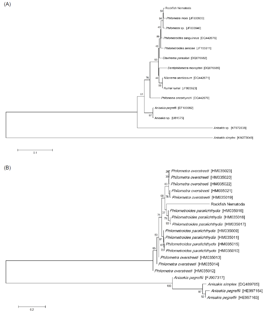
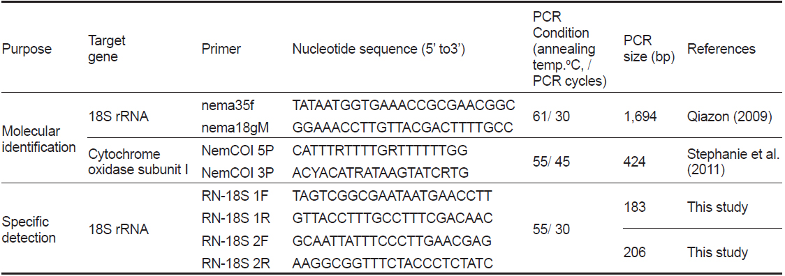
    선충의 종 동정을 위하여 18S rRNA와 COI 유전자를 분석하였다. PCR법은 Emerald Taq (Takara, Japan)을 사용하여 제조사의 매뉴얼에 따라 수행하였으며, 각 primer sequence와 PCR 조건은 Table 1에 표기하였다. PCR product는 1.5% agarose gel (Bioneer, Korea) 상에서 전기영동을 실시하여 band를 확인하였으며, PCR 증폭이 확인된 시료는 sequencing을 실시하여 염기서열을 확인하였다. 염기서열의 분석에는 BioEdit Ver. 7.2.3, MEGA Ver. 6를 사용하였으며, National Center for Biotechnology Information (NCBI)에서 제공되는 Basic Local Alignment Search Tool (BLAST)을 이용하여 기존에 보고된 선충 유전자와의 상동성 분석을 하였다. 또한 얻어진 염기서열을 바탕으로 실시한 유전자 계통분석은 BioEdit Ver. 7.2.3 program을 사용한 염기서열 정렬과 MEGA Ver. 6 program을 사용한 근린결합분석(NJ; neighbor-joining analysis, 1000 rounds of bootstrap)을 통하여 각 염기서열간의 유전적 거리와 계통도(phylogenetic tree)를 획득하였다. 본 연구에서 비교한 18S rRNA (1,695 bp) 염기서열은GenBank에 등록되어있는 Philometridae에 속하는 8종의 선충 18S rRNA 유전자(Philometra morii, Philometra sp., Philometroides sanguineus, Philometroides seriolae, Clavinema parasiluri, Dentiphilometra monopteri, Nilonema senticosum, Rumai rumai, Philonema oncorhynchi)와 Anisakidae 에 속하는 4종의 18S rRNA 유전자(Anisakis sp., A. pegreffi, A. simplex)의 염기서열을 대상으로 분석 하였다(Fig. 2A). 또한 COI (389 bp) 염기서열은 Philometra overstreeti, Philometroides paralichthydis, A. pegreffii, A. simplex의 유전자 염기서열을 대상으로 분석하였다(Fig. 2B).

    [Table 1.] Primer information and PCR condition for this study
    label

    Primer information and PCR condition for this study

      >  조피볼락 선충 검출 PCR법 개발

    위 연구에서 얻어진 조피볼락 선충의 18S rRNA 염기서열 중 다른 Philometridae 의 18S rRNA와 염기서열의 차이가 보이는 특이적인 영역에서 2개의 primer set를 제작하여 선충 검출을 위한 PCR법 개발에 사용하였다. PCR조건은 Table 1에 표기하였으며, PCR annealing 온도에 따른 검출률도 확인 하였다. 또한 조피볼락의 선충 검출에 사용된 primer의 특이성을 검증하기 위하여 선충을 포함한 다른 기생충 및 세균의 DNA를 추출한 뒤 PCR을 실시하였다. 실험에 사용된 기생충은 조피볼락에서 분리된 선충 이외에 어류에서 분리된 스쿠티카, 쿠도아 및 아니사키스 충을 사용하였으며, 세균은 어류에서 분리된 Edwardsiella tardaStreptococcus iniae를 사용하였다.

    넙치에서 유래된 스쿠티카충은 보유중인 CHSE-214 어류주화세포에서 배양중인 신선한 충을 사용하였으며 쿠도아충은 넙치의 근육에서 분리된 것을 사용하였다. 아니사키스충은 조피볼락과 넙치의 내장에서 각각 분리된 후 100% ET-OH에 고정된 것을 사용하였으며, 각각의 기생충은 상법과 동일하게 DNA를 추출한 뒤 PCR에 사용하였다. 또한 각 세균은 1.5% NaCl이 포함된 Brain Heart Infusion (BHI, Gibco, USA) broth 배지에서 25℃에서 12시간 동안 배양된 신선한 균을 사용하여 상법과 동일하게 DNA를 추출한 뒤 PCR에 사용하였다. PCR product는 1.5% agarose gel (Bioneer, Korea) 상에서 전기영동을 실시하여 band를 확인하였다.

    선충 검출 PCR primer의 감도 확인실험은 다음과 같은 방법으로 실시하였다. 조피볼락에서 분리된 선충의 PCR product는 1.5% agarose gel 상에서 전기영동을 실시한 뒤, PCR product purification kit (Roche, Germany)를 사용하여 Gel Purification을 실시하였다. 정제된 PCR product는 pGEM TEasy vector (Promega, USA)을 사용하여 cloning을 실시하였고, Plasmid Mini Extraction kit (Bioneer, Korea)를 사용하여 plasmid DNA를 획득하였다. 선충의 plasmid는 copy수를 계산한 뒤 Nuclease-free water (Bioneer, Korea)를 사용하여 1×10¹⁰copy부터 1×100copy까지 10배씩 단계희석 하였고 이를 template로 하여 PCR을 실시하였다. PCR product는 1.5% agarose gel 상에서 전기영동을 실시하여 band를 확인하였다.

    결과 및 고찰

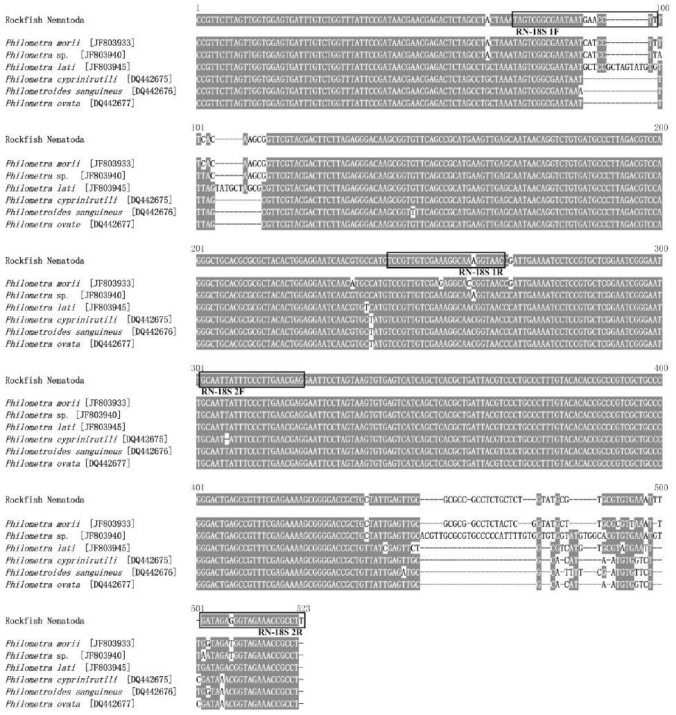
    본 연구에서는 조피볼락에서 분리된 선충의 분자생물학적 동정을 실시하기 위하여 선충류의 18S rRNA 및 COI 유전자를 증폭할 수 있는 universal primer를 각각 사용하여 PCR과 유전자 분석을 실시하였다. 18S rRNA의 유전자를 증폭하기 위하여 Quiazon (2009)의 논문에 인용된 PCR조건을 사용하였으나, PCR 반응이 일어나지 않아PCR 조건을 수정하여 설정하였다(Table 1). 조피볼락 선충의 genomic DNA를 template로 사용하여18S rRNA의 유전자를 증폭한 결과 1,695 bp의 염기서열을 획득하였으며(GenBank accession no. LC071530), 해당 염기서열에 대한 NCBI BLAST 결과 Philometra morii 18S rRNA gene (JF803933)과 98% 일치하여 상동성이 가장 높음을 확인하였다(Fig. 1). 조피볼락 선충의18S rRNA 유전자 계통발생학적 분석결과 어류에 기생하는 nematode 중에서도 Philometridae 과(family)에 속하는 선충으로 확인되었으며, Philometrodae중에서 Philometra, Philometroides, Clavinema의 속(genus)과 가장 근연한 것으로 확인되었다(Fig. 2A). 이전의Nyaku et al. (2013)은 식물에 감염되는 선충 유전자의 계통발생학적 분석을 위하여 18S rRNA를 사용하였으나 목(order) 단계까지만 분석이 가능하였다. 본 연구에서 실시한 조피볼락 선충의 경우에서도 18S rRNA 유전자를 통한 분석은 과(family) 단계까지만 정확한 분석이 가능하였으며, 종(species) 및 속(genus) 분류를 위해서는 다른 유전자의 분석이 필요하다고 생각하였다.

    하지만 조피볼락 선충 COI 유전자(PCR product size 389bp; GenBank accession no. LC073304)의 경우 NCBI BLAST 결과 소나무에서 분리되는 선충류인 Bursaphelenchus sinensis cytochrome c oxidase subunit I (mtCOI) gene (GenBank accession no. AB232163)과 84%로 가장 높은 상동성을 보였으며, 넙치에서 분리되는 선충류인 Philometra overstreeti의 COI 유전자(GenBank accession no. HM035020.1)와는 82%의 상동성이 확인되었다. 현재까지 등록된 어류를 숙주로 하는 선충류의 COI 유전정보를 바탕으로 phylogentic tree를 작성한 결과 (Fig. 2B), Philometra, Philometroides 의 속(genus)와 가장 근연하였으나, 선충의 COI 유전자의 경우 타종간 유전적 차이가 크고(Stephanie et al., 2011), GenBank에 등재되어 있는 어류 유래의 선충의 경우 Philometra overstreetiPhilometroides paralichthydis 종(species) 뿐이어서 정확한 분류가 어려운 것으로 확인되었다. 본 연구 결과를 통해 현재까지 COI 유전자의 경우 조피볼락 선충 분류를 위해는 유용성이 없는 것으로 확인되었으며, 앞으로 정확한 선충 동정을 위해서는 어류 기생성 선충류를 비롯한 다른 선충의 COI 유전자 정보가 추가적으로 연구되어야 할 것으로 판단된다.

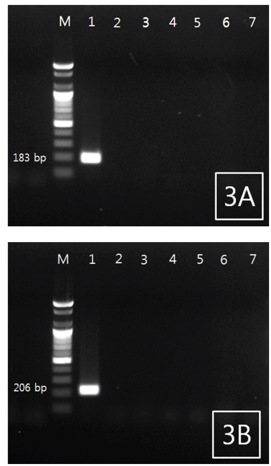
    조피볼락 선충을 특이적으로 검출하기 위한 PCR법을 위하여 18S rRNA 염기서열 중에서 2쌍의 primer set를 새로 디자인하였다(Fig. 1). 조피볼락 선충 검출 Primer 제작을 위해서, 조피볼락 선충의 18S rRNA와 상동성이 비교적 높은 다른 어종에서 분리되는 Philometrodae 기생충의 18S rRNA유전자 염기서열을 비교하여 조피볼락 선충 특이 primer 제작에 활용하였다. 선충의 DNA를 추출한 뒤 PCR을 실시한 결과 2종의 primer set 모두에서 양성을 나타내었으며, agarose gel 상에서 183 및 206 bp의 band를 각각 나타내었다. 추가적으로 선충 유전자의 검출을 위한 PCR 조건의 최적화를 위하여 BIO-RAD 회사의 Peltier Thermal Cycler (PTC-0220)을 사용하여 gradient PCR을 실시하였다. 조절된 annealing temp는 42-62℃이었으며 2종의 primer 모두 55℃에서 최적의 조건을 나타내었다(data not shown).

    조피볼락 선충 특이적 primer의 검증을 위하여 5종의 기생충(조피볼락 유래 선충 및 아니사키스충, 넙치 유래 스쿠티카충, 아니사키스충 및 쿠도아충)과 2종의 세균(Edwardsiella tarda, Streptococcus iniae)을 사용한 PCR 결과 조피볼락 선충 DNA를 제외한 모든 시료에서는 증폭이 확인되지 않았다(Fig. 3A, B). 위의 결과로 볼 때 본 연구에서 제작된 RN-18S 1 및 2 primer sets는 조피볼락 상피 선충의 유전자를 특이적으로 증폭시킬 수 있는 것으로 확인되었다. 다른 시기에 다른 양식장에서 분리된 선충들간의 유전자 분석결과 차이점이 확인되지 않았으나(data not shown), 추가적으로 타 어종에서 분리되는 Philometrid nematodes를 확보하여 검증할 필요가 있을 것으로 사료된다.

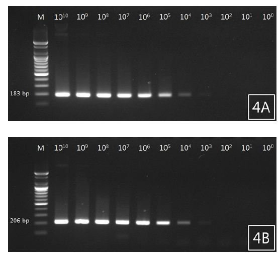
    조피볼락 선충 검출용 primer를 사용하여 선충 유전자가 증폭된 plasmid DNA를 제작하였고, 검출감도를 확인하였다. 그 결과 18S rRNA plasmid DNA는 1×10¹⁰copy부터 1×10³copy까지 band가 확인되었으며 두 primer에서 비슷한 검출감도를 확인할 수 있었다(Fig. 4A, 4B).

    Philometrid nematodes의 생활사에서 자충은 중간숙주에 의해 먹히게 되고, 중간숙주를 어류가 먹어서 종숙주에 감염이 된다. 중간숙주는 대개 요각류(copepodes)로서 유영생활을 하며 어류의 먹이가 된다(Wang, 2002). 체내에 새끼를 가지고 있는 암컷 선충의 경우 숙주의 근육을 통해 피부를 관통하여 구멍을 생성한다. 그 상처를 통하여 자충을 수계로 배출하며, 그 자충을 중간숙주가 섭취하며 최종적으로 어류에 감염이 된다고 한다(Gonzalez et al., 2007). Seo et al. (2014)은 2013년 8월, 조피볼락의 피부에서 선충이 감염된 후 어체 외부로 빠져나가면서 생긴 다수의 상처를 발견하였다. 또한 2013년 10월, 선충의 감염이 확인되지 않았기에 선충은 1년 주기의 생활사를 가지며, 고수온기 어체의 피부에 상처를 내어 자충을 방출한다고 생각된다. 선행연구와 마찬가지로 성충의 경우에 육안으로 검사가 가능한 선충은 자충의 경우 육안으로 확인이 불가능하며 이는 중간숙주도 마찬가지이다. Seo et al. (2014)에 의하면 선충류의 특성상 숙주로부터 선충을 감염이후에 치료는 매우 어려우므로, 생활사 중 선충의 감염을 차단하여야 하며, 선충의 중간숙주를 탐색하여 감염경로를 제거하는 방법에 대한 연구가 필요하다고 하였다. 따라서 본 연구에서 개발한 선충의 유전자 검출을 위한 PCR법을 사용한다면 어류 내 또는 생사료 등에 존재 가능한 자충을 포함한 중간숙주의 탐색이 가능하며 생활사의 규명에 도움이 될 것이라 생각한다.

    본 연구에서는 양식 조피볼락에서 분리된 선충의18S rRNA 유전자 분석결과 PhilometridaePhilometra와 가장 유전적 유연관계가 높지만, 정확한 종 동정은 하지 못하였기에, 현재 형태학적 분석을 통한 동정을 수행하고 있다. 하지만, 본 연구에서는 조피볼락 선충 유전자를 검출할 수 있는 새로운 PCR법을 개발하였으며, 이는 향후 어류 조직 내에 위치한 선충 자충의 검출 및 중간숙주의 탐색 등에 유용하게 사용될 수 있을 것이라 생각한다.

참고문헌
  • 1. Edmands S, Moberg PE, Burton RS 1996 Allozyme and mitochondrial DNA evidence of population subdivision in the purple sea urchin Strongylocentrotus purpuratus [Mar Biol] Vol.126 P.443-450 google cross ref
  • 2. Edward JN 1996 Diagnosis and Treatment, Fish Disease P.166-170 google
  • 3. Gonzalez-Solis D, Moravec F, Paredes VM 2007 A new species of Dentiphilometra (Nematoda: Philometridae) from the musculature of the gray snapper Lutjanus griseus (osteichthyes) off the Caribbean coast of Mexico [J Parasitol] Vol.93 P.1132-1135 google cross ref
  • 4. Han SH, Lee YD, Baek HJ, Oh HS, Noh CH 2014 Genetic structure and phylogenetic relationship of red spotted grouper (Epinephelus akaara) based on the haplotypes and polymorphisms of mitochondrial COI gene sequences [J Life Sci] Vol.24 P.626-632 google cross ref
  • 5. Herbert PDN, Cywinska A, Ball SL, deWaard JR 2003 Biological identifications through DNA barcodes [Proc Biol Sci] Vol.270 P.313-321 google cross ref
  • 6. Kang JH, Lee MH, Lee KB, Choi CS 2008 Comparison of macroscopic inspection and polymerase chain reactionrestriction fragment length polymorphism (PCR-RFLP) for the detection of Anisakis simplex complex [J Fd Hyg Safety] Vol.23 P.314-318 google
  • 7. Korean Statistical Information Service google
  • 8. Moravec F, Harrison FS 1989 Paraphilometroides nemipteri gen. et sp. N. (nematoda:philometridae) from the marine fish nemipterus peronei (valenciennes) from Malaysia [Folia Parasitol] Vol.36 P.345-350 google
  • 9. Moravec F, Wang GT 2002 Dentiphilometra monopteri n. gen., n. sp. (nematoda: philometridae) from the abdominal cavity of the ricefield eel monopterus albus in china [J Parasitol] Vol.88 P.961-966 google cross ref
  • 10. Nyaku ST, Sripathi VR, Kantety RV, Gu YQ, Lawrence K, Sharma GC 2013 Characterization of the two intra-individual sequence variants in the 18S rRNA gene in the plant parasitic nematode, Rotylenchulus reniformis [PLoS ONE] Vol.8 P.e60891 google cross ref
  • 11. 2009 Studies on philometrid and anisakid nematodes infecting marine fishes in Japanese waters. Ph.D. Thesis google
  • 12. Seo HG, Seo JS, Ryu MK, Lee EH, Kwon SR, Kang JS, No YS, Choi HS, Jung SH, Han HJ 2014 A nematoda infection in the Epithelial Tissue of Cultured Rockfish Sebastes schlegeli in Cheonsu Bay, Western Korea [Korean J Fish Aquat Sci] Vol.47 P.603-610 google cross ref
  • 13. Stephanie P, Weatherly AM, Isaure DB, William AR, Allan ES 2011 Use of molecular tools in identification of philometrid larvae in fishes: technical limitations parallel our poor assessment of their biodiversity [Parasitol Res] Vol.109 P.1725-1730 google cross ref
  • 14. Wang GT 2002 Seasonal dynamics and distribution of Philometroides fulvidraconi (Philometridae) in the bullhead catfish, Pseudobagrus fulvidraco (Richardson) [J Fish Dis] Vol.25 P.621-625 google cross ref
  • 15. Wijova M, Moravec F, Horak A, Lukes J 2006 Evolutionary relationships of Spirurina (Nematoda: Chromadorea: Rhabditida)with special emphasis on dracunculoid nematodes inferred from SSU rRNA gene sequences [Int J Parasitol] Vol.36 P.1067-1075 google cross ref
이미지 / 테이블
  • [ Table 1. ]  Primer information and PCR condition for this study
    Primer information and PCR condition for this study
  • [ Fig. 1. ]  Partial 18S rRNA gene sequences (position 1,173 through 1,695) of philometrid nematode from rockfish Sebastes schlegeli of Korea, indicating position of primer (boxes) used for detection PCR.
    Partial 18S rRNA gene sequences (position 1,173 through 1,695) of philometrid nematode from rockfish Sebastes schlegeli of Korea, indicating position of primer (boxes) used for detection PCR.
  • [ Fig. 2. ]  Neighbor-joining analysis of philometrid nematode from rockfish Sebastes schlegeli (A) 18S rRNA and (B) cytochrome c oxidase subunit I sequences from GenBank.
    Neighbor-joining analysis of philometrid nematode from rockfish Sebastes schlegeli (A) 18S rRNA and (B) cytochrome c oxidase subunit I sequences from GenBank.
  • [ Fig. 3. ]  PCR results obtained using (A) RN-18S1 primer and (B) RN-18S2 primer sets, with lanes M to 7 as follows: 100 bp DNA ladder, Nematode with rockfish Sebastes schlegeli, Scutica with flounder Paralichthys olivaceus, Kudoa with flounder, Anisakis with rockfish, Anisakis with flounder, Edwardsiella tarda and Streptococcus iniae.
    PCR results obtained using (A) RN-18S1 primer and (B) RN-18S2 primer sets, with lanes M to 7 as follows: 100 bp DNA ladder, Nematode with rockfish Sebastes schlegeli, Scutica with flounder Paralichthys olivaceus, Kudoa with flounder, Anisakis with rockfish, Anisakis with flounder, Edwardsiella tarda and Streptococcus iniae.
  • [ Fig. 4. ]  Analytical sensitivity of PCR for detection of philometrid nematode from rockfish Sebastes schlegeli. Plasmid DNA composing 18S rRNA gene of philometrid nematode was serially diluted from 1010 to 100 was used for PCR. Lanes 1 to 11: DNA ladder, 1010, 109, 108, 107, 106, 105, 104, 103, 102, 101 and 100 copy of plasmid DNA/tube. A, RN-18S1; B, RN-18S2.
    Analytical sensitivity of PCR for detection of philometrid nematode from rockfish Sebastes schlegeli. Plasmid DNA composing 18S rRNA gene of philometrid nematode was serially diluted from 1010 to 100 was used for PCR. Lanes 1 to 11: DNA ladder, 1010, 109, 108, 107, 106, 105, 104, 103, 102, 101 and 100 copy of plasmid DNA/tube. A, RN-18S1; B, RN-18S2.
(우)06579 서울시 서초구 반포대로 201(반포동)
Tel. 02-537-6389 | Fax. 02-590-0571 | 문의 : oak2014@korea.kr
Copyright(c) National Library of Korea. All rights reserved.