Two pleurostomatid ciliates, Loxophyllum perihoplophorum
Pleurostomatids are bilaterally compressed ciliates with a slit-like cytostome on the ventral side and commonly found in various habitats all over the world (Foissner, 1984; Fenchel, 1987; Lynn 2008). They have been accepted as a monophyletic taxon based on morphological and molecular studies (Corliss, 1979; Lipscomb and Riordan, 1990; Strüder-Kypke et al., 2006; Lynn, 2008; Pan et al., 2010).
>
Morphological taxonomy procedures
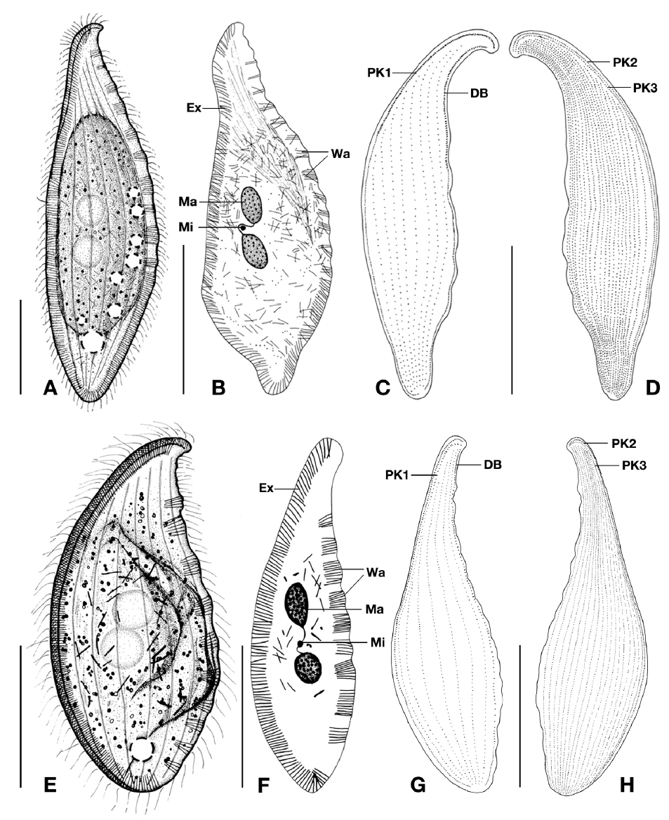
Samples were collected from coastal areas in the East Sea, Korea, using PEF-S (Xu et al., 2009) and then transferred to a laboratory. They were continually maintained in petri dishes at 17°C (light-dark 12 : 12 cycles) with rice grains as a food source to enhance bacterial growth. Living specimens were observed using phase contrast and differential interference microscopes at different magnifications. The protargol staining (Foissner, 1991) was performed to reveal ciliary pattern, nuclear apparatus, and extrusomes. Enumeration and measurements of stained specimens were performed under ×1,000 magnification (Leica DM2500; Wetzlar, Germany). Drawings of specimens were made using a lucida camera. The classification scheme used here was based on Lynn (2008). Terminology followed Corliss (1979), Lin et al. (2005), and Lynn (2008). Abbreviations were as follows: CV, contractile vacuole; DB, dorsal brush kinety; Ex, extrusome; Fu, furrow; FV, food vacuole; LSK, left somatic kinety; Ma, macronuclear nodule; Mi, micronucleus; Nd, nematodesmata; PK, perioral kinety; RSK, right somatic kinety; Wa, wart.
>
Molecular taxonomy procedures
Each living individual was isolated using a micropipette under a dissecting microscope (Leica MZ 12.5), and DNA extraction, amplification of the SSU rDNA, and sequencing were performed according to Kim and Min (2009). Sequences were aligned using CulstalX (Thompson et al., 1997) and further modified manually using Bioedit 7.0.9 (Hall, 1999). Nucleotide diversity within the species was calculated using MEGA 6.06 (Tamura et al., 2013) with the
Phylum Ciliophora Doflein, 1901
Class Litostomatea Small and Lynn, 1981
Subclass Haptoria Corliss, 1974
Order Pleurostomatida Schewiakoff, 1896
Family Litonotidae Kent, 1882
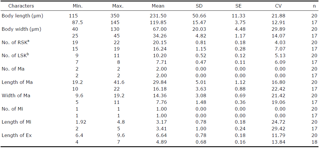
Morphological characterization of Loxophyllum perihoplophorum (1st line) and L. rostratum (2nd line) from protargol stained specimens
Material examined. Specimens were collected from the coastal waters of Hajeo-ri (36°23′N and 129°24′E), Gyeongsangbuk-do in the East Sea on 28 Apr-24 Jun 2008.
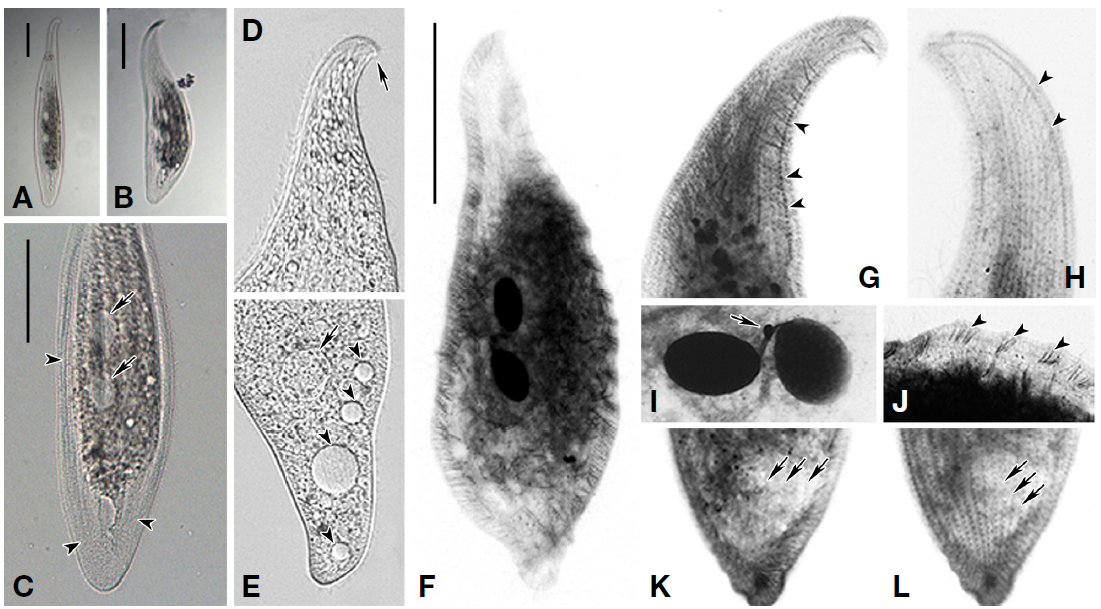
Description. Live cell size 200-650×50-100 μm, slender leaf-shaped in outline, anterior end hooked toward dorsal side; body flexible and contractile, length ratio of fully extended to most contracted bodies about 2-3 : 1, a thin and wide Ex-belted zone enclosed; highly shrunken after protargol staining, about 115-350 μm in length (Figs. 1A, B, 2A-D, F). Laterally compressed at a ratio of about 2-3 : 1, right side flat, left slightly vaulted; 4-5 longitudinal Fu on left side (Fig. 1A).
Two Ma connected by thread-like funiculus, dumbbell-shaped, located in body center, usually detectable in live specimens; each nodule egg-shaped, about 30×15 μm after fixation (Fig. 2C, I). One Mi positioned between two Ma; globule-shaped, ca. 2-5 μm in length (Fig. 2I). Approximately 7-9 CV settled along dorsal region, size variable; a few FV recognized
Ex thin, bar-shaped, about 6-10 μm long, recognizable in live cells under optimal conditions; evenly distributed along outline of entire body, and especially clustered into ca. 10 Wa along anterior 2/3 of dorsal margin; some scattered in cytoplasm (Figs. 1A, B, 2F, J).
Cytoplasm grayish; central portion of the body opaque, with numerous tiny shiny globules; cortical granules not observed (Fig. 2C). Movement by crawling along substrates or swimming with fast rotation and twisting.
Ciliary pattern typical of
Distribution. Germany (North Sea), China (Mangrove wetland in Techeng Island), and Korea (East Sea, this study).
Remarks.
[Table 2.] Comparison among populations of Loxophyllum perihoplophorum and L. rostratum
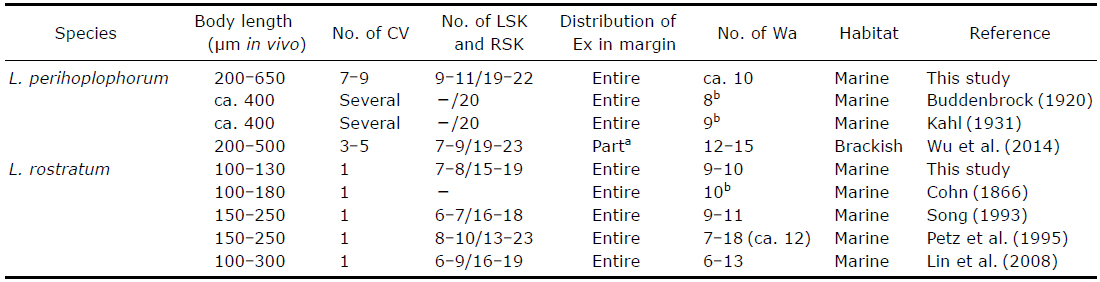
Comparison among populations of Loxophyllum perihoplophorum and L. rostratum
Material examined. Specimens were collected from the coastal waters of Munam 2-ri (38°17ʹN, 128°33ʹE), Gangwon-do in the East Sea on 12-19 Mar 2008. Environmental conditions of the sampling site were 9.2°C, 34.2 psu, and pH 8.3.
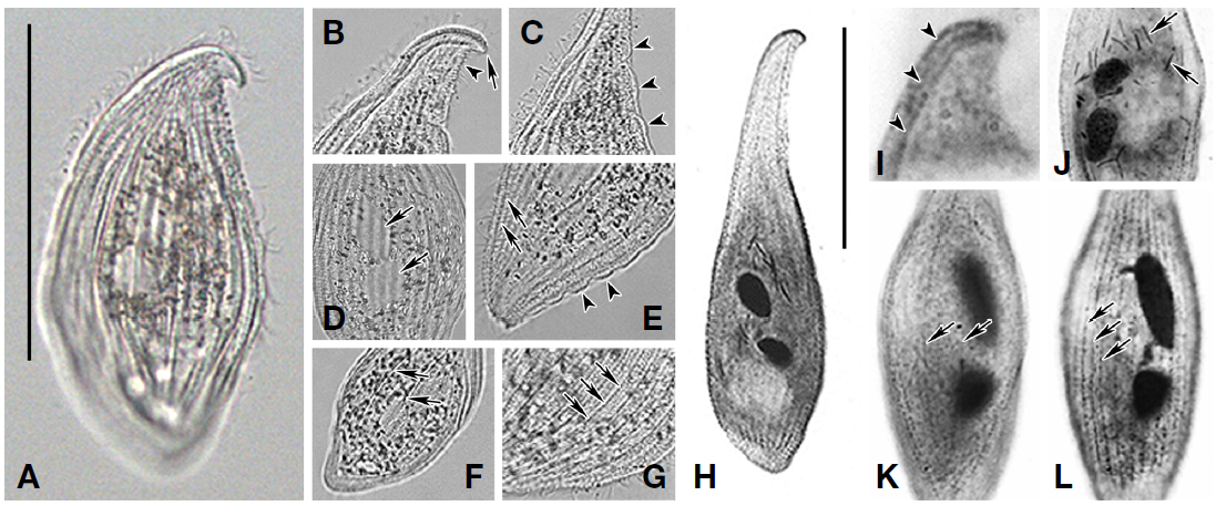
Description. Live cell size 100-130×45-65 μm; oblate leaf-shaped in outline; convex ventral side and S-shaped dorsal side; beak-like anterior end hooked toward dorsal side; winding dorsal-neck region, about 15-25 μm in length; body contractile, slightly shrunken after protargol staining (Figs. 1E, 3A, B). Laterally compressed at a ratio of about 2-3 : 1, right side flat, left side notably vaulted; 4-5 conspicuous longitudinal Fu on left side (Fig. 3A, F).
Two Ma connected by thread-like funiculus located in body center, usually detectable in live specimens; each nodule ovoid to elongate, about 10-22×5-11 μm after fixation (Fig. 3D, H). One Mi positioned between two Ma; globule-shaped, ca. 2-5 μm in length. One CV recognized at terminal region.
Ex thin, bar-shaped, about 4-7 μm long, recognizable in live cells under optimal conditions; evenly distributed along entire ventral margin, and clustered into 9-10 Wa on dorsal margin; some scattered in cytoplasm (Figs. 1E, F, 3C, E, J).
Cytoplasm slightly grayish colored, with numerous shining lipid globules and particles; dot-shaped cortical granules detected on pellicle, densely placed between rows of RSK (Fig. 3A-G). Movement by slow gliding, flexible crawling along substrates, or swimming with fast rotation and twisting.
Ciliary pattern typical of
Distribution. Germany (North Sea), Antarctica (Weddell Sea), China (Yellow Sea) and Korea (East Sea, this study).
Remarks.
SSU rDNA sequences of the Korean population were deposited in Genbank under accession number KT880229-KT880230. The sequences are identical, and are 1,592 bp in length. They show 99.7% similarity with known