For centuries, traditional herbal medicine has been administered orally in Asia for the treatment of various conditions [1]. Recently, various routes of administration, such as pharmacopuncture, have been used to deliver herbal medicine.
Research has been performed on
In Western medicine, the use of dextrose water containing glucose differs based on the concentration of glucose in the water. Generally, 5% dextrose water is used to treat dehydration, and in adults, one injection of 500 ─ 1000 mL via an IV route is used [8]. Thus, if an IV injection of
Therefore, this study was designed to evaluate the safety of administering a single-dose of GWG5 through an IV route, even though the lethal dose of GWG20 administered via an IV route, as determined in a previous study, is considered to be higher than 1.0 mL/animal.
The GWG5 used in the experimental groups was prepared in the facility at the Korean Pharmacopuncture Institute under the Good Manufacturing Practice guidelines. According to Bangyakhappyeon [4],
Forty-six (46)-week-old SD rats were used in this experiment: 20 male rats (body weights: 166.5 ─ 189.7 g) and 20 female rats (body weights: 133.9 ─ 148.5 g). Visual inspections and measurements of body weights by using electronic scales (CP3202S, Sartorius, Germany) were conducted on all animals. The general symptoms were observed once a day prior to the start of the experiments, and general symptoms and body weight changes were recorded on the last day of acclimatization.
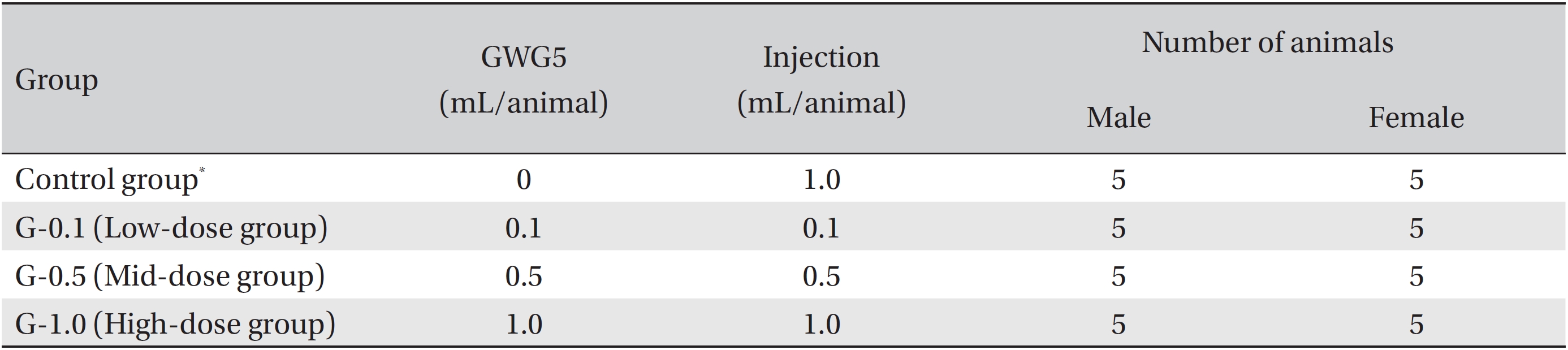
Group separation was conducted on the last day of acclimation. All animals were randomly distributed into four different groups with five individuals of each sex per group. The four different groups were labeled as follows: Control: normal saline solution, G-0.1: low-dose group, G-0.5: middose group, and G-1.0: high-dose group (Table 1). In a pilot test (Biotoxtech Study No.: B13482P), 1.0 mL/animal was administered through an IV route to one male and one female rat, which resulted in no deaths. From this result, the doses for GWG5 in this study were set as follows: 1.0 mL/ animal as the high dose (G-1.0), 0.5 mL/animal as the middose (G-0.5), and 0.1 mL/animal as the low dose (G-0.1). The same amount of normal saline solution (Choongwae Pharma Corp., Korea) as that of GWG5 for the high dose group was injected into the animals in the control group, and the results were observed and compared with those of the experimental groups. All injections were administered at a caudal vein at a rate of 2 mL/minute. This experiment was conducted at Biotoxtech, an authorized institution for non-clinical studies, under the regulation of Good Laboratory Practice (GLP) of the Korea Food and Drug Administration’s (KFDA’s) Notification No. 2012-61 (Guidelines for non-clinical laboratory studies, Aug 24, 2012) [10].
The general symptoms and mortality were observed after 30 minutes, and 1, 2, 4, and 6 hours on the day of injection (day 0). From the next day to the 14th day after the injection, the general symptoms were examined once a day. The body weights were measured on the day of the injection and on the 3rd, 7th, and 14th day after the injection.
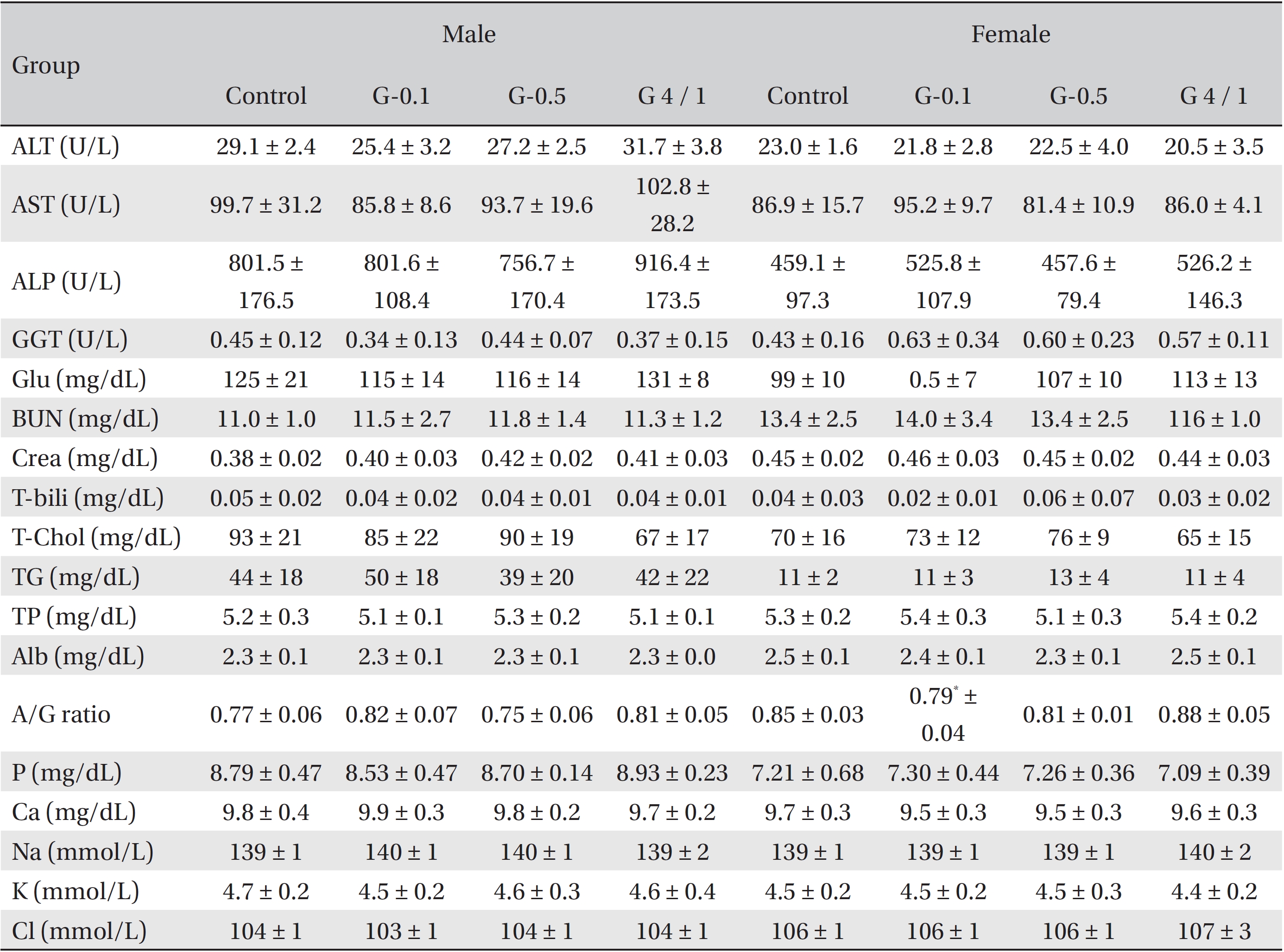
All rats had been fasting for more than 18 hours before necropsy. They were then anesthetized with isoflurane, after which blood was collected from the abdominal aorta. For the hematological test, about 1 mL of the collected blood was placed in an ethylene diamine tetra acetic acid tube (EDTA tube) and was analyzed with a hematology analyzer (ADVIA® 120, Siemens, Germany). For the coagulation test, about 2 mL of the collected blood was placed in a tube with 3.2% sodium citrate and centrifuged at 3,000 rpm for 10 minutes, after which blood plasma was collected. Different laboratory tests were conducted using a coagulation time analyzer (Coapresta® 2000, Sekisui, Japan). For the biochemical test, the blood remaining after carrying out the hematological tests was centrifuged at 3,000 rpm for 10 minutes, and the serum was collected. Tests were done using a biochemistry analyzer (7180, Hitachi, Japan) and an electrolyte analyzer (AVL9181, Roche, Germany).
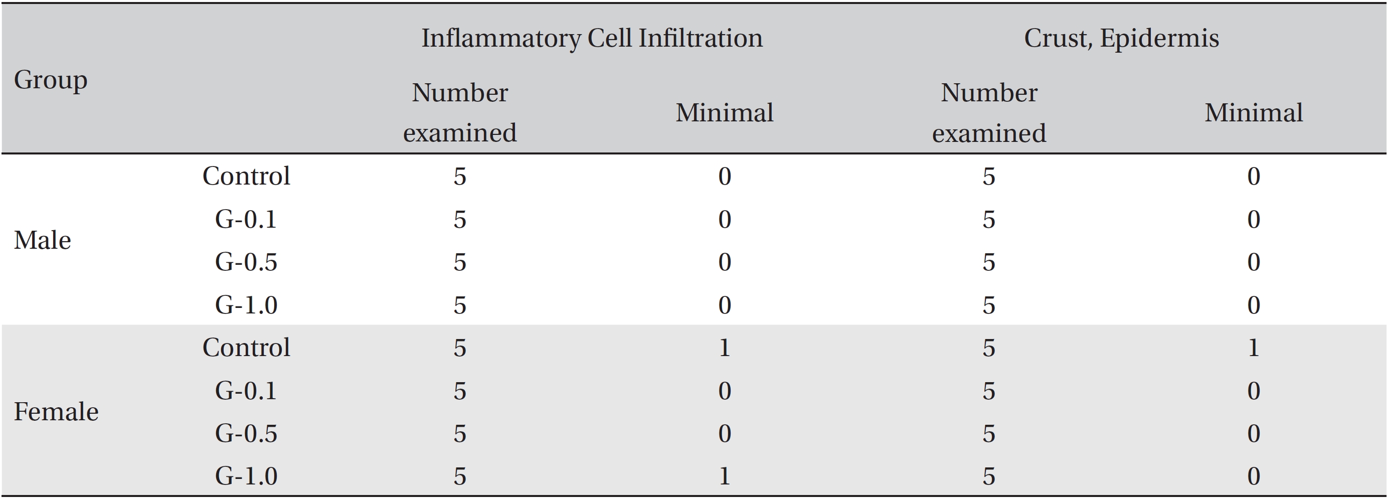
Visual inspections of all body organs and tissues were performed on all animals after necropsy. Body organs and tissues were extracted and fixed in 10% neutral buffered formalin. Routine histological methods, such as trimming, dehydration, and paraffin embedding, were conducted on the fixed organs and tissues. These were then sliced using a microtome and stained with hematoxylin & eosin (H&E).
All the results obtained were analyzed by using STATA/ SE 9.2 for Windows (Stata Corp LP, College Station, TX, U.S.A.). The equal variance was tested by using Bartlett’s test. If the sample variances were equal, significant results were obtained by using the one-way analysis of variance. Dunnett’s multiple range
During the observation, no mortalities or adverse clinical signs were observed in any of the control or the experimental groups. Weight gains were observed in both the experimental and the control groups, but no significant changes were observed in the comparisons of the experimental and control groups. In the hematological test, by Dunnett’s
Studies of IV pharmacopuncture injection have been conducted in Korea on wild ginseng [14, 15], Water-soluble
In this study, IV injection of 1.0 mL of normal saline solution per animal was administered to the control group, and IV injections of 0.1, 0.5, and 1.0 mL of GWG5 per animal were administered to the experimental groups (G- 0.1, G-0.5, and G-1.0). Observations of clinical signs and body weight measurements were carried out for 14 days following the injections. During the observation, no mortalities or adverse clinical signs were observed in any of the groups. Weight gains were observed in all groups, but no significant differences between the experimental and the control groups were observed.
In the hematological test, by Dunnett’s
Based on the above data, we consider the approximate lethal dose of GWG5 to be higher than 1.0 mL/animal. Therefore, GWG5 is a relatively safe pharmacopuncture that can be used for treatment. However, further studies should be performed and repeated IV injection tests, as well as sub-acute and chronic toxicity tests, should be conducted before GWG5 is used for clinical applications.
This study was performed to analyze the single-dose IV toxicity of GWG5. No meaningful changes were observed in general symptoms, body weights, hematological and biochemical test results, and necropsy histopathological observations. Therefore, the approximate lethal dose of GWG5 IV injection is considered to be more than 1.0 mL/animal in both male and female rats.
[Table. 1] Compositions of the experimental groups

Compositions of the experimental groups
[Table. 2] Hematological values of SD rats
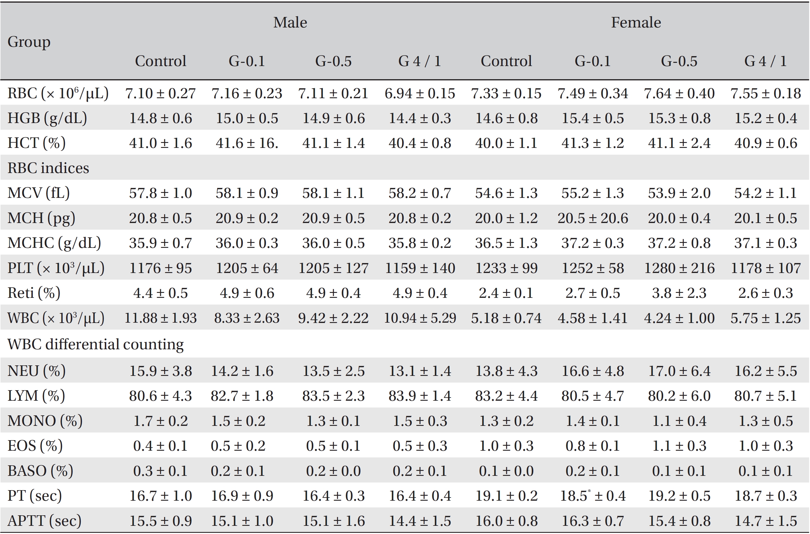
Hematological values of SD rats
[Table. 3] Biochemical values of SD rats

Biochemical values of SD rats
[Table. 4] Summary of histopathological findings

Summary of histopathological findings