Mountain ginseng pharmacopuncture (MGP) is a pharmacopuncture made by distilling extract from mountain cultivated ginseng or mountain wild ginseng. This pharmacopuncture is injected intravenously, which is a quick, lossless way of strongly tonifying Qi function. The present study was undertaken to evaluate a 4-week, repeated, intravenous injection, toxicity test of MGP in Sprague-Dawley (SD) rats.
Twenty male and female 6-week-old SD rats were used as subjects. We divided the SD rats into 4 groups: the high-dosage (10 mL/kg), medium-dosage (5 mL/kg), low-dosage (2.5 mL/kg) and control (normal saline) groups. MGP or normal saline was injected intravenously into the caudal vein of the rats once daily for 4 weeks. Clinical signs, body weights, and food consumption were monitored during the observation period, and hematology, serum biochemistry, organ weight, necropsy, and histological examinations were conducted once the observations had been completed.
No mortality was observed in any of the groups during the observation period. No changes due to MGP were observed in the experimental groups regarding clinical signs, body weights, food consumption, hematology, serum biochemistry, organ weight and necropsy. No histological changes due to MGP were observed in any of the male or female rats in the high-dosage group.
During this 4-week, repeated, intravenous injection, toxicity test of MGP in SD rats, no toxic changes due to MGP were observed in any of the male or female rats in the high-dosage group. Thus, we suggest that the high and the low doses in a 13-week, repeated test should be 10 mL/kg and 2.5 mL/kg, respectively.
Mountain ginseng pharmacopuncture (MGP) was developed using the aromatic substances found in mountain ginseng, and those substances serve to provide a mechanism for survival in a harsh environment. This pharmacopuncture is injected intravenously and is used in treating patients with intractable disorders such as terminal-stage cancer, patients undergoing chemotherapy, patients with immune disorders, and other patients not responding to conventional treatments. Also, injections of MGP at high doses ranging from 20 mL to 60 mL are possible [1].
Mountain wild ginseng refers to ginseng (Panax ginseng C. A. Meyer) of the Araliaceae family that is naturally grown in the mountains. Ginseng has been artificially cultivated from the 14th century, and ginseng used prior to that time is referred to as mountain ginseng. Mountain cultivated ginseng is defined as a young root of mountain wild ginseng that has been replanted in the mountain s or as ginseng cultivated from the seeds of mountain wild ginseng [2]. Because of the high cost of mountain wild ginseng and problems verifying its authenticity, mountain cultivated ginseng that has been grown for about 10 years in an environment similar to that of mountain wild ginseng is used for pharmacopuncture [1].
Groups treated with MGP have been reported to show increased immunoprotein CR2-C3d, which counteracts pathogenic organisms; anti-trypsin, which plays a role in anti-oxidation of proteins; proapolipoprotein and apolipoprotein of high density lipoprotein (HDL) cholesterol, which prevents arteriosclerosis; and vitamin D binding protein, which protects the lungs against inflammation. Also, 20 types of proteins with actions similar to those of ginseng were more than doubled after MGP treatment while proteins that induce mental impairment were suppressed [3, 4].
No significant changes were observed in a randomized, controlled trial of MGP [5], and an intravenous, single-dose toxicity test of MGP in Sprague-Dawley (SD) rats showed no side effects [6]. Thus, we conducted a 4-week, repeated, intravenous dose, toxicity test of MGP in SD rats as part of a safety evaluation of using MGP in SD rats.
Twenty-four 5-week-old male and female SD rats were purchased from Orientbio Inc. (Gyeonggi, Korea). At the time of purchase, the male and female SD rats had weights in the ranges of 121.3 ─ 134.17 g and 105.3 ─ 124.6 g, respectively. Twenty SD rats of each gender were selected based on average weights and were used as the subjects of this test after a week of quarantine and acclimatization. The Twenty 6-week-old male and female SD rats had weights in the ranges of 192.1 ─ 207.9 g and 153.2 ─ 174.4 g at the first injection. The animals were housed in a room maintained at 20.4 ─ 24.5℃ under a relative humidity of 31.9% ─ 57.9%. The room was illuminated with artificial lighting from 07:00 to 19:00 hours and had 10 ─ 15 air changes per hour. The animals were housed in suspended stainless-steel wire-mesh cages and were allowed sterilized tap water and commercial rodent chow (Teklad Certified Irradiated Global 18% Protein Rodent Diet 2918C, Harlan Laboratories, Inc., U.S.A.). This study’s protocol was approved by the Institutional Animal Care Committee at Biotoxtech Co (No. 110027, Ohchang, Korea).
The MGP was manufactured in a pathogen-free facility at the Korean Pharmacopuncture Institute, Seoul, Korea, following a previously-described procedure [7]. Mountain ginseng (MG) that had been cultivated from the seeds of mountain wild ginseng for over 10 years was selected for use in producing the MGP. First, we washed the MG with running water and dried it in air. Second, the dried MG was ground into a powder with a particle size below 10 ㎛ and was then decocted for 2 hours in distilled water. Residues were then removed, and the decoction was distilled, yielding the desired herbal acupuncture. Then, the pharmacopuncture was filtered using 0.1-㎛ filter paper and kept in a container. The pharmacopuncture was sterilized before being used.
Five healthy male and five healthy female rats were assigned to each of four groups: the control (normal saline), the low-dosage (2.5 mL/kg), the medium-dosage (5 mL/ kg), and the high-dosage (10 mL/kg) groups. No toxicity had been observed during 10- or 20-mL/kg intravenous- injection, single-dose tests of MGP in SD rats [6], so in this 4-week, repeated study, we set the new maximum single dose at 10 mL/kg. MGP was administered to the rats by intravenous injection in the caudal vein at a rate of 2 mL/min once daily for 4 weeks. The control group was administered an equivalent volume of normal saline (Lot No. GAJ0071, GAJ0086, Choogwae Pharma Corp., Seoul, Korea).
All animals were observed daily for clinical signs for 4 weeks from the first injection day. The body weight of each rat was measured at the initiation of treatment and twice a week during the treatment period. The amounts of food and water ingested by the rats prior to MGP injection were measured from the day the rats were divided into groups until the day of the first injection while the amounts food and water ingested during the treatment period were averages for a week. At the 4th week after the initiation of treatment, the amounts food and water ingested were the average amounts for 6 days.
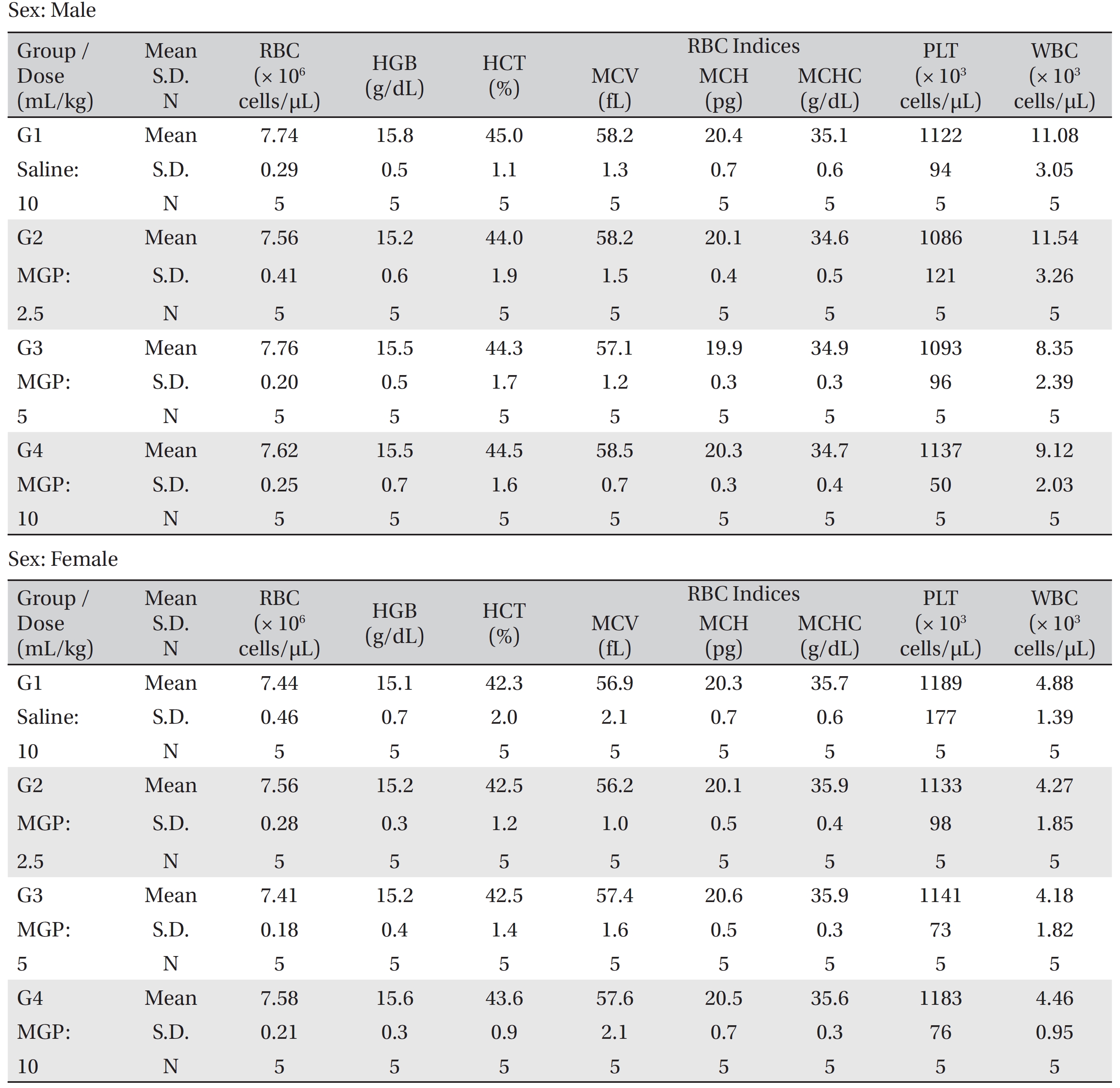
The animals were fasted for 18 hours prior to necropsy and blood was collection. Blood samples were drawn from the abdominal aorta by using a syringe needle under isoflurane anesthesia. Blood samples were collected into tubes containing ethylene diamine tetraacetic acid (EDTA), and were analyzed using a blood counting analyzer (ADVIA 120, SIEMENS, Eschborn, Germany) to determine the red blood cell count (RBC), hemoglobin concentration (HGB), hematocrit (HCT), mean corpuscular cell volume (MCV), mean corpuscular hemoglobin (MCH), mean corpuscular cell hemoglobin concentration (MCHC), platelet count (PLT), and white blood cell count (WBC).
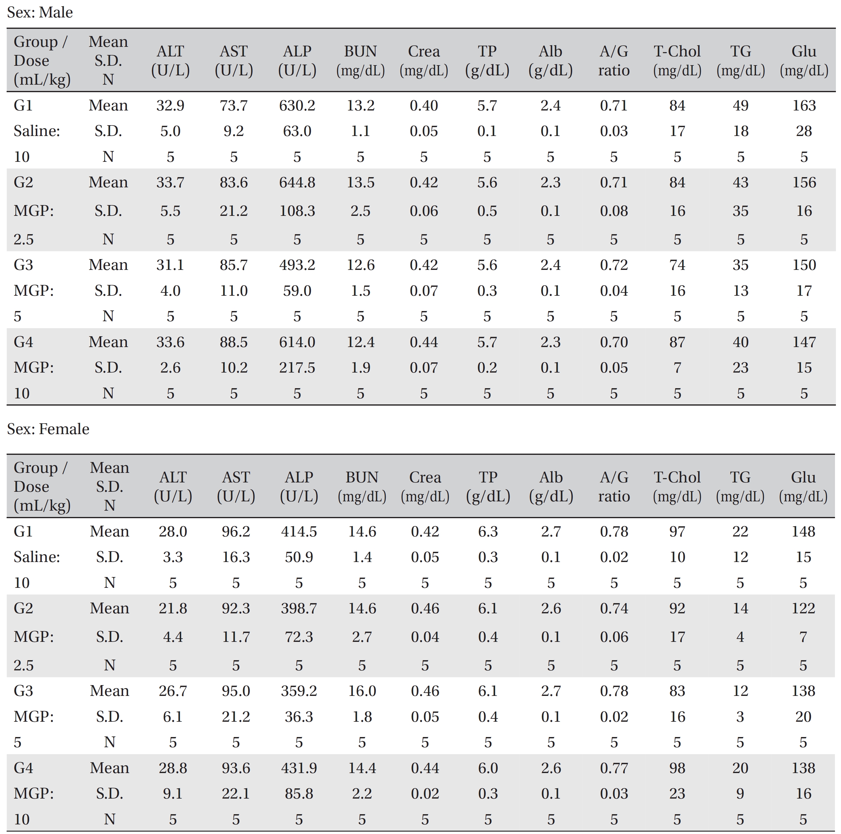
The serum biochemistry analysis was performed using an auto-analyzer (7180, HITACHI, Tokyo, Japan). Blood samples were centrifuged at 3,000 rpm for 10 minutes. Serum biochemistry parameters, including alanine aminotransferase (ALT), aspartate aminotransferase (AST), alkaline phosphatase (ALP), blood urea nitrogen (BUN), creatinine (Crea), total protein (TP), albumin (Alb), albumin/globulin ratio, (A/G ratio) total cholesterol (T-Chol), triglycerides (TG), and glucose (Glu) were examined.
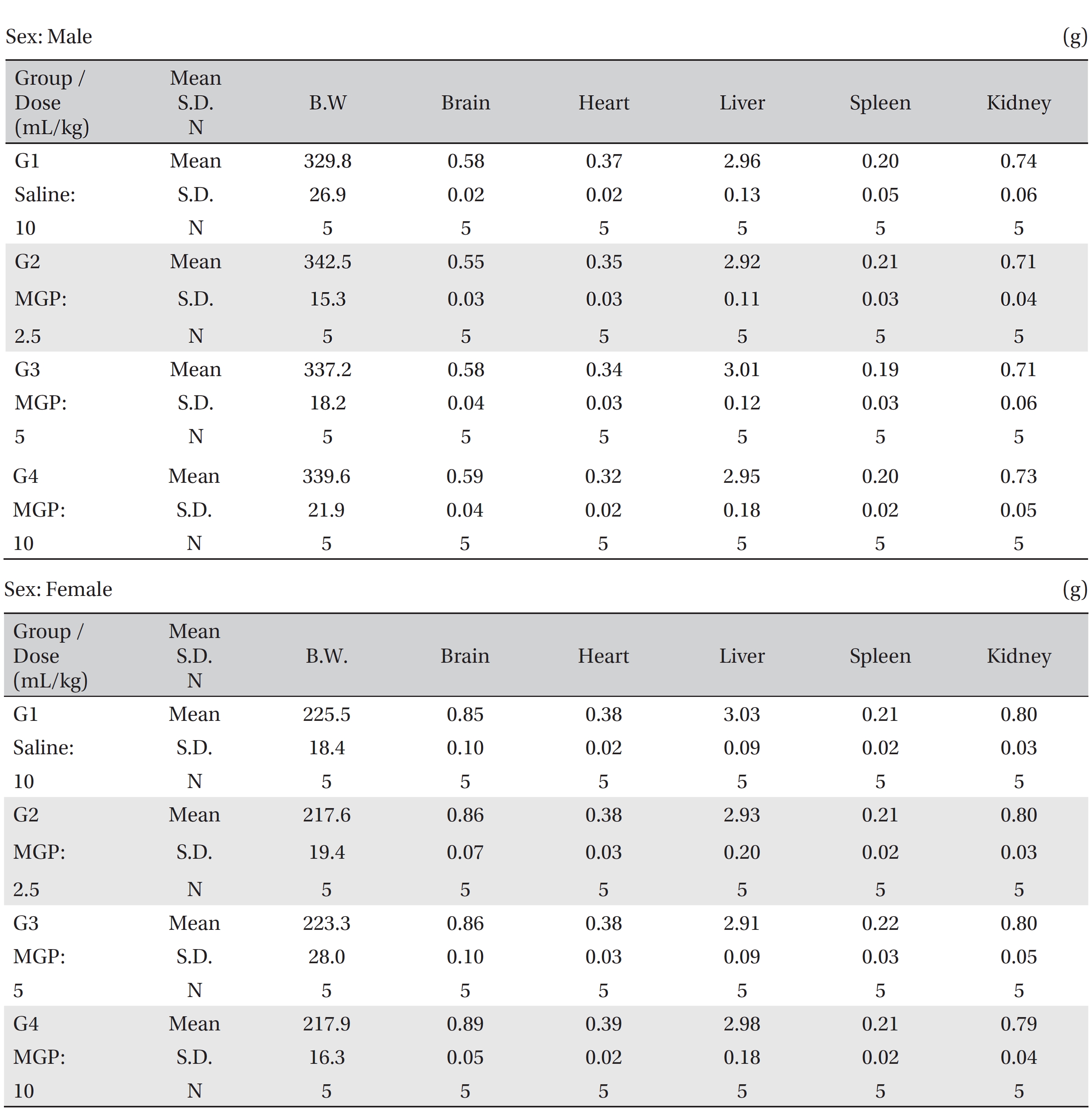
The organs and tissues of all the animals were visually examined. Net weights were measured for the following organs: the brain, heart, liver, spleen, and kidneys. A relative weight ratio was calculated based on the fasting weight. The following tissues were obtained from all the animals and were fixed with 10% neutral buffered formalin solution: the brain, thymus, thyroid and parathyroid, heart, lungs with bronchi, heart, liver, spleen, kidneys, adrenals, stomach, duodenum, jejunum, ileum, cecum, colon, rectum, pancreas, epididymis, ovaries, uterus, and spinal nerves. Testis was fixed with Davidson solution. These tissues were routinely processed, embedded in paraffin, and sectioned. The sections were stained with hematoxylin- eosin (H&E) dye for microscopic examination, and bone tissue was decalcified with Calci-Clear-RapidTM (National Diagnostics, Atlanta, U.S.A.) for microscopic examination. All organs and tissues taken from all animals were examined microscopically.
Data on the weights, food consumption, hematology, serum biochemistry, and organ weights were tested using SAS (version 9.2, SAS Institute Inc, Cary, USA). The variance was checked by using the Bartlett test. If the variance was homogeneous, the data were subjected to a one-way analysis of variance (ANOVA,
[Table. 1] Hematological parameters (group summary)

Hematological parameters (group summary)
[Table. 2] Blood chemistry values (group summary)

Blood chemistry values (group summary)
No mortality was observed in any of the groups of SD rat during the 4-week observation period. No clinical signs occurred in the male and the female SD rats, and no significant differences in weights or food consumptions were observed between the experimental groups and the control group during the 4 weeks. No significant differences in hematology, serum biochemistry and organ weights were observed between the experimental groups and the control group (Table 1)-4). No abnormal macroscopic features were observed during necropsy on the rats in the control and the experimental groups (Table 5).
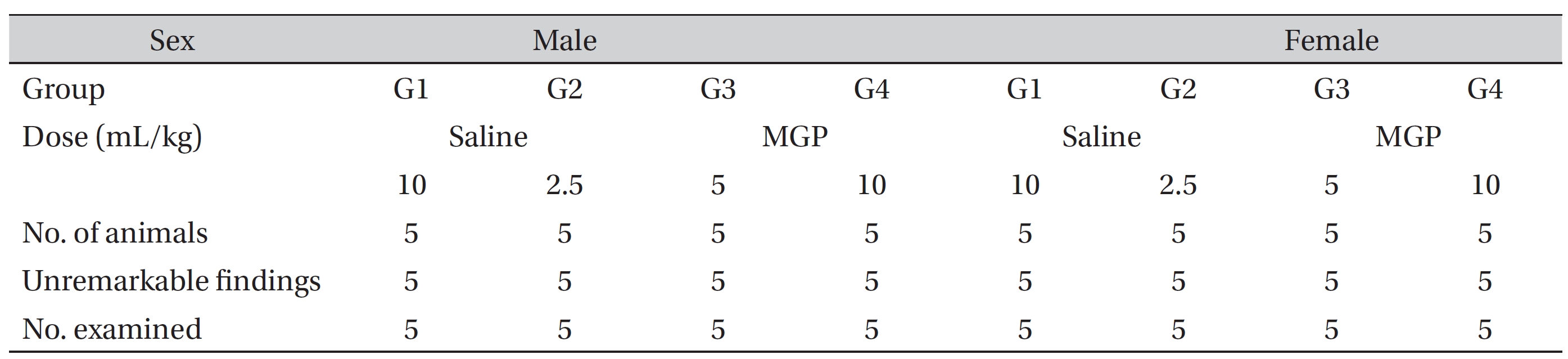
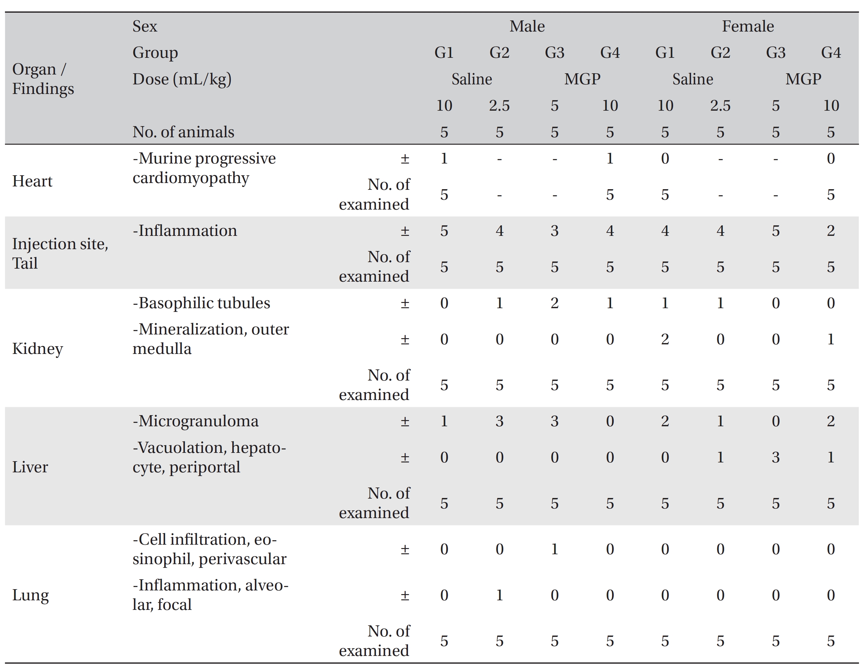
No effects due to MGP were observed in any of the animals. All groups, including the control group, showed microgranuloma, and female rats in the experimental group showed vacuolation in periportal hepatocytes at the tissue of the liver. In addition, all groups showed basophilic tubules and mineralization in the outer tissue of the kidney, but those changes were minimal and had occurred naturally. No changes were observed in the lungs, hearts, brains, spinal nerves and spleens of the control and the experimental groups. Some organs showed changes, and an inflammatory lesion was observed at an injection site: such changes are common in SD rats of similar age and seem to have occurred naturally or sporadically (Table 6).
[Table. 3] Absolute organ weights (group summary)
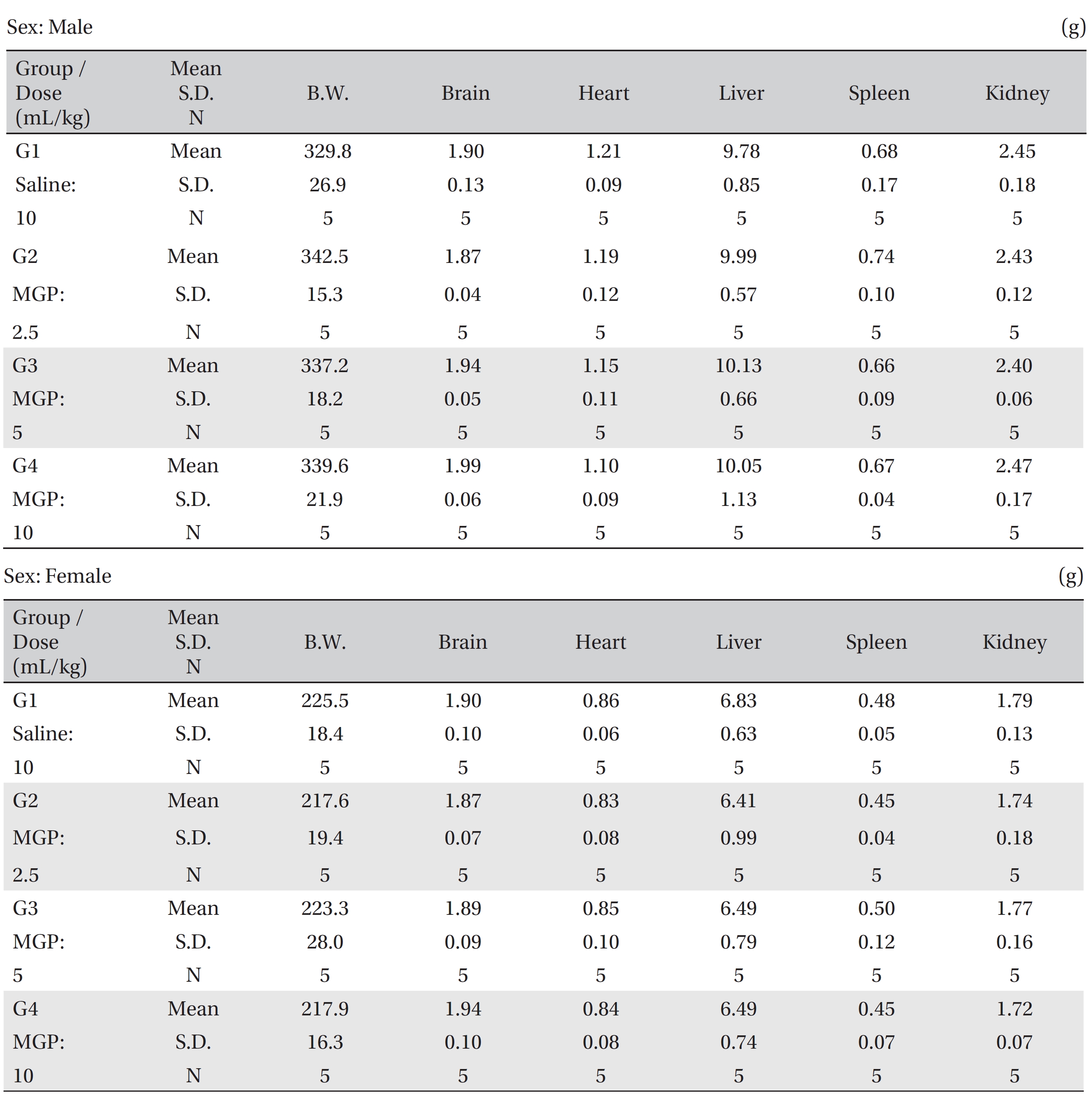
Absolute organ weights (group summary)
[Table. 4] Relative organ weights (g/100 g of body weight), (group summary)

Relative organ weights (g/100 g of body weight), (group summary)
[Table. 5] Necropsy findings (group summary)

Necropsy findings (group summary)
[Table. 6] Summary of histopathological findings

Summary of histopathological findings
In cancer, MGP has been an effective treatment for maintaining tumor size and enhancing the quality of life of cancer patients [8]. These effects of MGP are thought not to result from the MGP directly attacking the cancer cells but rather from an increase in physical stamina and the stimulation of immune cells, which help the body to suppress the growth of the cancer cells. MGP has a superior efficacy in treating general deficiency patterns, such as cancer, compared to other treatment methods, because of the strong tonifying Qi function of mountain ginseng [1]. Intravenous injection is a quick, lossless method of administering MGP with its strong tonifying Qi function [9].
MGP, in
The present study was designed to evaluate a 4-week, repeated, intravenous toxicity test of MGP in SD rats. Our study showed no significant changes in the weights, food consumptions, hematology, serum biochemistry, organ weights, necropsy, and histopathology of the rats. Also, no mortality was observed. Although some changes were observed in several organs and at an injection site, those changes seemed to have occurred naturally or sporadically. The results of this study are consistent with those of an earlier study, which will to be published in the near future; that study found intravenous injection of MGP to be a safe treatment in SD rats.
Treating patients by orally administering the pharmacopuncture to them presents difficulties that are not present when the pharmacopuncture, especially MGP, is administered by intravenous injection. Because MPG administered through intravenous injection can provide rapid and useful effects, additional studies, such as randomize, controlled trials, to validate the safety and the usefulness of MGP are necessary.
In summary, in this 4-week, repeated, intravenous injection, toxicity test of MGP in SD rats, no changes due to MGP were observed in high-dosage group. Consequently, we suggest that high dose should be 10 mL/kg and low dose should be 2.5 mL/kg in the upcoming 13-week, repeated test.