An ethanolic extract from
체내의 염증 반응은 미생물 감염, 내독소, 조직 손상과 같은 위해성 자극에 대한 방어기능으로, 이는 조직의 구조와 기능을 정상적으로 회복하기 위해 필수적으로 일어나는 반응이다. 정상적인 염증반응은 시간이 지남에 따라 염증촉진성 매개체(proinflammatory mediators)의 생성은 감소되고, 항염증성 매개체(anti-inflammatory mediators)는 증가됨으로써 스스로 염증반응이 제한되는 조절과정을 가지고 있다(Lawrence et al., 2002). 체내의 염증반응에 관여하는 세포 중 하나인 대식세포(macrophages)는 이러한 염증반응에 중요한 역할을 하고 있다. 대식세포는 interferon-γ (IFN-γ), interleukin (IL)-1β, IL-6, tumor necrosis factor-α (TNF-α)와 같은 염증촉진성 사이토카인(cytokines), 그리고 세균 세포막성분인 lipopolysaccharides(LPS) 등의 자극에 노출됨으로써 활성화된다(Xie et al., 1993). 활성화된 대식세포는 염증촉진성 사이토카인 이외에 inducible nitric oxide synthase (iNOS) 및 cyclooxygenase-2 (COX-2)와 같은 효소의 발현을 통해 nitric oxide (NO) 및 prostaglandin E2(PGE2)와 같은 다양한 염증매개 분자들을 생성하게 되고(Nathan, 1992; Zhang and Ghosh, 2000), 이들 매개체들의 과도한 생성은 다양한 만성염증성 질환의 발병에 기여하고 있는 것으로 알려져 있다. 따라서 염증성 매개체들의 생성을 억제하는 천연화합물의 발견은 보다 부작용이 적은 항염증 치료제 개발을 위한 방편으로 관심을 끌고 있다.
대식세포에 있어 염증촉진성 사이토카인 및 단백질들의 발현은 nuclear factor kappa-B (NF-κB)에 의해 전사수준에서 조절된다(Pahan et al., 2001). 자극이 없는 상태에서 NF-κB는 inhibitor of kappa B (IκB)와 결합한 상태로 불활성 형태로 세포질에 존재한다(D'Acquisto et al., 1997; Makarov, 2001). 그러나 LPS와 같은 자극이 주어지는 경우 IκB는 IκB kinase (Ikk)에 의해 인산화되어 proteasome에 의해 분해되고, 유리된 NF-κB는 핵으로 이동하여 다양한 염증성 매개체와 같은 표적유전자의 발현을 유도하게 된다(Chen et al., 1995). 한편 NF-κB의 활성화는 extracellular signal-regulated kinase (ERK), c-Jun N-terminal kinase (JNK), p38 kinase를 포함하는 mitogen-activated protein kinases (MAPKs) 그리고 PI3K/ Akt (PKB)와 같은 kinase에 의해 조절되는 것으로 알려져 있다(Marks-Konczalik et al., 1998; Jang et al., 2005).
Nuclear factor erythroid 2–related factor 2 (Nrf2)는 산화적 스트레스에 대응하는 세포 방어기전을 위한 일차적인 전사인자로 알려져 있다(Chen and Kunsch, 2004). 정상적인 상태에서 Nrf2는 Keap1에 의해 비활성화 상태로 세포질에 존재하지만, 산화적 손상을 받으면 Keap1과 해리되어 핵내로 이동하여 antioxidant response element (ARE)에 결합함으로써 HO-1과 같은 항산화 효소의 발현을 조절한다(Nguyen et al., 2000). 최근의 연구에 의하면, Nrf2는 패혈증을 유발시킨 생쥐의 모델실험에서 세포보호효과를 나타내었으며, 특히 Nrf2 knock-out 생쥐는 LPS로 유도된 염증반응에 과도하게 반응하는 것으로 보고되고 있다(Ha et al., 2011). Nrf2 활성화에 의해 생성된 HO-1은 RAW 264.7 세포에서 LPS에 의해 생성된 염증촉진성 사이토카인의 생성을 억제함으로써 항염증 효과를 나타낸다고 보고되고 있다(Lee et al., 2014).
연안에 널리 자생하는 해조류는 비타민과 무기질이 다량 함유되어 있으며 식이섬유가 풍부하다. 현재 많은 연구들이 해조류의 항균효과, 항진균효과, 그리고 항바이러스 활성을 가진다는 것을 밝혔으며(Del et al., 2001), 고지혈증 억제(Awad et al., 2003) 및 항암(Lee et al., 1992)과 같은 생리활성을 가진다는 결과들이 발표되었다. 그 중 갈조류는 저분자 생리활성 물질로써 fucoxanthin, fucosterol, phlorotannin 과 같은 다양한 생리활성 물질들을 가지고 있는 것으로 보고되고 있다(Kim et al., 2010b; Kim et al., 2013a; Lee et al., 2013; Jung et al., 2014). 특히 모자반류는 항염증효과를 가지고 있는 것으로 나타났고(Joung et al., 2012a; Joung et al., 2012b; Gwon et al., 2013; Kim et al., 2013b), phlorotannin, sargachromenol 및 sargaquinoic acid과 같은 다양한 화합물들이 함유되어 있는 것으로 보고되고 있다 (Kang et al., 2013; Kim et al., 2014).
다양한 해조류들 주에서 애기외톨개모자반(
부산 기장에서 채취한
RAW 264.7 세포(ATCC, Rockville, MD, USA)는 10% fetal bovine serum (FBS)와 penicillin (100 units/mL), streptomycin sulfate (100 μg/mL)을 첨가한 Dulbecco’s modified Eagle’s medium (DMEM)을 사용하였고, 5% CO2, 37°C 배양기에서 배양하였다. Cell culture plate에 RAW 264.7 cell이 70~80% 정도 채워지면 phosphate-buffered saline (PBS)로 한번 씻어낸 후, 계대 배양하였다. MYE는 100% dimethyl sulfoxide (DMSO)에 녹여 사용하였고, 배양세포 처리 전에 배지에 희석하여 처리하였다.
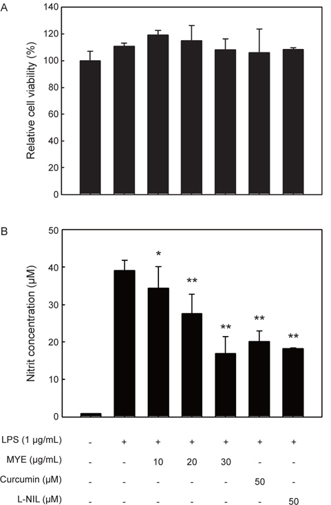
RAW 264.7 cell을 96-well plate에 5×10⁵ cells/well로 분주하고 37°C에서 24시간 동안 배양한 후, MYE가 0, 10, 20, 30μg/mL 농도로 희석된 DMEM 배지로 교체하여 1시간 배양 하였고, 이후에 LPS (1 μg/mL)를 함유한 DMEM 배지에 다시 24시간 배양하였다. 이후 CellTiter96ⓇAqueous 3-(4,5-dimethylthiazol- 2-yl)-5-(3-carboxymethoxyphenyl)-2-(4-sulfophenyl)- 2H-tetrazolium (MTS) 시험 키트(Promega, Madison, WI, USA)를 사용하여 제조사의 방법에 따라 세포생존율을 측정하였다. MTS 용액은 FBS-free DMEM에 5% (v/v)의 농도로 섞어 100 μL씩 처리하였다. 1시간 후에 microplate reader (Glomax Multi Detection System, Promega, Madison, WI, USA)를 이용하여 490 nm의 파장에서 흡광도를 측정하여 세포독성을 분석하였다.
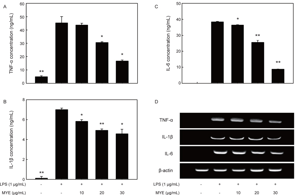
MYE의 항염증효과 비교를 위해 RAW 264.7 세포에서의 NO 생성에 대한 억제효과를 분석하였다. MYE를 1시간동안 전처리 한 다음 LPS (1 μg/mL)로 24시간동안 자극하고, 그 배지를 원심분리(2,000 g, 4°C, 10분)하여 회수하였다. NO의 농도는 배지(100 μL)와 Griess 시약(0.1% naphthylethylene diamine dihydrochloride + 1% sulfanilamide + 5% phosphoric acid)을 동일한 비율로 반응시켜 microplate reader 로 540 nm 의 파장에서 흡광도를 측정하였다(Kim et al., 2009). 배지 중의 IL-1β, IL-6, TNF-α의 양은 enzyme-linked immunosorbent assay kit (ELISA, R&D Systems, Minneapolis, MN, USA)를 이용하여 제조사의 방법에 따라 측정하였다.
RAW 264.7 세포(1×10⁶ cells/well)를 0, 10, 20, 30 μg/mL의 농도로 MYE를 1시간 동안 처리한 후, LPS 1 μg/mL의 농도로 6시간 동안 자극시켰다. 이후 Quiazol 시약(Quiagen Science, Valencia, CA, USA)을 이용하여 total RNA를 분리하였다(Kim et al., 2009). Total RNA로부터 역전사 중합효소 연쇄반응(RT-PCR) 분석에 의한 mRNA 발현양의 분석은 이전의 보고(Kim et al., 2009)에서 사용한 방법을 이용하였고, 유전자 발현량의 상대적인 비교를 위해서 housekeeping gene인 glyceraldehye-3-phosphate dehydrogenase (GAPDH)를 함께 분석하였다. PCR 반응에 이용된 각각의 primer는 Table 1에 나타내었다. 전기영동상 DNA 밴드의 정량 분석은 cooled CCD camera system EZ-Capture Ⅱ (ATTO & Rise Co., Tokyo, Japan) 과 CS analyzer ver. 3.00 software (ATTO)를 이용하여 최소 3번의 반복 실험을 통해 얻었다.
[Table 1.] Primer sequences used in this study
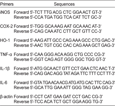
Primer sequences used in this study
NF-κB의 활성화 정도를 분석하기 위해 MYE 및 LPS를 각각 처리한 RAW 264.7 세포로부터 세포질 및 핵 단백질 추출물을 각각 분리 제조하였다(Kim et al., 2009). 즉, MYE로 처리된 세포(2×10⁶ cells/dish)를 PBS로 세척하여 회수하고, 180 μL의 hypotonic buffer [10 mM Tris-HCl, 10 mM NaCl, 3 mM MgCl2, 0.02% NaN3, 0.5 mM dithiothreitol (DTT), 1mM phenylmethanesulfonyl fluoride (PMSF), pH 7.4]를 넣고, 20 μL의 5% nonidet NP-40을 첨가하여 5분 동안 반응시켰다. 이후 원심분리(1,800 g, 4°C, 5분)한 후 상층액을 세포질 추출물로 이용하였다. 침전물은 hypotonic buffer로 한번 세척하고, hypertonic buffer [20 mM 4-(2-hydroxyethyl)-1-piperazineethanesulfonic acid, 25% glycerol, 420 mM NaCl, 1.5 mM MgCl2, 0.2 mM ethylenediaminetetraacetic acid, 0.02% NaN3, 0.5 mM DTT, 1 mM PMSF, pH 7.4]를 넣고 1시간 동안 얼음 위에 방치시킨 다음 원심분리(13,000 g, 4°C, 10분)하여 상층액을 회수하여 핵단백질 추출물로 이용하였다.
염증관련 단백질과 신호전단 단백질의 인산화 정도는 세포를 MYE 및 LPS로 처리한 이후 whole cell lysate를 제조하여 시료로, NF-κB 및 IκB의 활성화 및 인산화 정도는 상기의 핵 및 세포질 추출물을 시료로 이용하였고, 단백질의 양은 이전의 보고(Kim et al., 2009)와 마찬가지로 sodium dodecyl sulfatepolyacrylamide gel electrophoresis (SDS-PAGE)분리된 단백질을 nitrocellulose membrane에 이전시켜 Western blot으로 분석하였다. 검출된 밴드의 정량 분석은 mRNA 분석과 마찬가지로 cooled CCD camera system EZ-Capture Ⅱ와 CS analyzer ver. 3.00 software를 이용하여 최소 3번의 반복 실험을 통해 얻었고, 그 결과를 각 blot의 하단에 수치로 표기하였다. 그리고, Western blot에 사용된 각각의 1차 항체들은 다음과 같다: iNOS (sc-650), β-actin (sc-47778), phospho-Akt (sc- 4060), Akt (sc-1618), phospho-ERK (sc-7883), ERK (sc-94), phospho-JNK (sc-6254), JNK (sc-7345), NF-κB/p65 subunit (sc-8008), HO-1 (sc-7696), Nrf2 (sc-722), poly (ADP-ribose) polymerase (PARP, sc-7150)는 Santa Cruz Biotechnology (Santa Cruz, CA, USA)에서 구입하였고, phospho-IκB-α (4814), IκB-α (9246), phospho-p38 (4511), p38 (9212)는 Cell Signaling Technology (Danvers, MD, USA)에서 각각 구입하였다. Horseradish peroxidase (HRP)가 conjugate되어 있는 각각의 2차 항체들[rabbit anti-goat IgG (LF-SA5004), goat antimouse IgG (LF-SA5001), goat anti-rabbit IgG (LF-SA5002)]은 AbFrontier (Seoul, Korea)에서 구입하였고, Enhanced chemiluminescence (ECL) detection kit은 GE Healthcare Bio-Science (Piscataway, NJ, USA)를 사용하였다. β-actin과 PARP는 각각 세포질과 핵의 control 단백질로서 분석에 포함시켰다.
RAW 264.7 세포를 glass coverslips (SPL Lifesciences Co., Gyeonggi-do, Korea) 위에 24시간 배양한 뒤, MYE로 1시간 전처리 하고, LPS (1 μg/mL)로 30분 자극시켰다. 세포를 4.0% paraformaldehyde가 첨가된 PBS로 실온에서 15분 동안 반응시켜 고정시키고, 0.5% Triton X-100이 첨가된 PBS를 넣어 10분 동안 반응시켰다. PBS로 세척한 뒤에 3% BSA/PBS를 넣고 30분 동안 blocking시킨 후, anti-NF-κB polyclonal antibody가 희석된 3% BSA/PBS를 넣어 2시간 동안 반응시켰다. 그 다음, Alexa FluorⓇ 488-conjugated secondary antibody (Invitrogen, Carlsbad, CA, USA)가 희석된 3% BSA/PBS를 넣고 1시간 동안 반응시킨 뒤, 2 μg/mL의 4,6-diamidino-2-phenylindole (DAPI)로 핵을 염색하고 LSM700 laser scanning confocal microscope (Carl Zeiss, Oberkochen, Germany)로 관찰하였다.
>
NF-κB Promoter/Luciferase assay
RAW 264.7 세포(2×10⁵ cells/well)가 들어있는 24-well plate의 각 well에 1 μg의 pNF-κB firefly luciferase DNA와 20 ng의 pRL-TK renilla luciferase DNA를 lipofectamine/plus reagent (Invitrogen, Carlsbad, CA, USA)와 함께 처리하여 40시간 동안 transfection시켰다. 그 다음, MYE를 1시간 전처리 하고, LPS (1 μg/mL)로 6시간 자극시켰다. 이후 PBS로 세척하고 100 μL의 lysis buffer (0.5 mM HEPES, pH 7.8, 1% Triton N-101, 1 mM CaCl2, and 1 mM MgCl2)로 용해물을 만들고, luciferase assay kit를 사용하여 firefly luciferase activity와 renilla luciferase 활성을 측정하였다. Renilla luciferase의 발현은 지속적으로 일어나는 반면, firefly luciferase는 NF-κB에 의해서만 발현이 되므로 세포 수에 의한 오차를 보정할 수 있다.
본 연구의 모든 실험은 세 번 이상 반복하였으며, 얻어진 결과들을 평균값과 표준편차(mean±SD)를 계산하여 나타내었다. 실험군 간의 유의성 검증은 Student’s t-test로 검증하였다.
>
LPS로 유도되는 NO 생성 및 iNOS 발현에 대한 MYE의 억제 효과
LPS로 자극된 RAW 2645.7 세포에서 생성되는 NO에 대한 MYE의 억제효과를 알아보기 위해서 세포를 다양한 농도(0-30 μg/mL)의 MYE로 1시간 전처리하고, LPS로 24시간 자극하여 상층의 배지로 방출되는 nitrite 생성량을 측정하여 NO 생성량을 분석하였다. LPS 처리에 의해 증가된 NO는 MYE의 전처리에 의해 농도의존적으로 현저하게 감소하는 것으로 나타났다(Fig. 1A,
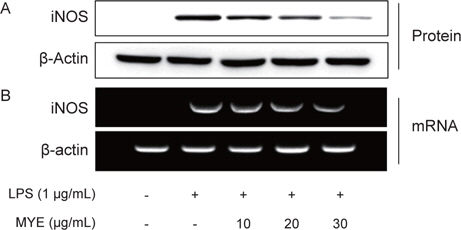
다음은 NO를 생성하는 효소인 iNOS의 발현에 대한 MYE의 효과를 알아보고자 하였다. 전술과 같이 RAW 264.7 세포에 MYE를 0-30 μg/L의 농도로 전처리한 이후 LPS로 자극하였고, 이후 세포 단백질과 total RNA를 분리하여 각각 iNOS의 단백질과 유전자의 발현수준을 분석하였다. Fig. 2에 나타내었듯이 iNOS 단백질의 발현양은 LPS 처리에 의해 현저하게 높게 유도되는 것을 알 수 있었고, MYE 처리에 의해 농도의존적으로 감소하는 경향을 나타내었다. iNOS mRNA 발현양도 단백질과 유사한 경향으로 MYE 처리에 의해 현저하게 감소하는 것으로 나타났으며, 그 억제효과는 전사수준에서 효과적으로 조절되고 있음을 나타낸다(Fig. 2).
NO는 NOS에 의해 L-arginine으로부터 생성된다. iNOS는 세균의 내독소 및 염증성 사이토카인에 의해 강하게 유도된다(Guha and Mackman, 2001). 병리적인 조건 하에서 iNOS에 의한 NO의 현저한 증가는 다른 염증성 매개체들과 함께 과도한 염증을 유발하게 되고 조직의 손상을 유발하는 것으로 알려져 있어 염증성 손상의 주요 매개체이다(Nathan, 1992; Pan et al., 2011). 따라서 iNOS의 발현 또는 활성을 억제함으로써 NO의 생성을 억제할 수 있는 화합물은 항염증 물질로 이용될 수 있을 것이다.
>
LPS로 유도되는 염증성 사이토카인의 생성에 대한 MYE의 억제효과
LPS로 자극된 RAW 2645.7 세포에서 생성되는 염증촉진성 사이토카인의 생성에 대한 MYE의 효과를 ELISA 방법으로 분석하였다. LPS 자극에 의해 TNF-α (Fig. 3A), IL-1β (Fig. 3B) 및 IL-6 (Fig. 3C)와 같은 염증촉진성 사이토카인의 생성량은 크게 증가하는 것으로 나타났고, 이러한 증가는 다소 차이는 있지만 MYE 처리에 의해 현저하게 감소하는 것으로 관찰되었다 (
염증촉진성 사이토카인들은 체내에서 다양한 면역 및 염증반응을 조절하는 역할을 한다. 세균의 LPS에 의해 자극된 대식세포는 TNF-α를 생성하고 분비된 TNF-α 및 LPS는 IL-1β와 IL-6의 생성을 유도함으로써 염증반응을 지속시키게 된다(Beutler and Cerami, 1989). LPS에 의해 유도된 TNF-α는 염증반응의 개시를 촉진하며 지속적인 생성은 만성염증을 유발하며 결국에는 패혈성 쇼크, 염증, 세포상해성 등의 다양한 생리학적 과정에 관여하고 있다(Balkwill, 2006; Dinarello, 1999). IL-1β는 대식세포에서 생성되는 주요 염증촉진성 사이토카인으로서, 세균 감염에 대한 염증성 응답의 개시 및 강화에 중요한 사이토카인이다(Lebovic et al., 2000). IL-6도 대식세포에서 생성되는 중요한 염증촉진성 사이토카인으로서 급성 면역응답에 작용한다(Yoshimura, 2006). 본 연구에서 관찰된 결과는 MYE가 LPS-자극에 의해 유도되는 IL-1β, IL-6, TNF-α의 생성을 억제시키는 것으로 나타났고, 이는 MYE가 LPS-자극에 의한 염증성 응답의 초기 단계를 억제하고 있음을 의미한다.
>
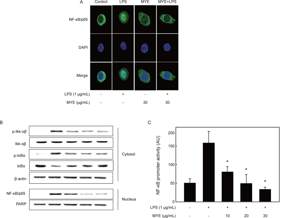
LPS로 유도되는 NF-κB의 활성화에 대한 MYE의 억제 효과
앞에서 서술하였듯이 여러 염증촉진성 사이토카인 및 iNOS의 발현은 주요 전사인자인 NF-κB에 의해 조절된다(Zhang and Ghosh, 2000). 따라서 LPS로 자극한 RAW 264.7 세포를 이용하여 NF-κB의 활성화에 대한 MYE의 처리 효과를 분석하였다. 먼저 면역형광법으로 염색을 하고 confocal microscopy로 분석한 결과를 Fig. 4A에 나타내었다. 아무런 자극이 가해 지지 않은 상태에서 NF-κB/p65 subunit (녹색)는 DAPI로 염색된 핵(청색) 주변에 대부분 분포하는 것이 관찰되지만, LPS로 자극한 경우 녹색인 NF-κB p65의 대부분은 청색인 핵과 함께 분포하는 것으로 나타났고 이는 NF-κB가 활성화되어 핵으로 이동했음을 보여주는 결과이다. 이러한 세포에 MYE (30 μg/mL)를 전처리한 경우 NF-κB/p65는 다시 핵 주변의 세포질에 대부분 분포하는 것으로 관찰되었고, 이는 MYE에 의해 NF-κB의 활성화가 현저하게 억제되고 있음을 보여주고 있다(Fig. 4A).
NF-κB의 활성화에 따른 핵으로의 이동은 이를 억제하는 단백질인 IκB-α의 인산화에 의한 분해에 의한다(Chen et al., 1995). 따라서 MYE로 처리된 세포의 세포질과 핵을 분획하여 이들에서 NF-κB 및 IκB-α의 양 및 인산화 정도를 Western blot으로 분석하였다. Fig. 4B에 나타낸 것과 같이, LPS 자극에 의해 세포질의 p-Ikk-αβ의 증가로 인하여 p-IκB-α의 양은 증가하는 반면 IκB-α 양은 유의적 감소하는 것으로 나타남으로써 인산화 증가에 의한 IκB-α의 분해가 일어났음을 나타내고 있다. MYE 처리에 의해 p-IκB-α의 경우 그 양이 현저하게 감소하는 것으로 나타남으로써 IκB-α의 인산화가 억제되었고, 이러한 결과는 핵에서 NF-κB의 수준이 농도의존적으로 감소하는 결과를 초래하였다. 이상의 결과는 Fig. 4A의 면역형광염색 결과와도 일치하는 것이다.
다음은 LPS로 자극된 대식세포주에 있어 NF-κB의 promoter 활성에 대한 MYE의 효과를 분석하였다. 이를 위해 RAW 264.7 세포에 NF-κB promoter를 가진 luciferase construct를 일시적으로 transfection하고, 이 세포를 다양한 농도의 MYE로 2시간 전처리하고 이어서 LPS로 6시간동안 자극하였다. Fig. 4C에 나타내었듯이 luciferase 활성은 LPS 자극에 의해 현저하게 증가하였으며, 이는 LPS 자극에 의해 활성화된 NF-κB가 NF-κB의 promoter를 가진 luciferase의 발현을 크게 증가시켰음을 의미한다. 이에 대한 MYE의 억제효과는 10 μg/mL의 낮은 농도뿐만 아니라 30 μg/mL 농도에 이르기까지 유의적으로 감소하는 것으로 나타났다. 이들 결과는 대식세포주에서 LPS 자극에 의해 유도되는 NF-κB의 활성화가 MYE에 의해 효과적으로 억제되고 있음을 보여주고 있고, 이는 MYE에 의한 상기의 염증성 사이토카인 및 iNOS의 발현 억제는 부분적으로 NF-κB 활성화 경로에 의해 조절되고 있음을 의미하는 것이다.
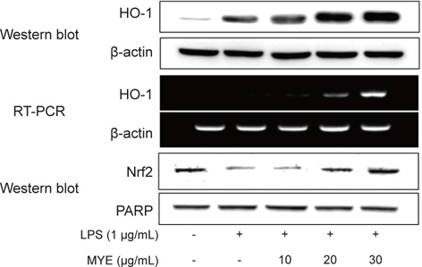
최근의 많은 연구를 통하여 HO-1는 뛰어난 항염증 효과가 있는 것으로 보고되고 있다. 이러한 사실을 입증하기 위하여 LPS로 처리된 대식세포주에 MYE를 처리하여 HO-1와 HO-1의 발현을 조절하는 전사인자인 Nrf2의 발현을 분석하였다. Fig. 5에 나타난 바와 같이 MYE 처리에 의하여 HO-1의 단백질과 mRNA는 농도 의존적으로 증가함을 알 수 있었다. 또한 HO-1의 발현을 조절하는 전사인자인 Nrf2 또한 농도의존적으로 발현량이 증가함을 알 수 있었다.
HO-1은 산화적 스트레스에 대응하는 주요한 방어효소이다. HO-1은 헴(heme)을 빌리베르딘(billiverdin), 유리 철분 및 일산화탄소로 분해하며 속도제한 효소로써 작용한다. 빌리베르딘은 빌리베르딘 환원효소에 의하여 빌리루빈(bilirubin)으로 전환된다. 헴의 대사산물인 빌리루빈은 항산화 활성을 지니며 일산화탄소는 항염증 활성을 지닌다(Otterbein et al., 1999; Ryter et al., 2006). 따라서 HO-1은 염증반응의 억제는 물론 산화적 스트레스를 감소시킴으로써 세포의 항상성을 유지시키는 것으로 알려져 있다. 게다가 HO-1의 항염증 작용은 LPS에 의한 NF-κB의 활성화를 억제함으로써 TNF-α, IL-1β, IL-6 및 NO와 같은 염증성 인자들의 생성을 저해하여 나타낸다고 보고되고 있다(Park et al., 2010). 따라서 MYE의 항염증효과는 NF-κB 경로는 물론 Nrf2 경로를 통하여 나타내는 것으로 판단된다.
>
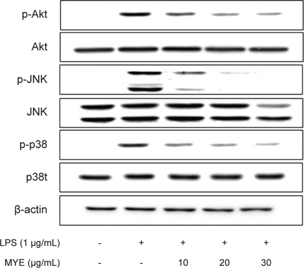
LPS로 유도되는 MAPKs와 Akt의 활성화에 대한 MYE의 억제 효과
MYE에 의한 NF-κB와 Nrf2 신호경로에 어떠한 신호단백질들이 관여하는지를 알기 위하여 MAPKs (ERK, JNK, p38 MAPK)와 Akt의 인산화를 Western blot으로 분석하였다. Fig. 6에 나타난 바와 같이, LPS로 유도된 RAW 264.7 세포에서 MYE 처리에 의하여 Akt, JNK, p38 MAPK의 인산화가 농도 의존적으로 감소하였으나, ERK의 인산화에는 영향을 미치지 않았다(결과 생략). 이러한 결과는 MYE가 IκBα의 인산화는 물론 NF-κB와 Nrf2 경로의 상위 단백질의 인산화를 조절함으로써 항염증 활성을 나타낸다는 것을 의미한다.
NF-κB와 Nrf2 경로는 세포내 다양한 신호전달에 관여하는 MAPKs나 Akt와 같은 단백질 인산기 전이효소들에 의해 조절된다(Pahl, 1999; Zhang and Ghosh, 2000; Kim et al., 2010a). 많은 연구들에서 p38, JNK, ERK와 같은 MAPKs가 설치류의 대식세포에서 LPS에 인한 NF-κB의 활성화에 중요한 역할을 한다고 밝히고 있다(Kim et al., 2010b; Guha and Mackman, 2001). 그리고 Akt 또한 NF-κB의 활성화를 조절함으로써 여러 염증 유전자의 발현을 조절하는 것으로 알려져 있다(Hattori et al., 2003). 본 연구의 결과에서 LPS 자극에 의해 유도되는 NF-κB의 활성화에 대한 MYE의 억제효과는 MAPKs 및 Akt와 같은 인산기 전이효소의 인산화 과정과 관련되어 있음을 나타낸다. 더불어 MYE에 의한 Nrf2 경로의 불활성화 또한 MYE의 항염증 작용 기전으로 작용하고 있음을 보여주고 있다.
앞서 설명한 것처럼 모자반류에서 분리된 fucoxanthin (Kim et al., 2010b), fucosterol (Jung et al., 2013), chromene (Kang et al., 2013; Kim et al., 2014)과 같은 화합물들의 항염증 활성에 대해 활발한 연구가 진행되었다. 본 실험에 사용된 MYE에 이들 화합물이 유효성분으로 포함되어 있는지, 또는 이외의 화합물에 의해 항염증활성이 나타나는가에 대해서는 후속적인 연구가 필요하다.
이상의 결과에서, LPS로 자극한 RAW 264.7 세포에 있어 NO와 같은 염증성 매개체뿐만 아니라 염증촉진성 사이토카인의 생성 및 발현이 MYE에 의해 억제된다는 것을 증명하였다. 이러한 MYE의 억제효과는 IκB의 분해를 저해함으로써 NF-κB 경로를 불활성화시키는 한편 Nrf2 경로는 활성화시키는 것과 관련이 있는 것으로 나타났다. 또한 MYE는 NF-κB 활성화의 상위 신호전달 경로인 MAPKs 및 Akt에도 영향을 미치는 것으로 확인되었다. 여러 만성염증성 질환에 과도한 염증성 매개체들의 발현 및 생성이 중요한 병인적 역할을 하고 있음을 고려했을 때, 본 연구 결과는 MYE가 항염증 효능을 가진 기능성 식품의 소재로 이용될 수 있다는 가능성을 제시하고 있다. 향후 MYE의 항염증 효과를 나타내는 유효화합물의 분리.동정 및 활성분석에 대한 향후 연구가 진행되어야 할 것으로 판단된다.