검색 전체 메뉴
PDF
맨 위로
OA 학술지
Initial Culture Conditions for Primary Cell Populations Derived from Radula Tissue in Abalone Haliotis discus hannai
  • 비영리 CC BY-NC
  • 비영리 CC BY-NC
ABSTRACT
Initial Culture Conditions for Primary Cell Populations Derived from Radula Tissue in Abalone Haliotis discus hannai
KEYWORD
Abalone , Haliotis discus hannai , Primary cell culture , Radula
  • Introduction

    Abalone is a valuable food source worldwide, and farming of abalone is common in numerous countries (Cook and Gordon, 2010). To improve the productivity and the quality of individuals, physiological properties of the abalone should be characterized at the cellular and molecular level. Moreover, the mechanism of diseases that can result in death en masse should be further examined. Establishment of immortalized cell lines can provide a good in vitro model to study various biological phenomenon including physiological properties and disease mechanisms of the organisms (Dauer and Przedborski, 2003; White et al., 2004). However, unlike vertebrate species in which cell line derivation techniques have been well established, only several reports have provided stable conditions for establishing cell lines from few aquatic invertebrate species including freshwater snail (Biomphalaria sp.; Hansen, 1976) and crayfish (Orconectes limosus; Neumann et al., 2000). In addition, progress has been limited for abalone cell culture despite significant efforts (Kusumoto et al., 1997; Suja and Dharmaraj, 2005; Suja et al., 2007; van der Merwe et al., 2010; Pichon et al., 2013). Thus, step-by-step optimization of the conditions for establishing abalone cell line should be conducted. The first issue to be addressed is to secure a sufficient number of primary cultured cell populations to be tested for further growth in vitro. For this reason, we examined the optimal initial culture conditions for the primary cell population from abalone (Haliotis discus hannai). Based on our preliminary experiments (data not shown), employing seven tissues from H. discus hannai including gill, gonad, heart, hepatopancreas, muscle, palpus, and radula, we noticed that the radula tissue-derived cell population is a good candidate for primary cell culture because radula was clearly separated from other tissues, its isolation was relatively easy, and a large number of cells could be retrieved from the tissue using a simple enzyme treatment. Therefore, we investigated the initial conditions for culturing a H. discus hannai radula tissue-derived cell population. Cell survival rate after the cell isolation procedure was determined, and subsequently, three experiments under different initial culture conditions that are varied depending on type of basal media, media salinity, and growth factor addition were conducted to examine the optimal conditions to induce initial culture of the isolated cell population.

    Materials and Methods

      >  Animals

    Abalones (H. discus hannai) were obtained from the Genetics & Breeding Research Center of the National Fisheries Research and Development Institute in Korea. Twenty-three abalones were sacrificed for this study, and the average weight and body length of the abalones were 73.80 ± 11.98 g and 8.61 ± 0.52 cm, respectively.

      >  Radula tissue collection and cell isolation

    For tissue collection, healthy adult abalones were sterilized using 70% ethanol (SK Chemicals, Sungnam, Korea) for 2 min. Radula tissues were removed from the bodies using sterilized surgical equipment and washed three times in Dulbecco’s phosphate-buffered saline (DPBS; Gibco, Grand Island, NY, USA). For cell isolation, radula tissues were placed in 35-mm petri dishes (SPL life Sciences, Pocheon, Korea) filled with digestive medium consisting of DPBS supplemented with 500 U/mL collagenase type I (Worthington Biochemical Corporation, Lakewood Township, NJ, USA) and 0.05% trypsin–EDTA (Gibco), minced using a surgical blade, and incubated for 30 min at room temperature. After enzyme inactivation by adding 3 mL of 10% (v/v) fetal bovine serum (FBS; Cellgro, Manassas, VA, USA)-containing Leibovitz’s L-15 medium (L15; Cellgro) or the high glucose Dulbecco’s modified Eagle’s medium (DMEM; Gibco), all tissue derivatives were filtered through a 40-μm cell strainer (SPL Life Sciences) and retrieved by centrifugation at 400 × g for 4 min. Cell number was counted with a hemocytometer (Paul Marienfeld GmbH and Co. KG, Lauda-Königshofen, Germany). To measure the cell survival rate after cell isolation, cell counting was conducted after Trypan Blue (Gibco) staining.

      >  Cell culture

    The isolated cells were seeded in a well of 0.1% gelatin (Sigma-Aldrich, St. Louis, MO, USA)-coated 48-well plates (Thermo Scientific, Vernon Hills, IL, USA) or 96-well microplates (Thermo Scientific) filled with culture media. The basic culture media were L15 (designated as L15-basic) or 386DMEM buffered with HEPES (designated as DMEM-basic) supplemented with 15% (v/v) FBS, 15% (v/v) H. discus hannai hemolymph, and 1% (v/v) mixed solution of penicillin and streptomycin (Gibco). To prepare abalone hemolymph, healthy adult abalones were anesthetized with 70% ethanol for 2 min, and the hemolymph was collected from the body cavity around heart using 3-mL syringes. Collected hemolymph was centrifuged at 3,500 × g for 15 min, and the supernatant was transferred into new tubes and filtered using a 0.2-μm syringe filter. To generate growth-promoting media (designated as L15-growth-promoting or DMEM-growth-promoting), additional components including 1% (v/v) nonessential amino acids (Gibco), 100 μM β-mercaptoethanol (Gibco), 2 nM sodium selenite (Sigma-Aldrich), 10 ng/mL recombinant human basic fibroblast growth factor (bFGF; Gibco), 25 ng/mL epidermal growth factor (EGF; Sigma-Aldrich), and 50 μg/mL Oryzias dancena embryo extract were supplemented to basic culture media. For DMEM growth-promoting, 1 mM sodium pyruvate (Gibco) was additionally supplemented. O. dancena embryo extract was prepared according to the method reported by Lee et al. (2013). For salinity tests in culture, each medium was adjusted to 25, 30, 35, 40, 45, 50, 60, 70, 80, or 90 psu by dissolving Red Sea salt (Red Sea, Houston, TX, USA). The cells were cultured in an 18ºC incubator under an air atmosphere. After 3 days of culture, unattached cells on the bottom of the plate were removed by two washes with DPBS, and initial cell attachment was visually investigated under an inverted microscope (TS-100F; Nikon, Tokyo, Japan).

    Results and Discussion

    The cell survival rate after the cell isolation procedure employing an enzymatic method was 9.95 ± 2.37% from four replicates [48/504 (live/total) × 104 cells = 9.52% in replicate 1, 48/612 (live/total) × 104 cells = 7.84% in replicate 2, 48/528 (live/total) × 104 cells = 9.09% in replicate 3, and 40/300 (live/total) × 104 cells = 13.33% in replicate 4]. Several factors may contribute to the low cell survival rate. For tissue digestion, enzymatic treatment is commonly used (Lian et al., 2003; Hinsch and Zupanc, 2006; McIntosh et al., 2006), but types of enzymes and their concentration may also affect cell survival. In addition, low salinity of the digestive medium may provoke cellular shock since H. discus hannai lives in a relatively high-salinity environment (Martello et al., 2000; Dmitrieva et al., 2001; Mahajan and Tuteja, 2005). Suboptimal conditions for enzymatic digestion may result in low cell survival after cell isolation procedures. Further experiments examining the enzyme type, enzyme concentration and salinity of the digestive medium will increase our understanding of these observations.

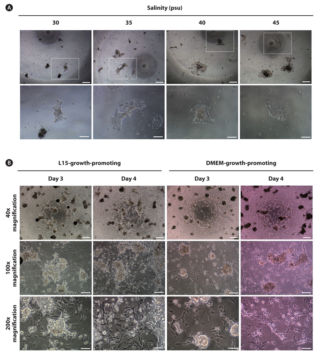
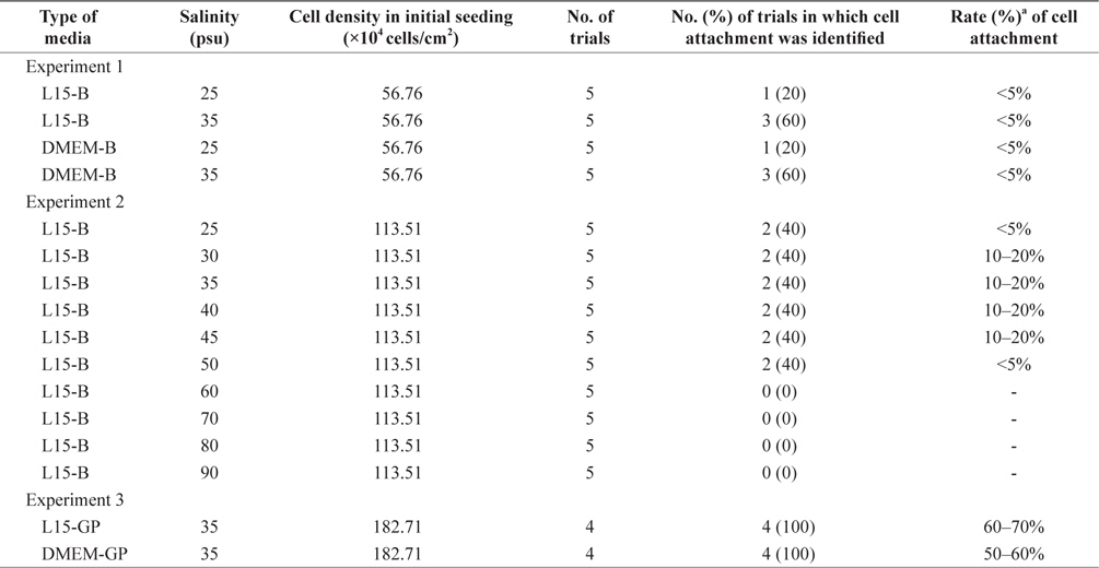
    Next, we cultured the isolated cells under three different experimental conditions to derive the primary cell population (Table 1). In experiment 1, L15-basic or DMEM-basic medium adjusted to either 25 or 35 psu were used for cell culture. No significant differences were detected among the treatment groups based on the number of trials in which the initial cell attachment was identified (p = 0.3909). Regardless of experimental treatments, 40% of trials (8/20) induced initial cell attachment, but the attachment rate was less than 5% in all cases. In this study, initial cell attachment and the rates were used as the final outcomes to evaluate primary culture success because cell attachment allows us to evaluate cell survival and growth based on simple microscopic observations. Additionally, because almost all cell types derived from animals have been cultured using an attachment-based culture method, the measures used in this study are deemed appropriate. Nevertheless, considering that several cell types grow well in a suspension manner (Bellamy et al., 2001; Sen et al., 2002; Gammell et al., 2007), another strategy to target nonattached cells in the initial culture is required. The basal salinity level of media was 25 psu, in which most animal and plant cells can grow normally. However, based on different physiological properties between marine invertebrates and terrestrial organisms, different conditions may be required for in vitro culture of the cells derived from marine invertebrates. Although our data in experiment 1 did not show a statistical difference, we further examined the salinity effect on the initial culture of H. discus hannai radula cells considering the improved results under 35 psu (20% in 25 psu vs. 60% in 35 psu in both types of media). In addition, a very low rate of cell attachment suggested the need for further optimization of other microenvironments in culture.

    [Table 1.] Culture outcome of primary cells derived from Haliotis discus hannai radula

    label

    Culture outcome of primary cells derived from Haliotis discus hannai radula

    In experiment 2, two factors including initial cell density and salinity were differentially applied to the initial culture of H. discus hannai radula cells in comparison with experiment 1. In this case, only L15-basic medium was used to simplify the experiment based on previous results showing no difference between DMEM-basic and L15-basic media. Initial cell density was doubled to account for the low rate of initial cell attachment, and a wide range of salinities (25, 39, 35, 40, 45, 50, 60, 70, 80, and 90 psu) was examined using the same parameters. Based on these results, the trials between 25 and 50 psu induced initial cell attachment, while trials at more than 60 psu did not. No statistical difference was observed in the number of trials in which initial cell attachment occurred corresponding to the salinities from 25 to 50 psu (40% in all), but the rate of initial cell attachment was at least more than twofold in the groups from 30 to 45 psu in comparison with two groups, including 25 and 50 psu (<5% in 25 and 50 psu groups vs. 10-20% in 30, 35, 40, and 45 psu groups). As shown in Fig. 1A, the cells were attached to the bottom of the plate as an aggregate, and slight growth without significant change was observed over time. The cells were retained for 8 days. The images in Fig. 1A are those at day 8 of culture. The results from experiment 2 indicate that the media adjusted from 30 to 45 psu could support initial attachment of the H. discus hannai primary cell population better than 25 psu and 50 psu media. This coincides with previous report that cultured H. midae hemocyte cells in artificial seawater media (van der Merwe et al., 2010). Although no difference was detected among the results from 30, 35, 40, and 45 psu groups, the optimal salinity for culturing H. discus hannai radula cells was 35 psu based on previous reports using artificial seawater media and considering that it corresponds to the average salinity of seawater. Improvement in the rate of cell attachment may have contributed to the initial cell density. Exposure of more viable cells to this supportive environment may increase cell attachment. Furthermore, an increased interaction between viable cells by increasing the cell density may result in a synergistic effect on cell survival in consideration of the role of cell density in culture, which is known to be an important factor to regulate cell survival and growth, as well as cellular function and differentiation (Young et al., 2000; Altman et al., 2002).

    As a final experiment, we used media containing growthpromoting factors under a fixed salinity of 35 psu. Two types of media including L15- and DMEM-growth-promoting media were used, and the initial cell density was further increased 1.6-fold compared with experiment 2. Significant improvement was derived from experiment 3. All eight trials (4/4 = 100% in L15-growth-promoting and 4/4 = 100% in DMEM-growth-promoting) successfully induced initial cell attachment with rates of 60–70% and 50–60% in L15- and DMEM-growth-promoting media, respectively. As shown in Fig. 1B, many cells were attached to the bottom of the plate as aggregates or a single cell in both media types at day 3 of culture. Based on visual observation, growth of these cell populations was detected at day 4 of culture regardless of the media type (Fig. 1B), which were continuously retained without significant change up to day 9 of culture. For one cell population cultured in L15-growth-promoting medium, the first subculture was successfully progressed at day 7 of culture, but the cells could not survive beyond 2 days after subculture. Under this experimental condition, the culture medium was additionally supplemented with various components to promote cell growth based on the composition of fish stem cell culture media (Yi et al., 2010; Lee et al., 2013). Specifically, bFGF, EGF, and embryo extracts are known to promote cell proliferation in culture (Collodi and Barnes, 1990; Raballo et al., 2000; Kimura et al., 2013). As expected, the initial culture outcomes of H. discus hannai radula-derived cells were significantly enhanced. Although the initial cell density increased under this experimental condition, which could contribute to the improvement of initial culture, it is not considered as a major factor for such improvement because a 1.6-fold increase in cell density resulted in at least a threefold increase in the rate of initial cell attachment. Therefore, growth factors or other components likely strongly affected the initial culture. This is supported by previous reports that identified the positive effects of growth factor addition in in vitro culture of different tissue cells from abalone species (Lebel et al., 1996; van der Merwe et al., 2010). In the future, the dependence of H. discus hannai radula-derived primary cell populations on each growth factor should be explored to identify factors that significantly affect the initial culture and to optimize the media composition together with the other microenvironments.

    In conclusion, initial culture for primary cell populations derived from H. discus hannai radula tissue can be supported by growth promoting media adjusted to 35 psu salinity. The results from this study will provide valuable information to derive immortal cell lines in abalone.

참고문헌
  • 1. Altman GH, Horan RL, Martin I, Farhadi J, Stark PR, Volloch V, Richmond JC, Vunjak-Novakovic G, Kaplan DL 2002 Cell differentiation by mechanical stress [FASEB J] Vol.16 P.270-272 google
  • 2. Bellamy WT, Richter L, Sirjani D, Roxas C, Glinsmann-Gibson B, Frutiger Y, Grogan TM, List AF 2001 Vascular endothelial cell growth factor is an autocrine promoter of abnormal localized immature myeloid precursors and leukemia progenitor formation in myelodysplastic syndromes [Blood] Vol.97 P.1427-1434 google
  • 3. Collodi P, Barnes DW 1990 Mitogenic activity from trout embryos [Proc Natl Acad Sci USA] Vol.87 P.3498-3502 google
  • 4. Cook PA, Gordon HR 2010 World abalone supply, markets, and pricing [J shellfish res] Vol.29 P.569-571 google
  • 5. Dauer W, Przedborski S 2003 Parkinson’s disease: mechanisms and models [Neuron] Vol.39 P.889-909 google
  • 6. Dmitrieva NI, Michea LF, Rocha GM, Burg MB 2001 Cell cycle delay and apoptosis in response to osmotic stress [Comp Biochem Physiol A Mol Integr Physiol] Vol.130 P.411-420 google
  • 7. Gammell P, Barron N, Kumar N, Clynes M 2007 Initial identification of low temperature and culture stage induction of miRNA expression in suspension CHO-K1 cells [J Biotechnol] Vol.130 P.213-218 google
  • 8. Hansen EL, Maramorosch K 1976 A cell line from embryos of Biomphalaria glabrata (Pulmonata): Establishment and characteristics P.75-97 google
  • 9. Hinsch K, Zupanc GK 2006 Isolation, cultivation, and differentiation of neural stem cells from adult fish brain [J Neurosci Methods] Vol.158 P.75-88 google
  • 10. Kimura H, Okubo N, Chosa N, Kyakumoto S, Kamo M, Miura H, Ishisaki A 2013 EGF positively regulates the proliferation and migration, and negatively regulates the myofibroblast differentiation of periodontal ligament-derived endothelial progenitor cells through MEK/ERK- and JNK-dependent signals [Cell Physiol Biochem] Vol.32 P.899-914 google
  • 11. Kusumoto K, Shirahata S, Katakuta Y, Murakami H, Kamei Y 1997 Establishment of an abalone digestive gland cell line secreting various glycosidases in protein-free culture [Cytotechnol] Vol.24 P.169-176 google
  • 12. Lebel JM, Giard W, Favrel P, Boucaud-Camou E 1996 Effects of different vertebrate growth factors on primary cultures of hemocytes from the gastropod mollusc, Haliotis tuberculata [Biol Cell] Vol.86 P.67-72 google
  • 13. Lee D, Kim MS, Nam YK, Kim DS, Gong SP 2013 Establishment and characterization of permanent cell lines from Oryzias dancena embryos [Fish Aquat Sci] Vol.16 P.177-185 google
  • 14. Lian ZX, Okada T, He XS, Kita H, Liu YJ, Ansari AA, Kikuchi K, Ikehara S, Gershwin ME 2003 Heterogeneity of dendritic cells in the mouse liver: identification and characterization of four distinct populations [J Immunol] Vol.170 P.2323-2330 google
  • 15. Mahajan S, Tuteja N 2005 Cold, salinity and drought stresses: an overview [Arch Biochem Biophys] Vol.444 P.139-158 google
  • 16. Martello LB, Friedman CS, Tjeerdema RS 2000 Combined effects of pentachlorophenol and salinity stress on phagocytic and chemotactic function in two species of abalone [Aquat Toxicol] Vol.49 P.213-225 google
  • 17. McIntosh K, Zvonic S, Garrett S, Mitchell JB, Floyd ZE, Hammill L, Kloster A, Di Halvorsen Y, Ting JP, Storms RW, Goh B, Kilroy G, Wu X, Gimble JM 2006 The immunogenicity of human adipose-derived cells: temporal changes in vitro [Stem Cells] Vol.24 P.1246-1253 google
  • 18. Neumann T, Kaiser HE, Rath FW 2000 A permanent cell line of the crayfish Orconectes limosus as a potential model in comparative oncology [In Vivo] Vol.14 P.691-698 google
  • 19. Pichon D, Cudennec B, Huchette S, Djediat C, Renault T, Paillard C, Auzoux-Bordenave S 2013 Characterization of abalone Haliotis tuberculata?Vibrio harveyi interactions in gill primary cultures [Cytotechnol] Vol.65 P.759-772 google
  • 20. Raballo R, Rhee J, Lyn-Cook R, Leckman JF, Schwartz ML, Vaccarino FM 2000 Basic fibroblast growth factor (Fgf2) is necessary for cell proliferation and neurogenesis in the developing cerebral cortex [J Neurosci] Vol.20 P.5012-5023 google
  • 21. Sen A, Kallos MS, Behie LA 2002 Passaging protocols for mammalian neural stem cells in suspension bioreactors [Biotechnol Prog] Vol.18 P.337-345 google
  • 22. Suja CP, Dharmaraj S 2005 In vitro culture of mantle tissue of the abalone Haliotis varia Linnaeus [Tissue Cell] Vol.37 P.1-10 google
  • 23. Suja CP, Sukumaran N, Dharmaraj S 2007 Effect of culture media and tissue extracts in the mantle explant culture of abalone, Haliotis varia Linnaeus [Aquaculture] Vol.271 P.516-522 google
  • 24. van der Merwe M, Auzoux-Bordenave S, Niesler C, Roodt-Wilding R 2010 Investigating the establishment of primary cell culture from different abalone (Haliotis midae) tissues [Cytotechnol] Vol.62 P.265-277 google
  • 25. White SM, Constantin PE, Claycomb WC 2004 Cardiac physiology at the cellular level: use of cultured HL-1 cardiomyocytes for studies of cardiac muscle cell structure and function [Am J Physiol Heart Circ Physiol] Vol.286 P.H823-H829 google
  • 26. Yi M, Hong N, Hong Y. 2010 Derivation and characterization of haploid embryonic stem cell cultures in medaka fish [Nat Protoc] Vol.5 P.1418-1430 google
  • 27. Young TH, Huang JH, Hung SH, Hsu JP 2000 The role of cell density in the survival of cultured cerebellar granule neurons [J Biomed Mater Res] Vol.52 P.748-753 google
OAK XML 통계
이미지 / 테이블
  • [ Table 1. ]  Culture outcome of primary cells derived from Haliotis discus hannai radula
    Culture outcome of primary cells derived from Haliotis discus hannai radula
  • [ Fig. 1. ]  Images after initial culture of primary cell populations derived from Haliotis discus hannai radula. (A) Images of the cells cultured under basic media (L15-basic) containing different salinities. The cells were grown as a form of aggregates and maintained up to day 8 of culture. Images were taken at day 8 of culture. (B) Images of the cells cultured under growth-promoting media. L15-basic or DMEM-basic was supplemented with various growth-promoting factors and adjusted to 35 psu salinity. Lots of cells were grown as a form of aggregates or a sing cell regardless of media type employed. At day 4 of culture, a little cell growth was identified from visual observation. Scale bar: 40x = 200 μm, 100x = 100 μm, 200x = 50 μm.
    Images after initial culture of primary cell populations derived from Haliotis discus hannai radula. (A) Images of the cells cultured under basic media (L15-basic) containing different salinities. The cells were grown as a form of aggregates and maintained up to day 8 of culture. Images were taken at day 8 of culture. (B) Images of the cells cultured under growth-promoting media. L15-basic or DMEM-basic was supplemented with various growth-promoting factors and adjusted to 35 psu salinity. Lots of cells were grown as a form of aggregates or a sing cell regardless of media type employed. At day 4 of culture, a little cell growth was identified from visual observation. Scale bar: 40x = 200 μm, 100x = 100 μm, 200x = 50 μm.
(우)06579 서울시 서초구 반포대로 201(반포동)
Tel. 02-537-6389 | Fax. 02-590-0571 | 문의 : oak2014@korea.kr
Copyright(c) National Library of Korea. All rights reserved.