검색 전체 메뉴
PDF
맨 위로
OA 학술지
Oxidative Stress and Antioxidant Defences in the Tasar Silkworm Antheraea mylitta D: Challenged with Nosema Species
  • 비영리 CC BY-NC
  • 비영리 CC BY-NC
ABSTRACT
Oxidative Stress and Antioxidant Defences in the Tasar Silkworm Antheraea mylitta D: Challenged with Nosema Species
KEYWORD
Lipid peroxidation , antioxidant defences , pebrine , tasar silkworm
  • Introduction

    Tropical tasar silkworm, Antheraea mylitta D. (Lepidoptera: Saturniidae) is a commercially important wild polyphagus sericigenous insect. It is an important component of Asian non-mulberry sericulture industry. Since tasar silkworms are reared outdoors, they are more vulnerable to various diseases such as pebrine, virosis, bacteriosis and mycosis. Among these, pebrine disease which is caused by Nosema sp. infects almost all stages and ecoraces of the tasar silkworm by both primary (transovarial) and secondary (peroral) infections. In insect, Nosema infection takes place after ingestion of mature spores that germinate in the midgut by polar tube extrusion and injection of the sporoplasm inside the epithelial cell cytoplasm (Higes et al., 2007). As ingestion is the main entry route of many pathogens, the intestinal epithelium is the first line of defense against invasion and dissemination of pathogenic microorganisms. In this view insect innate immune system plays a vital role in the defense against microorganisms (Medzhitov and Janeway, 1997). One of the most immediate epithelial responses in mammals to combat the pathogen is the generation of antimicrobial reactive oxygen species (Cohn et al., 1994; Geiszt et al., 2003). It is to be noted that ROS can affect both entomopathogens and the host tissues, and may lead to damages of cellular biomolecules such as lipids, proteins and nucleic acids.

    To protect against these toxic cellular environment, insect possess a suit of antioxidant defence mechanisms, comprised of both enzymatic as well as non-enzymatic components. The major enzymes involved in the process are superoxide dismutase (SOD), catalase (CAT), glutathione peroxidase (GPX), glutathione reductase (GR) and glutathione-S-transferase (GST). Superoxide radicals (O2•‾) are dismutated by SOD to hydrogen peroxide (H2O2) which is reduced to water and molecular oxygen by CAT. Further, GPX catalyzes the reduction of H2O2 to water and organic peroxide to alcohols using reduced glutathione GSH as a source of reducing equivalent. GR regenerates reduced glutathione (GSH) from oxidized glutathione (GSSG), which is a scavenger of ROS as well as a substrate for the other enzymes. GST conjugates xenobiotics with GSH for excretion. The non-enzymatic component consists of small organic molecules such as GSH, and vitamin C (Felton and Summers,1995; Krishnan and Kodrik,2006; Krishnan et al., 2009; Buyukguzel et al., 2010; Zhao and Shi 2009, 2010).

    Sufficiently large details are available on larval growth rate (Rath et al., 2003), cocoon characters Velide et al., 2013), total haemocyte count Madhusudhan et al., 2011) and excretory products (Renuka and Samitha, 2012) during infection. However very scant information exists regarding the damages caused by Nosema spores to the host tasar silkworm and the mechanism adopted by this insect to protect itself. The present study has been therefore designed to quantify the oxidative damages (LPX and total hydroperoxide) and antioxidant defence (SOD, CAT and GST) in midgut of tasar silkworm A.mylitta infected by Nosema spores.

    Materials and Methods

      >  Isolation of spores and Insect infection

    Nosema spores were isolated from diseased larvae of Daba ecoraces homogenized in 0.6% K2CO3, filtered and the filtrate was centrifuged at 3000 rpm for 15 min. Spores were purified on discontinuous sucrose gradient (25, 50 and 75%) by centrifugation at 4000 rpm for 10 min.. The spores were collected from the sediment and washed in distilled water thrice and stored as stock at 4℃ in 0.85% NaCl until use. They were then suspended in distilled water and counted using haemocytometer. The stock solution was diluted to obtain an inoculum dosage of 106 spore’s mL-1. Daba TV healthy fifth instar (after fourth moult) larvae were starved for 3-4 h to induce hunger and than fed on the Terminalia arjuna leaves smeared with inoculum dosage.

      >  Sample preparation

    The midguts were dissected out, thoroughly washed in ice-chilled phosphate buffer (50 mM, pH 7.0) to remove haemolymph, muscle tissues and fat body contamination. Tissue samples (midguts) were homogenized in ice-cold buffer (50 mM phosphate buffer, pH 7.0). Homogenization was carried out in an ice-chilled motor driven Teflon Potter-Elvejhen homogenizer and centrifuged at 8000 x g for 15 min at 4℃. The supernatant was used for biochemical analysis.

      >  Estimation of lipid peroxidation

    LPX level was assayed by measurement of malondialdehyde (MDA), a decomposition product of polyunsaturated fatty acids hydro peroxides were determined by the TBA reaction as described by Bar-Or et al. (2001). Briefly, the reaction mixture containing 0.1 mL of sample, 0.9 mL of 0.8 % aqueous solution of TBA (in 20% TCA). Then the mixture was heated at 95℃ for 60 min and cooled under room temperature. The supernatant was read at 532 nm after removal of any interfering substances by centrifuging at 4000 x g for 10 min. The amount of MDA formed was calculated by using an extinction coefficient of 1.56 × 10⁵M-1cm-1 (Wills 1969), and expressed as nmol MDA/mg protein.

      >  Estimation of total hydroperoxides

    Total hydroperoxides were determined spectrophotometrically according to the method of ferrous oxidation with xylenol orange (FOX1) (Wolff, 1994). Hydroperoxides oxidize ferrous to ferric ions selectively in dilute acid and the resultant ferric ions can be determined by using ferric sensitive dyes as an indirect measure of hydroperoxide concentration. Xylenol orange binds ferric ions with high selectivity to produce a coloured (blue-purple) complex. The absorbance was read at 560 nm after removal of any flocculated material by centrifugation at 4000 x g for 10 min. The signal was read against an H2O2 standard curve.

      >  Superoxide dismutase

    Super oxide dismutase activity was estimated by Kono, (1978). The reaction mixture consisted of 50 mM sodium carbonate, 25 μM NBT, 0.6% Triton X 100 and 0.1 mM EDTA. The reaction was initiated by addition of 1 mM hydroxylamine-hydrochloride. The rate of NBT reduction was recorded at 560 nm. The control was simultaneously run without tissue homogenate. One unit of SOD is defined as the amount required inhibiting the photoreduction of NBT by 50%. The specific activity of SOD was expressed as unit/mg protein.

      >  Catalase

    Catalase activity was determined according to Aebi, (1974). The method is based on the decomposition rate of H2O2 by the enzyme. The assay mixture contained 2.9 mL of 12 mM H2O2 and 0.1 mL of sample (100 μg protein). Absorbance was measured at 240 nm and CAT activity is expressed as nkat/ mg protein (1 katal = 1 mol sec-1).

      >  Glutathione-S-transferase

    Glutathione-S-transferase activity was measured according to Habig et al. (1974) using 1-chloro-2, 4-dinitrobenzene (CDNB) as a substrate. Assay mixture contained 2.7 ml of 100 mM phosphate buffer (pH 7), 0.1 mL of 30 mM GSH, 0.1 mL of 15 mM CDNB and 0.1 mL of sample (100 μg protein). The change in absorbance was recorded at 340 nm and enzyme activity was expressed as nmol CDNB conjugate formed/ min/ mg protein using a molar extinction coefficient of 9.6 mM-1 cm-1.

      >  Protein assay

    The protein content was estimated by the Bradford (1976) method using bovine serum albumin as standard.

      >  Statistical analysis

    Results were expressed as mean ± standard deviation (SD). Difference between control and treatment was analyzed by Student’s t-test. Differences were considered statistically significant when p<0.05. Further inter relationship was analysis by correlation.

    Results

      >  Lipid peroxidation

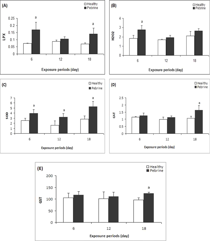
    A significant increase in LPX level was observed in the midgut of A.mylitta larvae on 6th and 18th day after the insects were inoculated with Nosema spore, compared to controls (Fig-1A, p < 0.05). Also an insignificant increase was also seen on 12th day of the treatment in comparison to respective controls (p > 0.05).

      >  Total hydroperoxides

    Total hydroperoxide level significantly increased on 6th day of treatment (Fig-1B, p < 0.05) while an insignificant increase was recorded on 12th and 18th day in relation to controls (Fig-1B, p > 0.05).

      >  Superoxide dismutase

    A significant increase in SOD activity to was seen up to (1.55 fold) on the 6th day, 1.98 fold on 12th day and 1.89 fold on the 18th day in the larvae exposed to Nosema spores (Fig-1C, p < 0.05) in relation to control (Fig-1C).

      >  Catalase

    CAT activity showed a significant increase on 18th day of exposure compared to control larvae (Fig-1D, p < 0.05). However, no significant change in CAT activity was recorded on 6th and 12th day of post inoculation.

      >  Glutathione-S-transferase

    GST activity in the midgut of infected larvae significantly increased on 18th day after inoculation in comparison to the control (Fig-1E, p < 0.05). However, on 6th and 12th day the GST activity no significant changes were seen in the infected larvae (Fig-1E).

    Discussions

    LPX serves as an indicator of oxidative damage in cells and tissues (Pampanin et al., 2005). An enhanced level of MDA (lipid peroxidation product) in the midgut of larvae was observed in response to Nosema infection (Fig-1A). Similar to this higher level of LPX was also detected in insects during viral (Wang et al., 2001) and bacterial infection in insects Dubovskiy et al., 2008). It is understood that one of the most immediate immune response of the gut involves the production of ROS to fight microbial infection both in mammals (Kinnula et al., 1992; Geiszt et al., 2003) and insects Ha et al., 2005a,b; Ryu et al., 2010). The increased immune response during infection could be the possible reason for the increase in the rate of ROS formation, resulting in oxidative stress. The observed higher level of total hydroperoxide (H2O2 and other water soluble hydroperoxides, Wolff, 1994) in infected larvae (Fig-1B) as compared to healthy may indicate increased formation of OH by Fenton reaction, thereby, enhancing the LPX level in midgut of larvae. This is also evident from a significant positive correlation between total hydroperoxide and LPX in midgut tissues samples (Fig-2A, p < 0.05). In A. mylitta reduction of larval weight (Madhusudan et al., 2012), cocoon weight (Velide et al., 2013) was also observed during Nosema infection. This may be due to an increase in the production of ROS and oxidative damage in response to diseases.

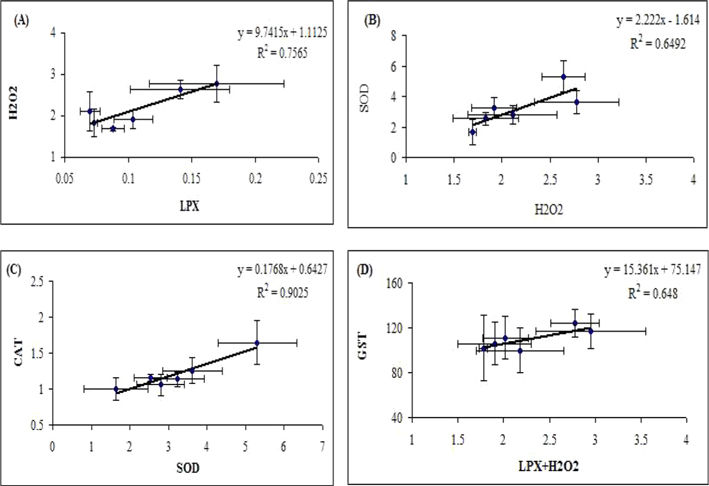
    Determination of antioxidant status of larva exposed to Nosema is important to understand the toxic mechanism and predict the damage potential in the organism. An increased oxidative stress suggests elevated activity of antioxidant enzymes, thereby protecting animals from oxidative stress (Halliwell and Gutteridge, 2001). SOD is a crucial antioxidant enzyme, which dismutates O2•‾ to H2O2. In the present study significant increase in SOD activity was observed in the midguts of infected larvae (Fig-1C), which suggests active production of O2•‾ radicals during pathogenic infection. Similarly, higher SOD activity was also observed in midgut of insect (Galleria mellonella) infection with bacterial pathogen (Dubovskiy et al., 2008). The induction of SOD during Nosema infection may indicate that it helps in inhibiting the oxygen radical accumulation.

    The principal H2O2 scavenging enzyme CAT showed increased level in midguts of A.mylitta in response to Nosema infection (Fig-1D). This suggests that during infection period cell might elevate the rate of H2O2 production in tissues, and as a consequence, an elevation of CAT may be seen. Similarly, upregulation of CAT activity was observed in honey bee Apis mellifera infected with Nosema ceranae (Dussaubat et al., 2012). However, in early stages (newly hatched larvae) a biphasic response of CAT activity was observed in whole body mass of pebrinised larvae (Madhusudhan et al., 2012). Further, CAT activity has been shown to be a key enzyme of the Drosophila defense system during pathogenic infection in the gut epithelia as reported by Ha et al. (2005b). The enhanced activity of CAT prevents the accumulation of ROS as evidenced by lesser total hydroperoxides in midgut tissues of Nosema infected larvae in comparison with 6th day.

    GSTs are involved in the detoxification of both reactive intermediates and oxygen radicals (Van der Oost et al. 2003). In the present study, increased GST activity (Fig-1E), suggests formation of oxidative damage products which might lead to increased GST expression in order to protect the tissues against oxidative stress generated by Nosema spores. In support of this observation a significant positive correlation observed between oxidative stress indices (such as LPX and total hydroperoxides) and GST activity (Fig-2D, p < 0.05). Induction of GST in insect after exposure of Nosema is well documented in honey bee (Dussaubat et al., 2012).

    The present study demonstrates that Nosema spore can significantly modulate LPX and ROS production in the midgut of tasar silkworm, which is a disorder of pebrine diseases. To protect against oxidative stress, SOD, CAT and GST activities get activated in midgut of Nosema infected larvae. This is indicated by a significant positive correlation observed between oxidative stress indices and antioxidant enzymes (Fig-2A-D). Despite this protective response, Nosema infected larvae have reduction of growth and development compared to healthy ones (Rath et al., 2003), suggesting that they are unable to cope with the physiological stress of Nosema spore over the long period.

참고문헌
  • 1. Aebi H, Bergmayer HU 1974 Catalase; Methods in Enzymatic Analysis P.673-678 google
  • 2. Bar-Or D, Rael LT, Lau EP, Rao NKR, Thomas GW, Winkler JV, Yukl RL, Kingstone RG, Curtis CG 2001 An analog of the human albumin N-terminus (Asp-Ala-His-Lys) prevents formation of copperinduced reactive oxygen species [Biochem Biophys Res Commun] Vol.284 P.856-862 google
  • 3. Bradford MM 1976 A rapid and sensitive method for the quantitation of microgram quantities of protein utlizing the principle of protein dye binding [Anal Biochem] Vol.72 P.248-254 google
  • 4. Buyukguzel E, Hyrsl P, Buyukguzel K 2010 Eicosanoids mediate hemolymph oxidative and antioxidative response in larvae of Galleria mellonella L [Comp Biochem Physiol] Vol.156 P.176-183 google
  • 5. Cohn LA, Kinnula VL, Adler KB 1994 Antioxidant properties of guinea pig tracheal epithelialcells in vitro [Am J Physiol] Vol.266 P.L397-L404 google
  • 6. Dubovskiy IM, Martemyanov VV, Vorontsova YL, Rantala MJ, Gryzanova EV, Glupov VV 2008 Effect of bacterial infection on antioxidant activity and lipid peroxidation in the midgut of Galleria mellonella L. larvae (Lepidoptera, Pyralidae) [Comp Biochem Physiol] Vol.148 P.1-5 google
  • 7. Dussaubat C, Brunet JL, Higes M, Colbourne JK, Lopez J, Choi JH, Martin-Hernandez R, Botias C, Cousin M, McDonnell C 2012 Gut Pathology and Responses to the Microsporidium Nosema ceranae in the Honey Bee Apis mellifera [PLoS ONE] Vol.7 P.37017 google
  • 8. Felton GW, Summers CB 1995 Antioxidant systems in insect [Arch Insect Biochem Physiol] Vol.2 P.187-189 google
  • 9. Geiszt M, Witta J, Baffi J, Lekstrom K, Leto TL 2003 Dual oxidases represent novel hydrogen peroxide sources supporting mucosal surface host defense [FASEB J] Vol.17 P.1502-1504 google
  • 10. Ha EM, Oh CT, Bae YS, Lee WJ 2005 A direct role for dual oxidase in Drosophila gut immunity [Science] Vol.310 P.847-850 google
  • 11. Ha EM, Oh CT, Ryu JH, Bae YS, Kang SW, Jang IH, Brey PT, Lee WJ 2005 An antioxidant system required for host protection against gut infection in Drosophila [Dev Cell] Vol.8 P.125-132 google
  • 12. Habig WH, Pabst MJ, Jakoby WB 1974 Glutathione-S-Transferases, The first enzymatic step in mercapturic acid formation [J Biol Chem] Vol.249 P.7130-7139 google
  • 13. Halliwell B, Gutteridge JMC 2001 Free Radicals in Biology and Medicine google
  • 14. Higes M, Garcia-Palencia P, Martin-Hernandez R, Meana A 2007 Experimental infection of Apis mellifera honeybees with Nosema ceranae (Microsporidia) [J Invertebr Pathol] Vol.94 P.211-217 google
  • 15. Kinnula VL, Adler KB, Ackley NJ, Crapo JD 1992 Release of reactive oxygen species by guinea pig tracheal epithelial cells in vitro [Am J Physiol] Vol.262 P.L708-L712 google
  • 16. Kono Y 1978 Generation of superoxide radical during auto oxidation of hydroxyl amine and an assay for superoxide dismutase [Arch Biochem Biophys] Vol.186 P.189-195 google
  • 17. Krishnan N, Kodrik D, KIudkiewicz B, Sehnal F 2009 Glutathione-ascorbic acid redox cycle and thioredoxin reductase activity in the digestive tract of Leptinotarsa decemlineata (Say) [Insect Biochem Mol Biol] Vol.39 P.180-188 google
  • 18. Krishnan N, Sehnal F 2006 Compartmentalization of Oxidative Stress and Antioxidant Defense in the Larval Gut of Spodoptera littoralis [Arch Insect Biochem Physiol] Vol.63 P.1-10 google
  • 19. Madhusudhan KN, Iresh-Kumar Nungshi-Devi C, Sing GP, Sinha AK, Kirankumar KP, Prasad BC 2012 Impact of pebrine infection on catalase activity in tropical tasar silkworm (Antheraea mylitta D.) [Int J Sci Nat] Vol.3 P.212-213 google
  • 20. Madhusudhan KN, Nungshi-Devi C, Lokesh G, Sing GP, Sinha AK, Kirankumar KP, Prasad BC 2011 Impact of Nosema mylitta (pebrine) infection on the larval parameters, protein concentration and total hemocyte level in Daba ecoraces of Antheraea mylitta D. (tropical tasar silkworm) [Microbiol J] Vol.1 P.97-104 google
  • 21. Medzhitov R, Janeway CA 1997 Innate immunity: the virtues of a nonclonal system of recognition [Cell] Vol.91 P.295-298 google
  • 22. Pampanin DM, Camus L, Gomiero A, Marangon I, Volpato E, Nasci C 2005 Susceptibility to oxidative stress of mussels (Mytilus galloprovincialis) in the Venice Lagoon (Italy) [Mar Pollut Bull] Vol.50 P.1548-1557 google
  • 23. Rath SS, Prasad BC, Sinha BR 2003 Food utilization efficiency in fifth instar larvae of Antheraea mylitta (Lepidoptera:Saturniidae) infected with Nosema sp. and its effect on reproductive potential and silk production [J Invertebr Pathol] Vol.83 P.1-9 google
  • 24. Renuka G, Shamitha G 2012 Studies on the excretory products of pebrine infected tasar silkworm, Antheraea mylitta Drury (Daba BV) [Int J Pharm Bio Sci] Vol.3 P.1054-1062 google
  • 25. Ryu JH, Ha EM, Lee WJ 2010 Innate immunity and gut-microbe mutualism in Drosophila [Dev Comp Immunol] Vol.34 P.369-376 google
  • 26. Van der Oost R, Beyer J, Vermeulen NPE 2003 Fish bioaccumulation and biomarkers in environmental risk assessment: a review [Environ Toxicol Pharmacol] Vol.13 P.57-149 google
  • 27. Velide L, Bhagavanulu MVK, Purushotham Rao A 2013 Study of impact of parasite (Nosema species) on characters of tropical tasar silkworm Anthereae mylitta drury [J Environ Biol] Vol.34 P.75-78 google
  • 28. Wang Y, Oberley LW, Murhammer DW 2001 Evidence of oxidative stress following the viral infection of two Lepidopteran cell lines [Free Rad Biol Med] Vol.31 P.1448-1455 google
  • 29. Wills ED 1969 Lipid peroxide formation in microsomes: General considerations [Biochem J] Vol.113 P.315-324 google
  • 30. Wolff SP 1994 Ferrous ion oxidation in presence of ferric ion indicator xylenol orange for measurement of hydro peroxides [Methods in Enzymol] Vol.233 P.182-189 google
  • 31. Zhao LC, Shi LG 2009 Metabolism of hydrogen peroxide between univoltine and polyvoltine strains (Bombyx mori) [Comp Biochem Physiol] Vol.152 P.339-345 google
  • 32. Zhao LC, Shi LG 2010 Metabolism of hydrogen peroxide between diapuse and non-diapuse eggs of the silkworm, Bombyx mori during chilling at 5℃ [Arch Insect Biochem Physiol] Vol.74 P.127-134 google
OAK XML 통계
이미지 / 테이블
  • [ Fig. 1. ]  (A) Lipid peroxidation (nmol TBARS/ mg protein), (B) total hydroperoxide (μmol/ mg protein), (C) catalase (μkat/ mg protein), (D) superoxide dismutase (Unit/mg protein) and (E) glutathione-S-transferase (nmol CDNB conjugate formed/min/mg protein) in midgut of tasar silkworm A. mylitta. Data expressed as mean ± SD (n = 3). Symbols a indicate significant difference between control and Nosema infected at p < 0.05.
    (A) Lipid peroxidation (nmol TBARS/ mg protein), (B) total hydroperoxide (μmol/ mg protein), (C) catalase (μkat/ mg protein), (D) superoxide dismutase (Unit/mg protein) and (E) glutathione-S-transferase (nmol CDNB conjugate formed/min/mg protein) in midgut of tasar silkworm A. mylitta. Data expressed as mean ± SD (n = 3). Symbols a indicate significant difference between control and Nosema infected at p < 0.05.
  • [ Fig. 2. ]  Correlation between (A) LPX vs H2O2, (B) H2O2 vs SOD, (C) SOD vs CAT, (D) LPX + H2O2 vs GST
    Correlation between (A) LPX vs H2O2, (B) H2O2 vs SOD, (C) SOD vs CAT, (D) LPX + H2O2 vs GST
(우)06579 서울시 서초구 반포대로 201(반포동)
Tel. 02-537-6389 | Fax. 02-590-0571 | 문의 : oak2014@korea.kr
Copyright(c) National Library of Korea. All rights reserved.