검색 전체 메뉴
PDF
맨 위로
OA 학술지
Redescription of Parabrachiella bera (Copepoda: Siphonostomatoida: Lernaeopodidae) Parasitic on Parajulis poecilepterus (Actinopterygii: Perciformes: Labridae) from Korea
  • 비영리 CC BY-NC
  • 비영리 CC BY-NC
ABSTRACT
Redescription of Parabrachiella bera (Copepoda: Siphonostomatoida: Lernaeopodidae) Parasitic on Parajulis poecilepterus (Actinopterygii: Perciformes: Labridae) from Korea
KEYWORD
Parasite , Redescription , Copepoda , Parabrachiella bera , Parajulis poecilopterus
  • Introduction

    The family Lernaeopodidae Milne Edwards, 1840 is the largest group of parasitic copepods, with a long history of synonymies, misidentifications, and status changes among its members (Kabata, 1979). Lernaeopodid copepods currently include about 267 valid species (Boxshall and Halsey, 2004; Boxshall and Walter, 2012); they are most widely adjusted to parasitism (Kabata, 1986) and are highly host-specific (Piaseckiet al., 2010). The genus Parabrachiella Wilson, 1915 was proposed to accommodate some species that were previously placed in the genus Neobrachiella Kabata, 1979 (Piasecki et al., 2010), and currently consists of 67 valid species (Piasecki et al., 2010; Boxshall and Walter, 2012).

    The copepod Parabrachiella bera (Yamaguti, 1939) was established by Yamaguti (1939) based on seven adult females and two attached males from the gills of multicolorfin rainbowfish, Halichoeres poecilopterus (valid name Parajulis poecilepterus Temminck and Schlegel, 1845) captured at Tarumi, Japan. After more than 50 years, P. bera was reported, but not taxonomically described, by Choi et al. (1996) from four fish specimes, P. poecilopterus, from Gamak Bay on the southern coast of Korea, but it was previously misidentified as Parabrachiella incurva Shiino, 1956. In this study, detailed redescriptions of female and male P. bera are provided based on samples collected from the type host landed at Wando fishery port on the southern coast of Korea to clarify the taxonomic standing of this species.

    Materials and Methods

    Parabrachiella bera samples were collected from P. poecilopterus landed at Wando fishery port on the southern coast of Korea and were fixed and preserved in 95% ethanol. Parasitic copepods were carefully removed from the gill rakers of hosts and preserved in 70% ethanol. Before microscopic observation and dissection, copepod specimens were immersed in lactic acid for at least 30 min. Dissection was done using the reversed slide method of Humes and Gooding (1964). Drawings were made with the aid of a drawing tube equipped on a Nikon Eclipse 80i microscope. The descriptive terminology follows Kabata (1979) and the common scientific names of host fishes follow Froese and Pauly (2012). Specimens were deposited in the National Institute of Biological Resources (NIBR), Incheon, Korea.

    Results and Discussion

    Order Siphonostomatoida Thorell, 1859Family Lernaeopodidae Milne Edward, 1840Genus Parabrachiella C. B. Wilson, 1915(new Korean genus name: Gyeot-dol-gi-teok-beol-re-sok)

      >  Parabrachiella bera (Yamaguti, 1939) (Figs. 1, 2)

      >  Material examined

    3♀♀ and 1♂ on a wall of gill rakers from P. poecilopterus collected at Wando fishery port, Jeollanam-do province, Korea (34°18′52″ N, 126°45′23″ E) on 21 Sep 2009 by Moon SY. 2♀♀ (NIBRIV0000266730) deposited at the NIBR, Incheon, Korea. Dissected specimens, 1♀ and 1♂, were deposited in the author’s collection.

      >  Description

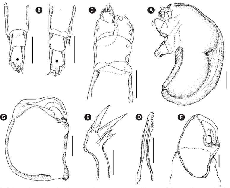
    Female. Cephalothorax (Fig. 1A) subcylindrical, 2.74 mm long, shorter than trunk with lateral curves, its anterior half expanded slightly in girth to from head, covered by subcircular, well delimited dorsal shield (Fig. 1B), with transversely truncated anterior and posteriorly converging lateral margins, slightly subdivided by mid-dorsal groove; at base of cephalothorax subspherical, lateral swellings. Trunk (Fig. 1C) cylindrical in ventral view, 1.33 times (1.55 × 1.21 mm) longer than wide with nearly parallel lateral margin and rounded posterior corners. Posterior part outfitted with a pair of digitiform processes (Fig. 1C). Antennule (Fig. 1D) incompletely 4-segmented; proximal and second segments indistinctly fused; proximal segment unarmed; second segment armed with seta (whip) on medioventral margin; third segment unarmed; distal segment with slightly tapering tip with prominent gibbous and apical armature consisting of five setae and two tubercles. Antenna (Fig. 1E and 1F) biramous, elongate; exopod prominent and longer than endopod, armed with prominent denticulate on rounded tip; endopod 2-segmented, armed apically with 11 spiniform setae. Mandible (Fig. 1G) with dental formular P1, S1, P1, S1, P1, S1, B5. Maxillule (Fig. 1H) biramous, with small endopod and prominent tripartite exopod; endopod composed of short digitiform process surmounted with two setae (small terminal and larger subterminal); exopod tripartite with mediolateral spinulose ornamentation, two large digitiform processes and a short third one, processes ending with conical elongate setiferous processes. Maxillae (Fig. 1I) completely fused together and forming “maxillary trunk”, about one third of cephalothorax length; bulla small with short manubrium. Maxilliped (Fig. 1J) subchelate with robust corpus, covered by thick, wrinkled cuticle and single seta on medial side; long slender subchela with single ventral seta and a row of teeth at base; claw large (constituting almost one-third of subchela), large auxiliary seta at base of claw medially. Thoracic appendages not observed.

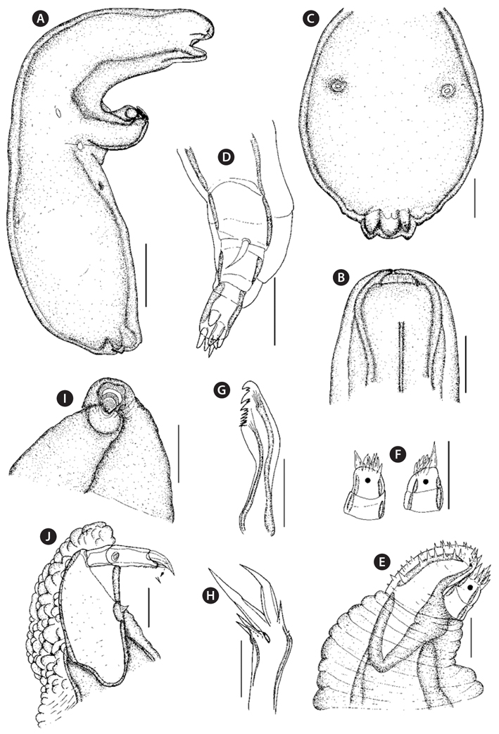
    Male. Body 0.73 mm long (Fig. 2A), representing male structural type A (Kabata, 1979). Cephalothorax about half of total length, oval in dorsal view and dorsal shield not visible. Trunk subcylindrical; lateral ends tapered and bent posteriorly. Pair of reduced caudal rami. Antennule (Fig. 2B) 3-segmented; proximal segment longest, with seta (whip); apical armature consisting of eight spiniform setae. Antenna (Fig. 2C) biramous, elongate; sympod cylindrical, unarmed; bulbous 1-segmented, exopod distinctly shorter than endopod, armed with two tubercles endopod 2-segmented with proximal segment with denticulate pad; distal segment with three spiniform setae and deticulate pad. Mandible (Fig. 2D) with dental formula P1, P1, S3, B5. Maxillule (Fig, 2E) similar to female although more slender; endopod terminating with two equal small setae. Maxilla (Fig. 2F) subchelate (made of strong thick cuticle) with robust corpus and strong subchela; corpus unarmed; subchela with well delimited, powerful claw and slightly shorter cylindrical shaft; closed subchela partly hiding tip of claw behind medial outgrowth of corpus with scattered denticles; claw with small seta and spine medioventrally. Maxilliped (Fig. 2G) subchelate, similar in structure to maxilla but stronger in appearance; subchela very robust with claw positioned at right angle to shaft, scattered spinules distolaterally; claw with broad base and slender tip. Thoracic appendages not observed.

      >  Distribution

    Parabrachiella bera has been reported from Japan (Yamaguti, 1939) and Korea (Choi et al., 1996; this study).

      >  Host

    Parabrachiella bera parasitic on Parajulis poecilepterus (Temminck and Schlegel, 1845).

      >  Site of infection

    Parabrachiella bera attached to the hosts’ gill rakers.

      >  Prevalence

    Prevalence was recorded as 27.2% .

      >  Remarks

    The number of species of Parabrachiella Wilson, 1915 described above should be compared with the size of species groups based on the number of posterior processes, which provides a key for grouping females within this genus (Castro Romero and Baeza Kuroki, 1987). Parabrachiella bera has four pairs of posterior processes in Japan (Yamaguti, 1939), but Korean specimens had a single pair of posterior processes. Parabrachiella bera has been assigned to Group II, which is the largest group, consisting of 35 members, in Parabrachiella (Piasecki et al., 2010). Careful comparison between the current materials and the original description of P. bera provided by Yamaguti (1939) revealed several omissions, in that the fine setulose ornamentation of the maxillule was not shown and the mandible was not described. Parabrachiella bera was characterized based on the following female features: 1) single pair of posterior processes; 2) exopod of maxillule with mediolateral spinulose ornamentation; 3) mandible with dental formula P1, S1, P1, S1, P1, S1, B5; and 4) maxilliped with row of teeth at base. Males were characterized by: 1) pair of reduced caudal rami; 2) endopod of antenna with denticle ornamentation; 3) mandible with dental formula P1, P1, S3, B5; 4) medial outgrowth of corpus of maxilla with scattered denticles; and 5) subchela of maxilliped with scattered spinules distolaterally.

    Choiet al. (1996) reported that the same lernaeopodids were collected from the gills of P. poecilopterus as P. incurva (= N. incurva). Parabrachiella incurva was originally illustrated and described using two females that were recovered from a kyphosid fish, Kyphosus lembus (= Kyphosus vaigiensis) at Nagatura, Izu Peninsula, Sizuoka Prefecture, Japan by Shiino (1956). In comparison, the closely related congener P. bera has a slender trunk and indistinct genital processes (Yamaguti, 1939), while P. incurva has a stout trunk and distinct genital processes. In Korean waters, P. bera was found to be parasitic on P. poecilopterus, however P. incurva has to date not been recorded. Members of Parabrachiella are sufficiently host-specific and the geography of their hosts was reported by Piasecki et al. (2010). All of these features clearly assure the identity of the redescribed specimens in relation to related species from the genus Parabrachiella.

참고문헌
  • 1. Boxshall GA, Halsey SH 2004 An Introduction to Copepod Diversity google
  • 2. Boxshall GA, Walter TC 2012 Parabrachiella C. B. Wilson, 1915. World Copepoda database [Internet] google
  • 3. Castro Romeo R, Baeza Kuroki H 1987 Four new species of Neobrachiella (Copepoda: Lernaeopodidae), parasitic on Sciaena genus (Teleostei: Sciaenidae) in the South Pacific [Estud Oceanol] Vol.6 P.1-24 google
  • 4. Choi SD, Suh HL, Hong SY 1996 Two species of parasitic copepods (Neobrachiella incurva and Peniculus ostraciontis) from the marine fishes, Halichoeres poecilopterus and Sebastes schlegeli, of the south coast of Korea [Korean J Aquac] Vol.9 P.117-123 google
  • 5. Froese R, Pauly D 2012 FishBase [Internet] google
  • 6. Humes AG, Gooding RU 1964 A method for studying the external anatomy of copepods [Crustaceana] Vol.6 P.238-240 google
  • 7. Kabata Z 1979 Parasitic Copepoda of British Fishes google
  • 8. Kabata Z 1986 Redescriptions of and comments on four little-known Lernaeopodidae (Crustacea: Copepoda) [Can J Zool] Vol.64 P.1852-1859 google
  • 9. Piasecki W, Mlynarczyk M, Hayward CJ 2010 Parabrachiella jarai sp. nov. (Crustacea: Copepoda: Siphonostomatoida) parasitic on Sillago sihama (Actinopterygii: Perciformes: Sillaginidae) [Exp Parasitol] Vol.125 P.55-62 google
  • 10. Shiino SM 1956 Copepods parasitic on Japanese fishes. 12. Family Lernaeopodidae [Rep Fac Fish Pref Univ Mie] Vol.2 P.269-311 google
  • 11. Yamaguti S 1939 Parasitic copepods from fishes of Japan. Part. 6. Lernaeopodoida, I P.529-578 google
OAK XML 통계
이미지 / 테이블
  • [ Fig. 1. ]  Parabrachiella bera, female: (A) habitus, lateral, (B) dorsal shield of cephalotorax, (C) trunk, ventral, (D) antennule, (E) antenna, (F) tip of antenna, (G) mandible, (H) maxillule, (I) maxilla, (J) maxilliped. Scale bars: A = 0.4 mm, B, C = 0.2 mm, D-J = 0.02 mm.
    Parabrachiella bera, female: (A) habitus, lateral, (B) dorsal shield of cephalotorax, (C) trunk, ventral, (D) antennule, (E) antenna, (F) tip of antenna, (G) mandible, (H) maxillule, (I) maxilla, (J) maxilliped. Scale bars: A = 0.4 mm, B, C = 0.2 mm, D-J = 0.02 mm.
  • [ Fig. 2. ]  Parabrachiella bera, male: (A) habitus, lateral, (B) antennule, (C) antenna, (D) mandible, (E) maxillule, (F) maxilla, (G) maxilliped. Scale bars: A = 0.02 mm, B-G = 0.01 mm.
    Parabrachiella bera, male: (A) habitus, lateral, (B) antennule, (C) antenna, (D) mandible, (E) maxillule, (F) maxilla, (G) maxilliped. Scale bars: A = 0.02 mm, B-G = 0.01 mm.
(우)06579 서울시 서초구 반포대로 201(반포동)
Tel. 02-537-6389 | Fax. 02-590-0571 | 문의 : oak2014@korea.kr
Copyright(c) National Library of Korea. All rights reserved.