The Dryopidae (Coleoptera) is composed of approximately 300 species worldwide belonging to 33 genera; however, members of the family have not been recorded in Korea. We collected and recorded adult specimens of Elmomorphus brevicornis Sharp (=E. brevicornis brevicornis Sharp) on piles of submerged twigs and branches of a decayed tree (Prunus sp.) from a mountain stream in southwestern Korea with a comparison of the type specimens of E. brevicornis amamiensis Nomura from Japan. Both of the subspecies are raised to the species level herein. Redescription, photographs, line drawings of diagnostic characteristics, distribution map, habitat, and taxonomic notes for Elmomorphus brevicornis are provided.
The long-toed water beetle family Dryopidae is composed of approximately 300 described species worldwide that belong to 33 genera (Jäch and Balke, 2008). Dryopid diversity is the greatest in the tropics, but several species prefer temperate regions. Most adults are mainly found in aquatic or riparian habitats, while the larvae are usually terrestrial or semiaquatic and inhabit moist decaying wood, humid soil, and leaf litter (Ulrich, 1986; Hayashi and Kadowaki, 2008).
The genus
The adults of
In this study, we report the family Dryopidae for the first time as part of the Korean fauna on the basis of the specimens of
Adult specimens of
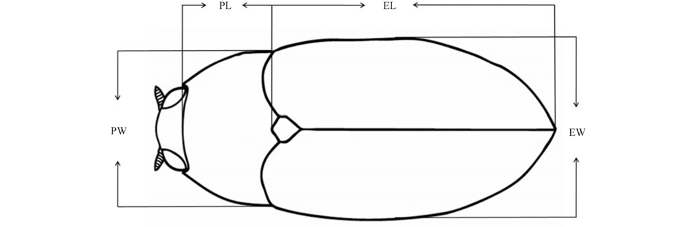
Morphological terminology for Dryopidae follows Kodada and Jäch (2005). The locality and dimensional abbreviations used in this study are as follows: JB, Jeollabuk-do; PL, pronotal length along midline in dorsal view; PW, maximum width of pronotum; EL, elytral length along the suture from the scutellum bases to the elytral apices; EW, maximum width of elytra; and TL, total length of PL and EL (Fig. 1).
Order ColeopteraFamily Dryopidae Billberg, 1820 (Korean name: Yeo-ulbeol-re-bu-chi-gwa)Genus Elmomorphus Sharp, 1888 (Korean name: Yeo-ulbeol-re-bu-chi-sok)Elmomorphus Sharp, 1888: 242; Satô, 1985: 255; Kodada et al., 2003: 455; Satô and Yoshitomi, 2005: 636.Dryopidius Grouvelle, 1896: 33.Elmomorphellus Chûjô and Satô, 1964: 193.Type species: Elmomorphus brevicornis Sharp, 1888: 243.
>
Elmomorphus brevicornis Sharp (
>
(Korean name: Yeo-ul-beol-re-bu-chi)
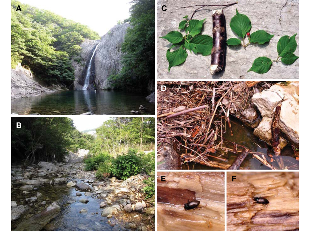
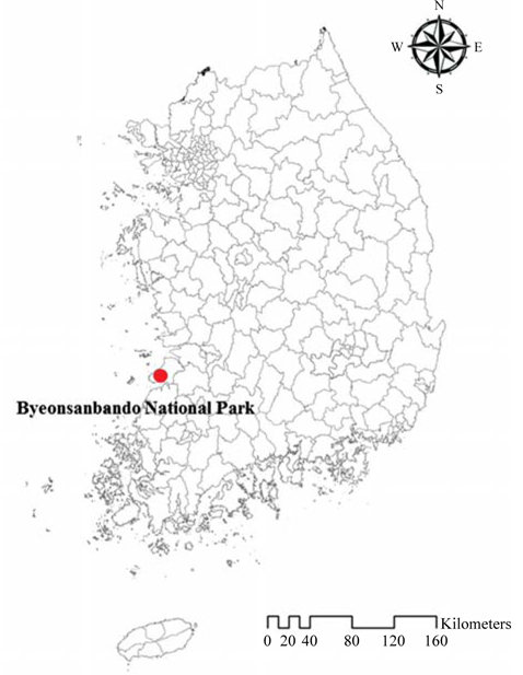
Material examined. ♂, Korea: JB, Buan-gun, Byeonsanmyeon, Junggye-ri (35°37′37.4′′N, 126°34′12.4′′E, 137 m a.s.l.), downstream of Jikso Falls, 4 Jul 2009, Hwang JM (KU); 9♂, 7♀, ditto but 15 Jun 2011, Jung SW, Park HR (KU); ♂, ♀, ditto but deposited in CNUIC.
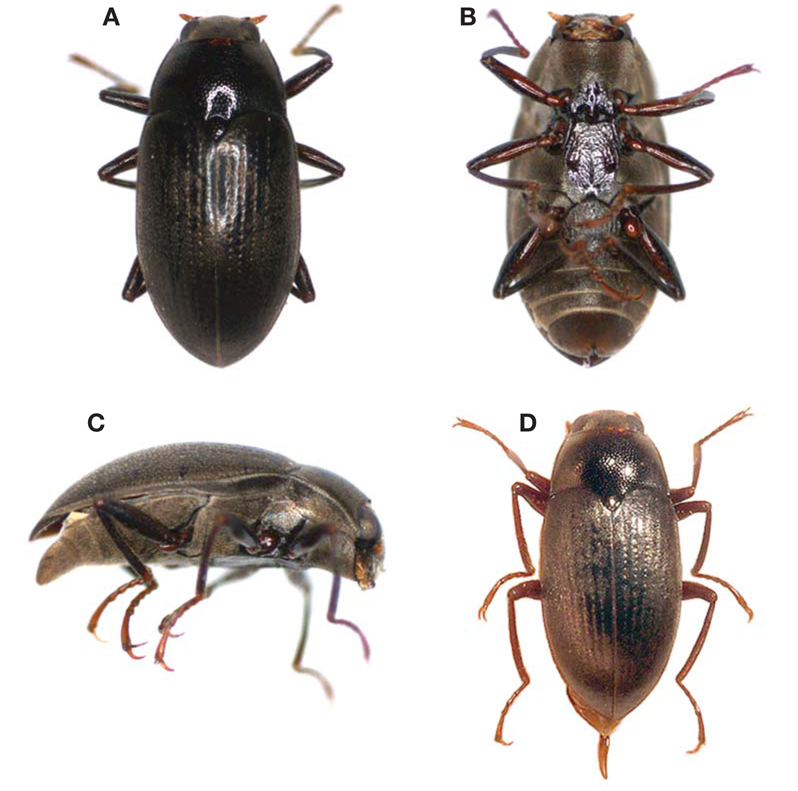
Redescription. Male adult: Body length (TL) 3.35-3.70mm; width (EW) 1.60-1.80 mm. Body elongate, oval, convex dorsally, and black color. Femora, tibiae, prosternal process, middle parts of meso- and metaventrite reddish brown. Antennae reddish brown (live specimen) or brown (in alcohol); mouth parts and tarsi brown.
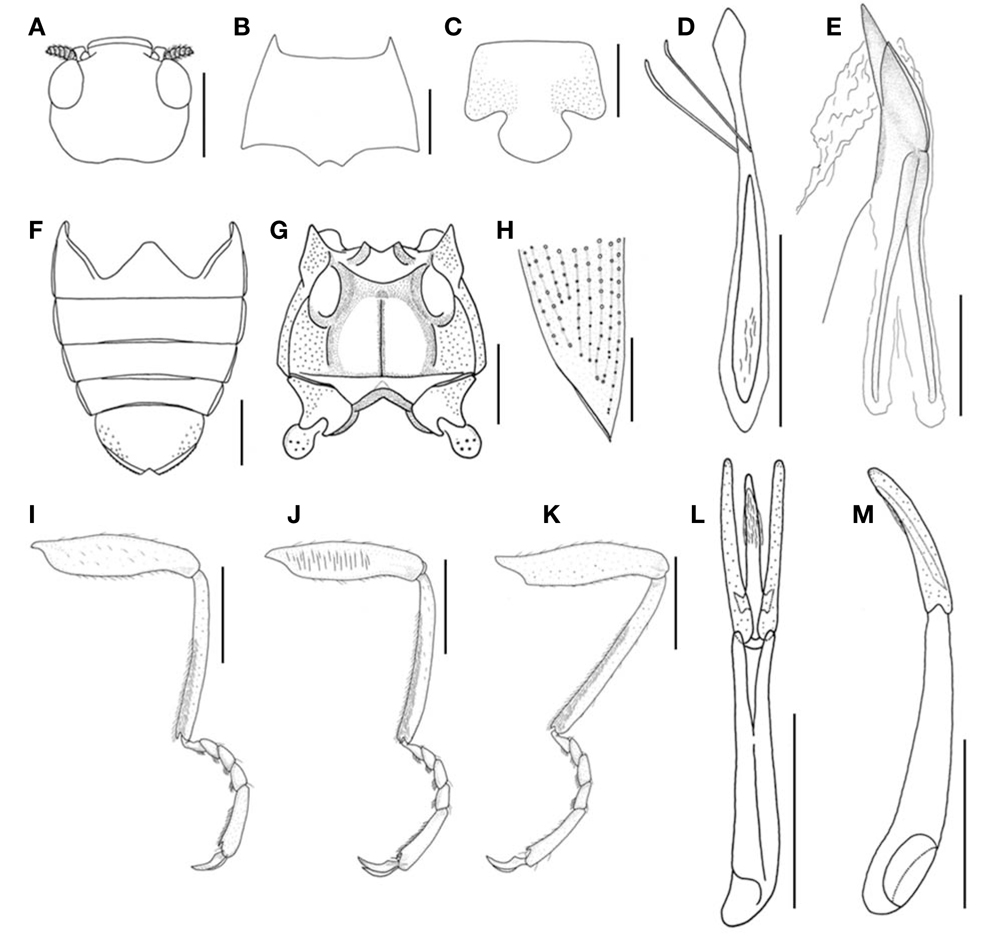
Head (Fig. 3A) retractable, hypognathous, with numerous punctures smaller than a facet, and with hair-like setae on dorsal surface. Fronto-clypeal suture absent. Labrum wider than long, with dense setae anteriorly, round in frontal angles. Antennae short, with dense hydrofuge pubescence, 9-segmented; antennomere 1 slender posteriorly and stout anteriorly; antennomere 2 wider, 1/2× length of antennomere 1; antennomere 3 short and small; antennomeres 4-9 wider than long, gradually reducing in length and width. Mandibles symmetrical, longer than wide; dark brown and setigerous punctures in lateral parts; apex strongly curved, with four teeth; two long teeth anteriorly, two small teeth posteriorly; prostheca with numerous short stout spines anteriorly, dense thin setae in middle parts. Galea flattened, 2-segmented; basal galea short; apical galea expanded with dense hair-like setae anteriorly. Lacinia long, narrower than terminal palpomere, with long setae. Maxillary palp 4-segmented; palpomere 1 short; palpomere 2 longer than 3; palpomere 3 longer than 1; terminal palpomere relatively long with lateral sensory field, elongate, more or less wide, about half length of terminal palpomere. Labial palp 3-segmented; palpomere 1 shortest; palpomeres 2 and 3 short.
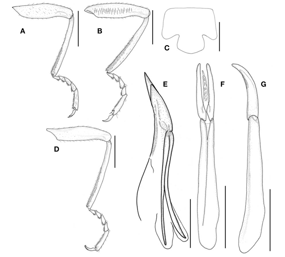
Pronotum (Fig. 3B) strongly convex, shiny at middle part, carinae absent, wider than long, widest posteriorly; punctures similar to head punctures on dorsal surface; anterior angle protruding, acute; posterior angle rectangular and acute. Scutellum shiny, longer and wide, with punctures, acute at apex. Prosternum punctate, with plastron; prosternal process (Fig. 3C) wider than long, without plastron, widest at middle part, shiny, round at apex. Hypomeron broadest posteriorly, narrowed anteriorly with plastron. Mesoventrite with plastron and punctate, deeply grooved for reception of prosternal process. Metaventrite (Fig. 3G) punctate, covered with plastron except narrow middle part, flat and shiny at middle part; median longitudinal suture; posterior part with transverse suture. Legs (Fig. 3I-K) long and slender; procoxae and mesocoxae globular; metacoxae transverse; trochanters of hind legs flat, largest and round; femora clavate, with small punctures; middle femora with row of long strong setae on inner surface; tibiae slender, tomented on inner surface, 1.11× as long as tarsi; tarsi 5-segmented, light brown; tarsomeres 1-4 short; tarsomere 5 longest, as long as tarsomeres 1-4 combined; two claws with one basal tooth, slender, sharp, and shiny.
Elytra (Fig. 3H) elongate oval, convex dorsally, widest near middle part, sparsely short setae laterally and posteriorly; lateral margin crenulate; pointed at apex. Each elytron with nine punctate striae; strial punctures moderately large and deep anteriorly, becoming gradually finer posteriorly; striae 6 and 7 obscure at posterior 1/4; 8th stria combined with 5th at posterior 1/6; 9th stria obscure at posterior 1/5; intervals 1-5 wide; intervals 6-8 narrow. Hind wings fully developed. Abdomen (Fig. 3F) with five ventrites, longer than wide, with plastron; ventrite 5 strongly granulate laterally, widely emarginate at apex; apical margin feebly crenulate. Sternite 9 (Fig. 3D) long, with anterior medial process narrow.
Aedeagus (Fig. 3L, M); penis shorter than parameres, gradually narrowed anteriorly, apex narrow and round, ventrally curved in lateral view; anterolateral apophyses short and acute; membranous ventral sac with fine longitudinal furrows; fibula absent; phallobase moderately long, 2.08× as long as parameres.
Female adult: Body length (TL) 3.70-3.95 mm; width (EW) 1.80-1.90 mm. Metaventrite with plastron except wide middle part. Abdominal ventrite 5 narrowly emarginate at apex; ovipositor (Fig. 3E) strongly sclerotized, elongate; coxite curved, pointed at apex; valvifer about twice as long as coxite.
Measurements. Males (n=5): TL 3.35−3.70 (3.51) mm, PL 0.75−0.80 (0.79) mm, PW 1.35−1.45 (1.40) mm, EL 2.60−2.90 (2.72) mm, EW 1.60−1.80 (1.71) mm. Females (n=5): TL 3.70−3.95 (3.83) mm, PL 0.85−0.90 (0.88) mm, PW 1.50−1.60 (1.56) mm, EL 2.85−3.05 (2.95) mm, EW 1.80−1.90 (1.83) mm.
Larva. Hayashi and Kadowaki (2008) firstly described the larva of
Distribution. Korea (new record) (Fig. 4), Japan.
Habitat. We found all the Korean specimens under piles of submerged twigs and branches of a tree belonging to
Taxonomic remarks. According to Sharp (1888), G. Lewis found two specimens of this species from Kobe, South Japan. Jan Kodada (Comenius University, Slovakia) confirmed deposition of the type specimen (1 dry specimen) in the British Museum of Natural History (Fedor Ciampor, personal communication).
This is the first record of the family Dryopidae in Korea. Two subspecies belonging to the genus
>
Elmomorphus amamiensis Nomura (
Material examined. ♀, Japan: Amami, Kawakami, Kasaricho, 16 Jul 2006, Kamite Y leg (KU); 2♂, 2♀, Okinawa, Genka-ôkawa, Nago-shi, 16 May 2007, Kamite Y leg (KU); ♀, Kumejima, Suhara-gawa, Kumejima-chô, 19 May 2007, Kamite Y leg (KU).
Description. Male adult: Body length (TL) 3.15-3.20 mm, width (EW) 1.55 mm. Body elongate, oval, and convex dorsally. Legs, prosternal process, and middle part of mesoventrite reddish brown. Antennae and mouth parts brown.
Head retractable, with numerous punctures smaller than a facet, and with hair-like setae on dorsal surface. Fronto-clypeal suture absent. Labrum wider than long, with dense setae anteriorly, round in frontal angles. Antennae short, with dense hydrofuge pubescence, 9-segmented; antennomere 1 slender posteriorly and stout anteriorly; antennomere 2 wider; antennomere 3 short and small; antennomeres 4-9 wider than long, gradually reducing in length and width. Mandibles dark brown, longer than wide; apex strongly curved, with four teeth; two long teeth anteriorly, two small teeth posteriorly. Maxillary palp 4-segmented; palpomere 1 short; palpomere 2 longer than 3; palpomere 3 longer than 1; terminal palpomere relatively long with lateral sensory field. Labial palp 3-segmented; palpomere 1 shortest; palpomeres 2, 3 short and stout.
Pronotum convex, shiny at middle part, carinae absent, wider than long, widest posteriorly; punctures similar to head punctures on dorsal surface; anterior angle protruding, acute; posterior angle acute. Scutellum shiny, longer and wide, with punctures. Prosternum punctate, with plastron; prosternal process (Fig. 6C) wider than long, without plastron, widest posteriorly, shiny. Hypomeron broadest posteriorly, narrowed anteriorly with plastron. Mesoventrite with plastron and punctate, deeply grooved for reception of prosternal process. Metaventrite punctate, covered with plastron except middle part, flat and shiny at middle part; median longitudinal suture; posterior part with transverse suture. Legs (Fig. 6A, B, D) long and slender; procoxae and mesocoxae globular; metacoxae transverse; trochanters of hind legs flat, largest and round; femora clavate, with small punctures; middle femora with row of long strong setae on inner surface; tibiae slender, tomented on inner surface, 1.32× as long as tarsi; tarsi 5-segmented, slender and small, light brown; tarsomeres 1-4 short; tarsomere 5 longest, as long as tarsomeres 1-4 combined; two claws with one basal tooth, slender, sharp, and shiny.
Elytra elongate oval, convex dorsally, widest near middle part, sparsely short setae laterally and posteriorly; lateral margin slightly crenulate; pointed at apex. Each elytron with nine punctate striae; strial punctures moderately large anteriorly, becoming gradually finer posteriorly; intervals 1-5 wide; intervals 6-8 narrow. Hind wings fully developed. Abdomen with five ventrites, longer than wide, with plastron; ventrite 5 widely emarginate at apex.
Aedeagus (Fig. 6F, G); penis slightly curved, shorter than parameres, gradually narrowed anteriorly; membranous ventral sac with fine longitudinal furrows; fibula absent; parameres strongly curved apically in lateral view; phallobase moderately long, 2.62× as long as parameres
Female adult: Body length (TL) 3.40-3.50 mm; width (EW) 1.60-1.65 mm. Metaventrite with plastron. Abdominal ventrite 5 narrowly emarginate at apex; ovipositor as in Fig. 6E.
Measurements. Males (n=2): TL 3.15-3.20 (3.17) mm; PL 0.80 mm; PW 1.30-1.35 (1.32) mm; EL 2.35-2.40 (2.37) mm; EW 1.55 mm. Females (n=4): TL 3.40-3.50 (3.46) mm; PL 0.80 mm; PW 1.35-1.40 (1.36) mm; EL 2.60-2.70 (2.65) mm; EW 1.60-1.65 (1.61) mm.
Larva. Unknown.
Distribution. Japan (Amami-ôshima, Kumejima, Okinawa, Tokuno-shima).
Habitat. All specimens were collected from the Ryukyu Islands, volcanic islands with subtropical climate.
Taxonomic remarks. This species is endemic to Japan. Male adult of
>
Key to the Korean and Japanese species of Elmomorphus
1. Mesofemur long; tarsi and claws small; parameres strongly curved apically in lateral view ………………………………………………………………………… E. amamiensis (Japan) - Mesofemur shorter; tarsi and claws large; parameres not strongly curved apically in lateral view ………………………………………………………… E. brevicornis (Korea, Japan)