The gastric ulcer is a common disorder of the stomach and duodenum. The basic physiopathology of a gastric ulcer results from an imbalance between some endogenous aggressive and cytoprotective factors. This study examined whether Ganoderma lucidum pharmacopuncture (GLP) would provide protection against acute gastric ulcers in rats.
Sprague-Dawley rats were divided randomly into 4 groups of 8 rats each: normal, control, normal saline (NP) and GLP groups. The experimental acute gastric ulcer was induced by using an EtOH/HCl solution and the normal group received the same amount of normal saline instead of ethanol. The NP and the GLP groups were treated once with injections of saline and GLP, respectively. Two local acupoints were used: CV12 (中脘) which is the alarm point of the Stomach Meridian, and ST36 (足三里), which is the sea point of the Stomach Meridian. The stomachs from the rats in each group were collected and analyzed for gross appearance and histology. Also, immunohistochemistry staining for BAX, Bcl-2 and TGF-β1 was performed.
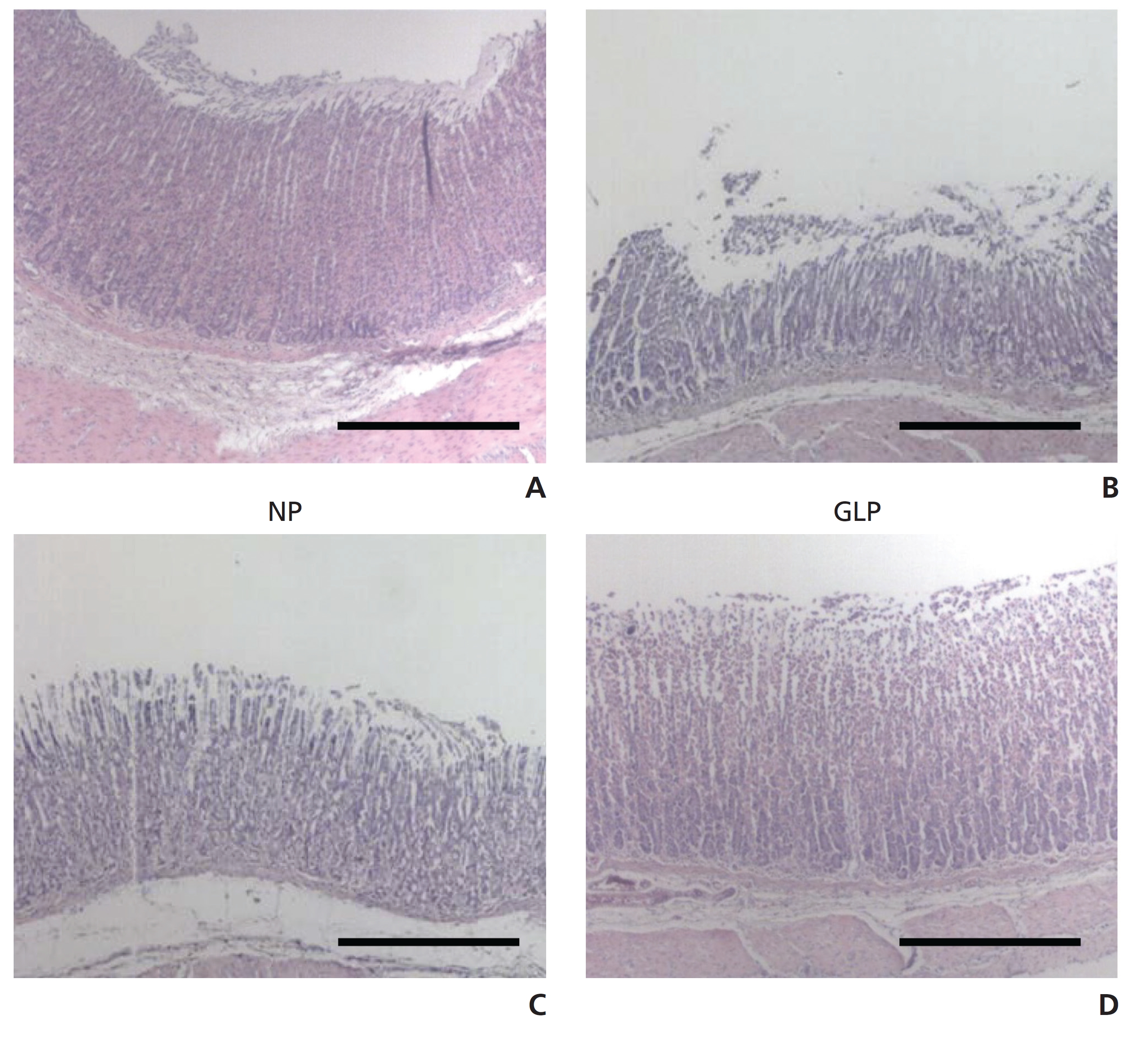
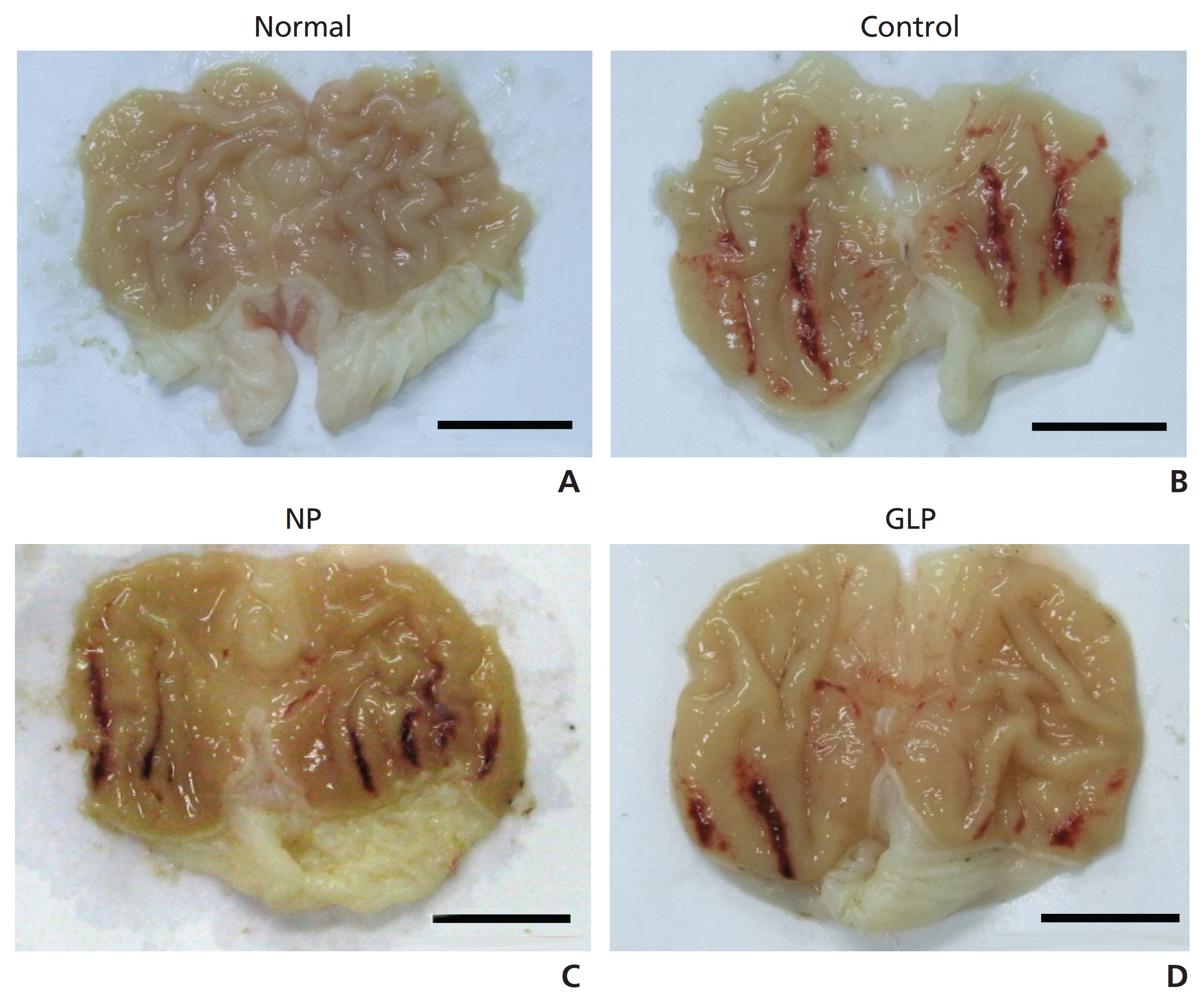
Histological observations of the gastric lesions in the control group showed comparatively extensive damage of the gastric mucosa and necrotic lesions had penetrated deeply into the mucosa. The lesions were long, hemorrhagic, and confined to the glandular portions. The lesions were measured microscopically by using the clear depth of penetration into the gastric mucosal surface. The length and the width of the ulcer were measured and the inhibition percentage was calculated. Wound healing of the acute gastric ulcer was promoted by using GLP, and significant alterations of indices in gastric mucosa were observed. Such protection was shown by gross appearance, histology and immunohistochemistry staining for BAX, Bcl-2 and TGF-β1.
These results suggest that GLP administered at CV12 and ST36 can provide significant protection to the gastric mucosa against an ethanol-induced acute gastric ulcer.
Gastric disease is common, affecting millions of people yearly. Among these diseases, the gastric ulcer is defined as a disturbance of the integrity of the gastric mucosa that causes a local defect or excavation due to active inflammation [1]. Nowadays, aggressive and defensive factors are widely known to play a role in the development of a gastric ulcer. The aggressive factors are gastric acid, pepsin, bile reflux, non-steroidal anti-inflammatory drugs (NSAIDs), Helicobacter pylori bacteria and alcohol, while the defensive factors are mucosal blood flow, surface epithelial cells, prostaglandin, phospholipids or surfactants, mucus, bicarbonate secretion, gastric motility, mucosa impermeability against H+ ions, heat shock protein, and others [2-4].
Pharmacopuncture is an effective therapy in Oriental medicine. A solution extracted from medicinal herbs is injected into acupuncture points according to the condition of the patient [8]. The present study was undertaken to determine whether
Adult male Sprague-Dawley (SD) rats (weighing 220 - 240 g, 15-weeks old and housed five rats per cage) were purchased from SAM-Taco Co. They were provided with standard food and water ad libitum and were maintained in an animal house at a controlled temperature (22 ± 2℃) with a 12-hours light/dark cycle. The rats were divided randomly into 4 groups of 8 rats each: normal, control, normal saline (NP) and GLP groups. The study was approved by the Ethics Committe for Animal Experimentation, Dong- Eui University.
The rats were fasted for 24 hours, but were allowed free access to drinking water until 2 hours before the experiment. A gastric injury model based upon a modification of the method described by Mizui and Doteuchi [9] was induced by using an acidified ethanol solution (150 mM HCl/absolute ethanol) 40 : 60 v/v, (HCl/ethanol solution).
The normal group was orally administered saline. The control, NP and GLP groups were orally administered EtOH/HCl (5 mL/kg). Immediately after EtOH/HCl administration, the NP and the GLP group were treated once with injections of saline and GLP, respectively. Two local acupoints were used: CV12 (中脘), which belongs to the Controlling Vessel, and ST36 (足三里), which belongs to the Stomach Meridian. A pharmacopuncture needle (29 gauge × 8 mm, 1 mL, disposal, insulin-injection syringe from HWAJIN Co. Busan, Korea) was used, and the amount of injection was 1 mL for each animal. One hour after this injection, the rats were sacrificed, and their stomachs were immediately excised.
The reactivity, the activity and the death of rats in each group were observed during the experiment. The animals were killed by cervical dislocation after administration of an overdose of ether at the end of the 2 week experiment. The abdomen was opened immediately. The whole stomach was cut and removed 1.5 cm away from the cardia and the pylorus. Dissection was done along the greater curvature for general specimen observation. The obviously- damaged gastric-mucosa specimen was rinsed in cold saline solution. Then, the specimen was placed in formaldehyde and glutaraldehyde, and stored in a liquid nitrogen solution for later observation.
The length and the width of the injured gastric mucosa region were measured with a vernier caliper. The gastric mucosa ulcer index (UI) was determined according to the Guth standard [10]: spot erosion was recorded as 1 point, erosion lengths < 1 mm were recorded as 2 points, erosion lengths from 1 to 2 mm were recorded as 3 points, those from 2 to 3 mm were recorded as 4 points, and those > 3 mm were recorded as 5 points; the score was doubled if the erosion width was > 1 mm.
Ulcers of the gastric mucosa appear as elongated bands of hemorrhagic lesions parallel to the long axis of the stomach. The gastric mucosa of each rat was examined in order to estimate damage. The length and the width of the ulcer (mm) were measured. The inhibition percentage (%) was calculated by using the following formula [11]:

Where UA = ulcer area
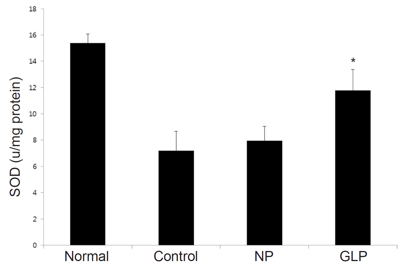
To investigate whether GLP affects the activity of radical scavenging enzymes, was measured the activity of superoxidase dismutase (SOD) in the gastric mucosa to the method of McCord and Fridovich [12]. The standard assay was performed in 3 mL of 50 mM potassium phosphate buffer at pH 7.8 containing 0.1-mM EDTA in a cuvette thermostated at 25℃. The reaction mixture contained 0.1-mM ferricytochrome c, 0.1-mM xanthine, and sufficient xanthine oxidase to produce a reduction rate of ferricytochrome c at 550 nm of 0.025 absorbance units per minutes. Tissue homogenate was mixed with the reaction mixture. A kinetic spectrophotometric analysis was started with the addition of xanthine oxidase at 550 nm. Under these conditions, the amount of SOD required to inhibit the reduction rate of cytochrome c by 50% was defined as 1 unit of activity. The results were expressed as units/mg of protein.
The opened stomachs were preserved in 10% buffered formalin overnight after they had been cut into small pieces. The tissues underwent automated tissue processing the next day. Next, the biopsy samples were embedded in paraffin wax, sectioned into 5-μm thicknesses by using a microtome, and then stained with hematoxylin and eosin (H&E). The tissue sections were assessed for histopathological changes such as congestion, edema, hemorrhage and necrosis by using a light microscope.
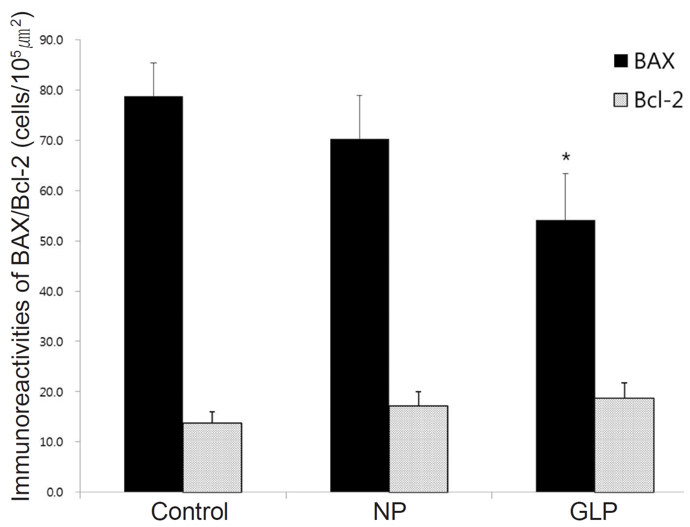
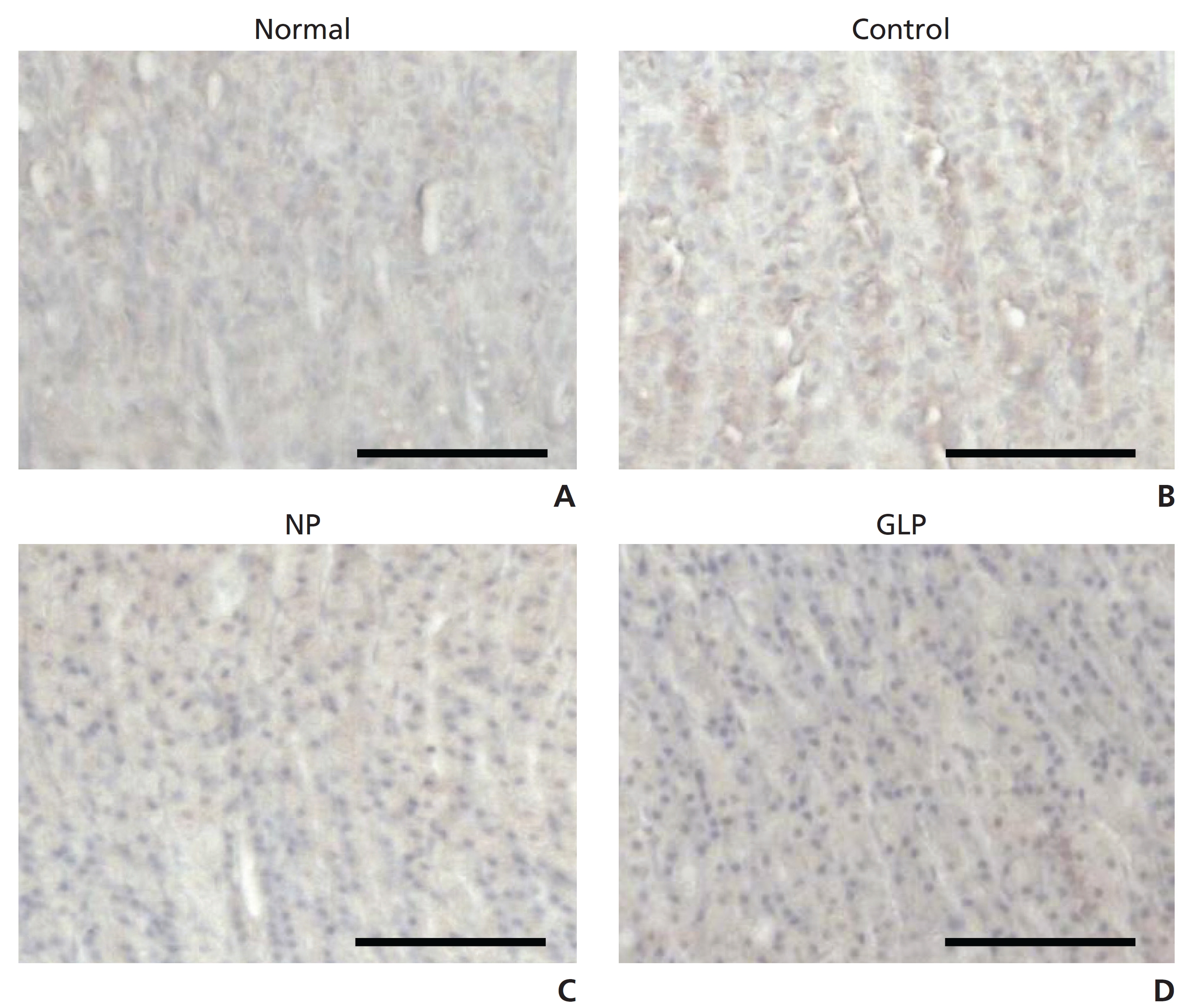
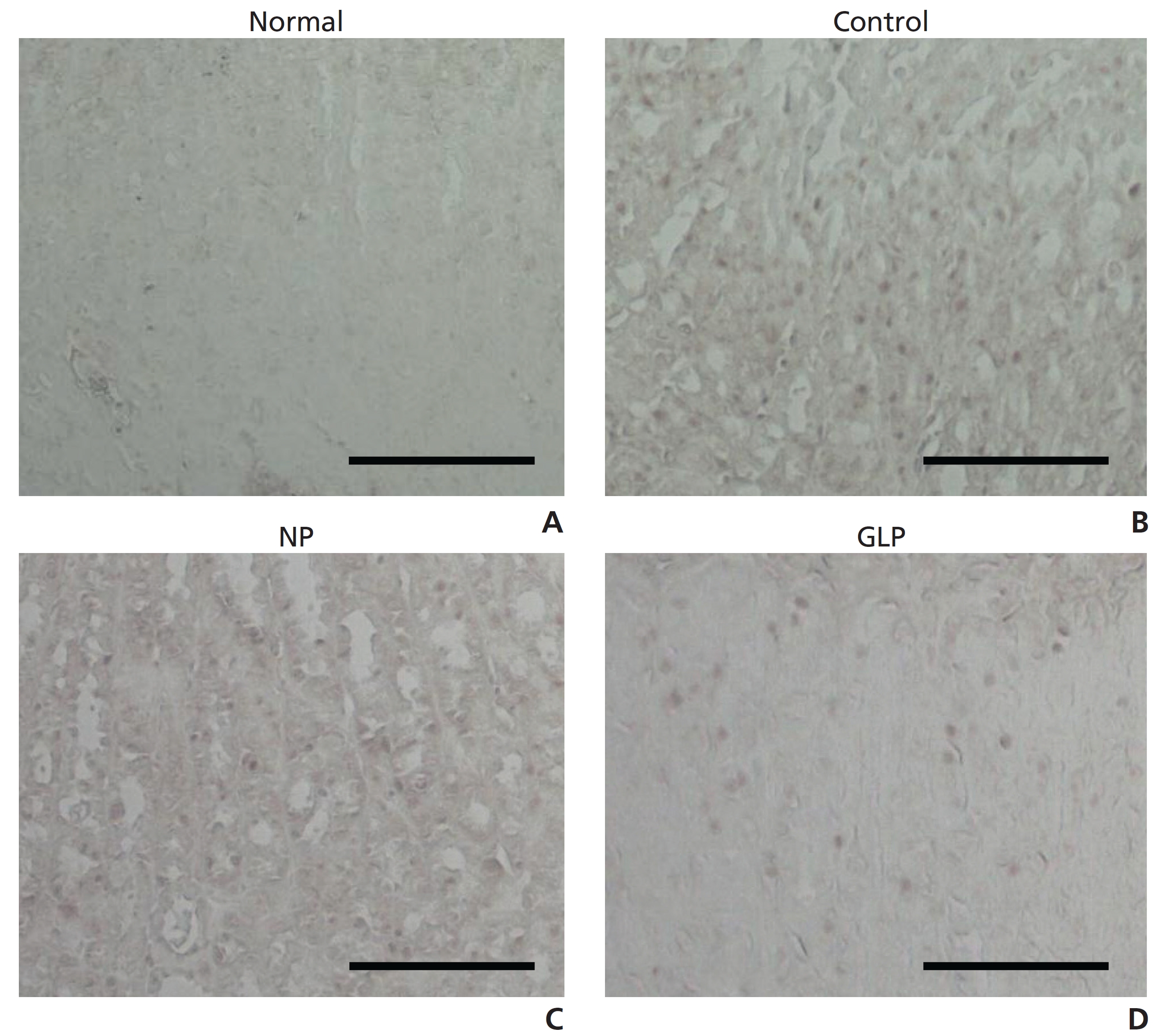
Tissue section slides were heated at 60℃ for approximately 25 minutes in a hot oven. The tissue sections were deparaffinized in xylene and rehydrated with graded alcohol. An antigen retrieval process was performed in a 10 mM sodium citrate buffer. Immunohistochemical staining was conducted according to the manufacturer’s protocol (Dakocytomation, USA). Briefly, endogenous peroxidase was blocked by using a peroxidase block (0.03% hydrogen peroxide containing sodium azide) for 5 minutes. Tissue sections were washed gently with a wash buffer and then incubated with Bcl-2-associated X protein (BAX) (1 : 200), Bcl-2 (1 : 200), and transforming growth factor-β1 (TGF-β1) (1 : 100) biotinylated primary antibodies for 15 minutes. The sections were rinsed gently with the wash buffer and placed in a buffer bath. The slides were then placed in a humidified chamber and incubated for 15 minutes. Then, the tissue sections were rinsed gently in the wash buffer and placed in buffer bath. Following washing and counterstaining with hematoxylin for 5 seconds, we added diaminobenzidine substrate – chromagen to the tissue sections and incubated them further for 5 minutes.
All photomicrographs were obtained using Infinity Capture imaging software (ver. 3, Lumenera, Canada) at 100 × magnification. For statistical analysis, the densities of immuno - positive cells were compared. The total immuno-positive cells per field (105 μm2) were counted in at least 15 randomly-chosen fields at 100 × magnification.
All values are reported as means ± standard errors (S.Es). The statistical significance of differences among groups were assessed with the one-way ANOVA (post-hoc analysis). A value of
The areas of gastric ulcer formation were reduced in the GLP group compared with control group (Fig 1). The GLP significantly (
Histological observation of gastric lesions in control group showed comparatively extensive damage of the gastric mucosa, and necrotic lesions penetrated deeply into the mucosa. Also, extensive edema and leukocytes infiltrations of the submucosal layer appeared microscopically (Fig 3B). The effect of NP was not significant (Fig 3C). However, GLP group had good protection for the gastric mucosa from reduction or absence of ulcer area, submucosal edema, and leukocytes infiltration (Fig 3D).
Ethanol was able to reduce SOD activity as shown in the control group compared with normal group. The treatment of GLP increased significantly (
BAX protein in the animals treated with EtOH/HCl was up-regulated. In contrast, the expression of BAX protein in the GLP group was down-regulated significantly (
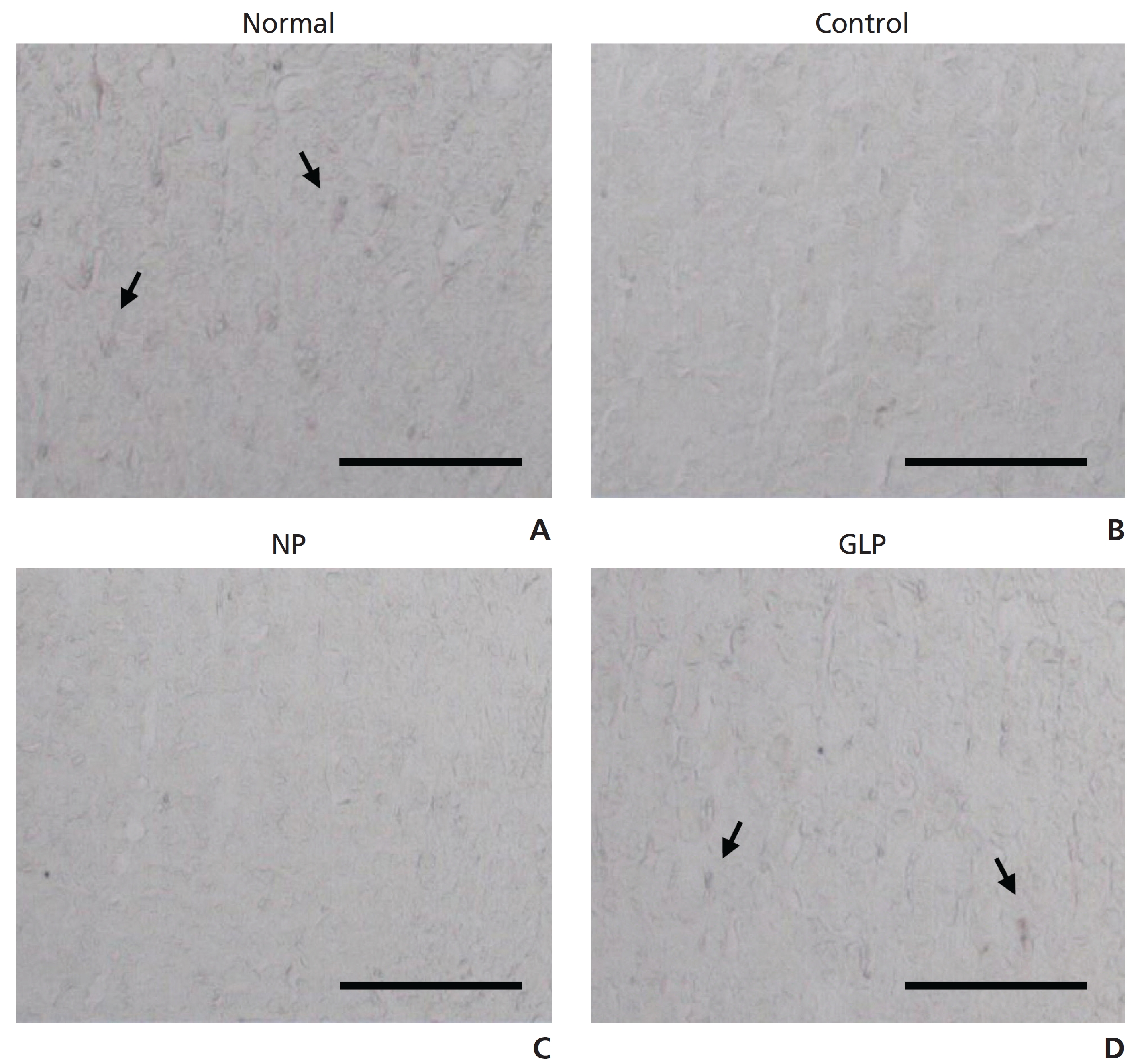
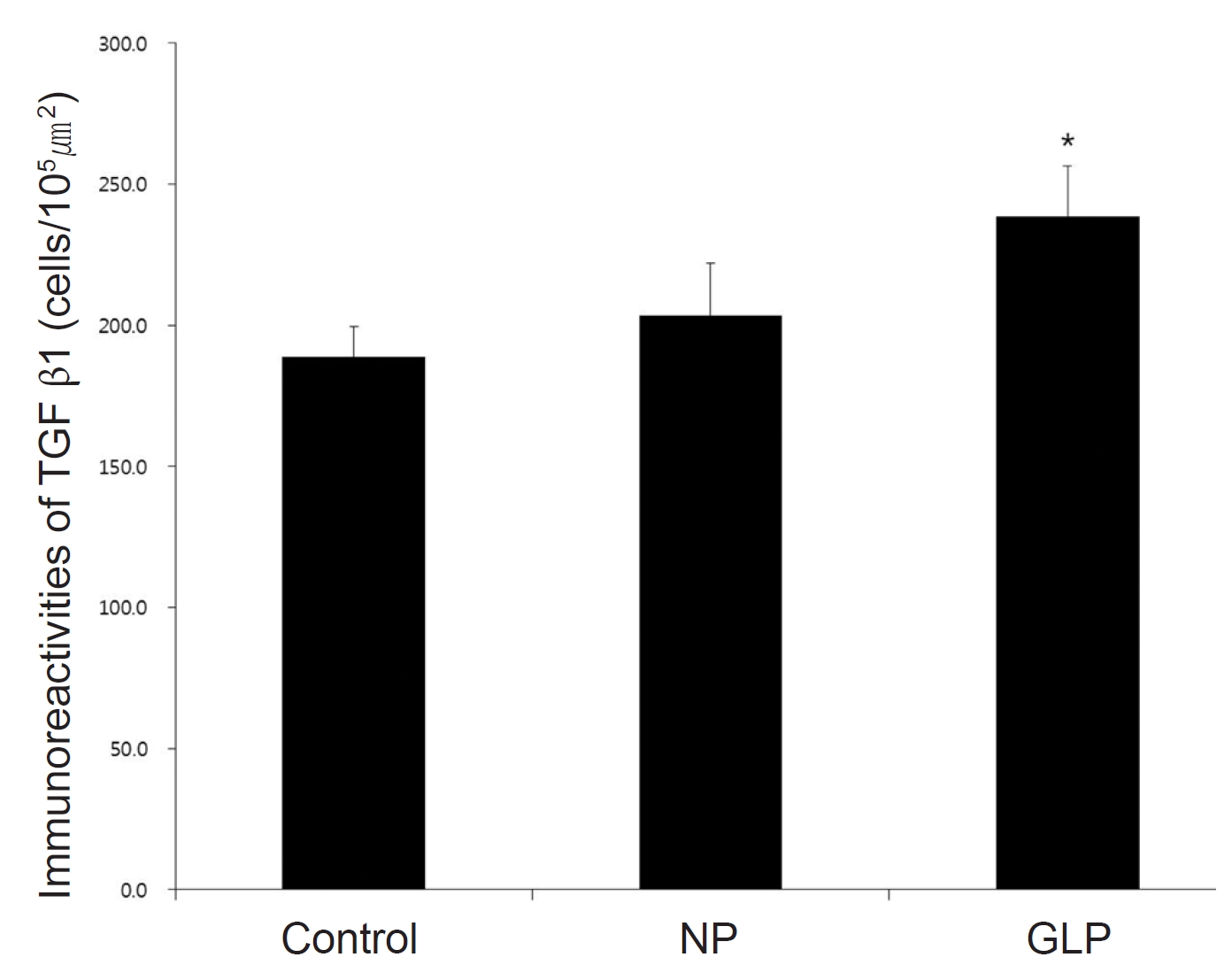
TGF-β1 immunoreactivities was weakly observed in normal group, control group showed slight increase compared with normal group. However, TGF-β1 activities of NP and GLP group were dynamically increased. GLP group was more significant (
Gastritis is an inflammation, irritation, or erosion that occurs when the endogenous defensive mechanisms of the mucosal barrier cannot properly protect the organ [13]. Usually, exposure to excess acid and pepsin causes insult to the gastrointestinal wall. Several factors, including Helicobacter pylori infection, increase the incidence of gastric ulcer disease. Some medications such as NSAIDs, potassium, and iron supplements, have been reported to have the potential for increasing the risk gastric ulcer disease [14]. Generally, an imbalance among aggressive chemical agents versus protective substances, such as SOD, catalase, and glutathione peroxidase, causes gastric mucosal lesions [15]. In experimental animals, the oral administration of ethanol rapidly induces gastric mucosal lesions, and these ethanol-induced lesions are commonly used to study both the pathogenesis of and the therapy for human ulcerative disease [16]. In this study also, the oral administration of ethanol was used for gastric-ulcer induction. Consequently, acute ethanol administration caused damage to the gastric mucosa in the control group, including the general view and microscopic structures, compared with the normal group.
Pharmacopuncture is a new form of therapy derived from combinations of two traditional therapeutic methods, herbal medicine and acupuncture therapy [8]. In general, pharmacopuncture treatment is performed by injecting small amounts of extracted medicinal materials at acupoints or in affected areas in order to obtain the combined efficacies of acupuncture and herbs. Although the first primitive trials with bee venom were recorded in old medical books from the Han Dynasty of China, acupuncture with injection started in the early 1950s in China and was referred to a aqua-puncture. Recently, pharmacopuncture therapy in Korea has developed into quite a unique and systematic framework for the diagnosis and the treatment of various diseases [17].
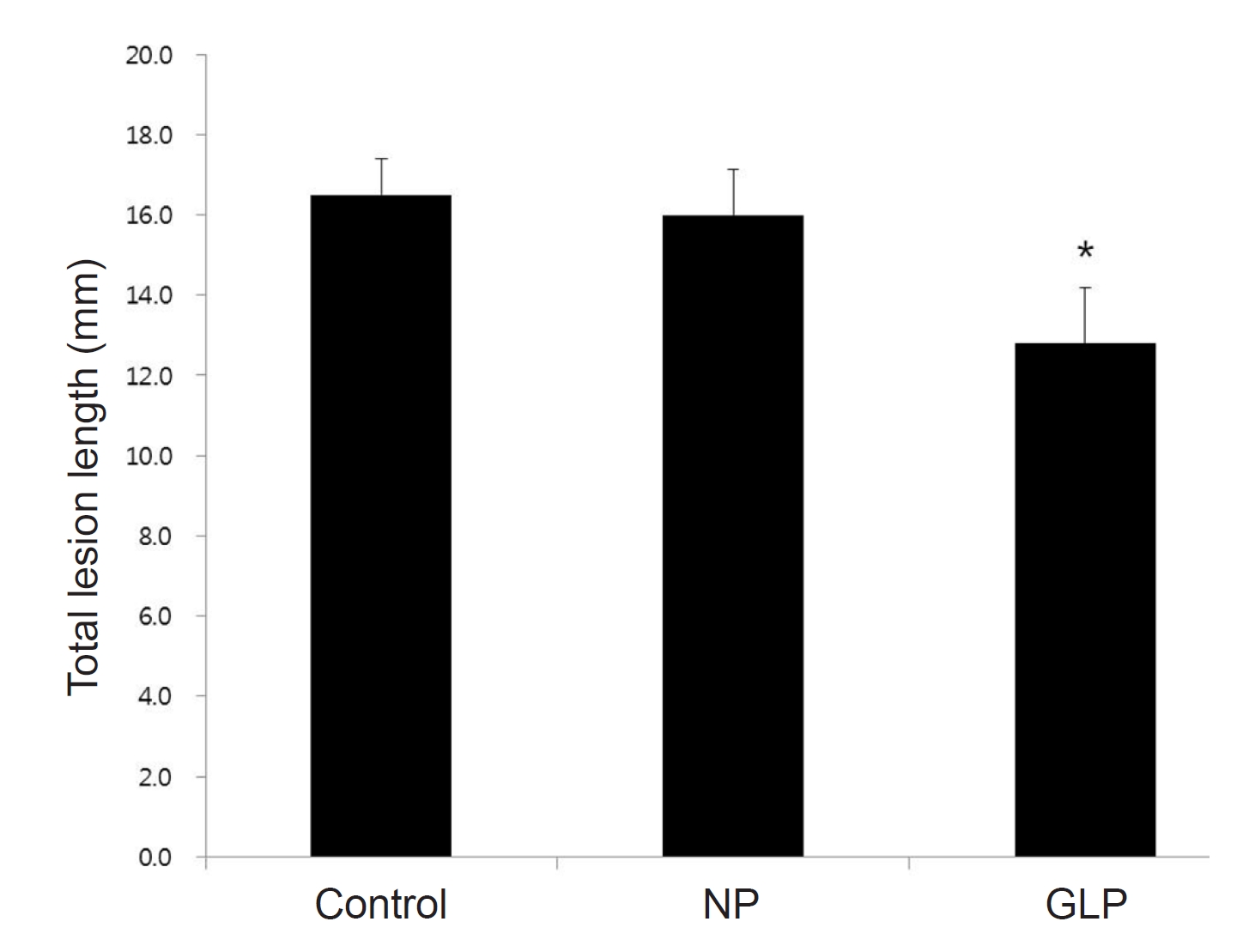
In this study, gastric mucosal lesions in rats were induced by using EtOH/HCl for an acute injury model. Animals were treated with GLP or NP and were kept under observation for 1 hour, during which time they remained alive. Gastric lesions were evaluated macroscopically by using the depth of penetration into the gastric mucosal surface. The rats treated with the GLP had significantly reduced areas of gastric ulcer formation when compared with the ulcer control group (Fig 1).
To confirm the effect of GLP against ethanol-induced acute gastric ulcers, gastric ulcer lesions were measured microscopically by using the clear depth of penetration into the gastric mucosal surface in all experimental groups except the normal group (Fig 3) The length and the width of the ulcer were measured, and the inhibition percentage was calculated. We hypothesized that GLP could inhibit ethanol-induced gastric mucosal lesions, and that such protective effects might directly involve its antioxidant property. We investigated the effect of GLP on the activities of the radical scavenging enzymes, SOD, in the gastric mucosa. As shown in (Fig 4), the control group had significantly reduced SOD activities in comparison to the normal group, but the activities of these enzymes were significantly increased in the GLP group compared to those in the control group.
In the present study, to investigate the role of GLP in mitochondrial regulation in ethanol-induced acute gastric ulcer, we carried out immunohistochemical analyses for BAX and Bcl-2. BAX immunoreactivities in the GLP group were down-regulated significantly compared with those in the control group, and Bcl-2 immunoreactivities in the GLP group were up-regulated compared with those in the control group. The NP group also showed a reduced expression of BAX and an enhanced expression of Bcl-2, but the effects of NP were not significant (Fig 6,7). Therefore, GLP can be assumed to suppress apoptosis by regulating the mitochondrial-damage-mediated endogenous pathway, which might be one of the important mechanisms for preventing gastric ulcer disease.
Cells of the digestive tract are well known to have a rapid turnover rate, which makes the gastrointestinal mucosa one of the most rapidly-proliferating tissues in the body [19]. The gastrointestinal mucosa has a remarkable ability to repair damage. Both the intensive proliferation rate and the remarkable ability to repair damage are supported by the coordinated actions of a variety of growth factors, one of which is transforming growth factor-β1 (TGF-β1) [20]. In this study, to investigate the effects of GLP on the ability to repair gastric ulcer damage, we carried out an immunohistochemical analysis of TGF-β1 in rats with ethanol- induced acute gastric ulcers (Fig 8,9). The level of TGF-β1 was significantly ameliorated in the control group, but it was significantly increased in the GLP group (Fig 8,9). This means that GLP stimulates TGF-β1 up-regulation, which induces angiogenesis through the induction of vascular endothelial growth factor to promote recovery in the injured mucosa of the gastric ulcer.
This study revealed that GLP can significantly protect the gastric mucosa against an ethanol-induced gastric ulcer. Such protection was shown by gross appearance, histology, and immunohistochemistry staining for BAX, Bcl-2 and TGF-β1.