


This study was performed to check for reversibility in the changes induced by a 13-week, repeated, dose toxicity test of Sweet Bee Venom (SBV) in Sprague-Dawley (SD) rats.
Fifteen male and 15 female SD rats were treated with 0.28 mg/kg of SBV (high-dosage group) and the same numbers of male and female SD rats were treated with 0.2 mL/kg of normal saline (control group) for 13 weeks. We selected five male and five female SD rats from the high-dosage group and the same numbers of male and female SD rats from the control group, and we observed these rats for four weeks. We conducted body-weight measurements, ophthalmic examinations, urinalyses and hematology, biochemistry, histology tests.
(1) Hyperemia and movement disorder were observed in the 13-week, repeated, dose toxicity test, but these symptoms were not observed during the recovery period. (2) The rats in the high-dose group showed no significant changes in weight compared to the control group. (3) No significant differences in the ophthalmic parameters, urine analyses, complete blood cell counts (CBCs), and biochemistry were observed among the recovery groups. (4) No changes in organ weights were observed during the recovery period. (5) Histological examination of the thigh muscle indicated cell infiltration, inflammation, degeneration, necrosis of muscle fiber, and fibrosis during the treatment period, but these changes were not observed during the recovery period. The fatty liver change that was observed during the toxicity test was not observed during the recovery period. No other organ abnormalities were observed.
The changes that occurred during the 13-week, repeated, dose toxicity test are reversible, and SBV can be safely used as a treatment modality.
Bee venom has strong analgesicand anti-infla-matory actions [1, 2], so bee venom pharmacopuncture (BVP) shows high efficacy in the treatment of degenerative arthritis [3-5], rheumatoid arthritis [6, 7], disc herniation [8-10] and whiplash injury [11, 12]. However, because one of the biggest obstacles in using BVP clinically is allergic reactions [13], Sweet Bee Venom (SBV) was developed to solve this problem. SBV is another name for pure melittin from the bee venom [14, 15].
A ‘single dose toxicity test in Sprague-Dawley (SD) Rats’ [16], a ‘4-week repeated intramuscular dose toxicity test in SD Rats’ [13], a ‘single dose toxicity test of SBV in beagle dogs’ [17], and a ‘4-week repeated intramuscular dose toxicity test in beagle dogs’ [18] were conducted. In addition, Kwon
Thus, we earlier performed a 13-week, repeated, dose toxicity test of SBV in SD rats using a high-dosage of 0.28 mg/kg, a medium dosage of 0.14 mg/kg and a low dosage of 0.07 mg/kg, and to identify any abnormalities from SBV treatment, we conducted clinical observations, body weight measurements, ophthalmic examinations, urinalyses, hematology and biochemistry tests, and histological observations using hematoxylin and eosin (H&E) staining.
The main results from our earlier research are as follows: The histological examination of the thigh muscle that had been treated with SBV showed that cell infiltration, inflammation, degeneration, and necrosis of muscle fiber, as well as fibrosis, had occurred in both the medium and the high-dosage groups. Fatty liver change was observed in the periportal area of rats receiving medium and high dosages of SBV. Thus, we wanted to know if these changes were reversible, so after 13-week, repeated, dose toxicity test, we performed a 4-week recovery test to ascertain the reversibility of any adverse changes that had occurred during the SBV treatment.
In the 13-week, repeated, dose toxicity test, 50 healthy male and 50 female rats had been selected by average weight, and assigned to 4 groups: the control (normal saline, 0.2 mL/kg), the low (0.07 mg/kg SBV, 0.2 mL/kg)-, the medium (0.14 mg/kg SBV, 0.2 mL/kg)-, and the high (0.28 mg/kg SBV, 0.2 mL/kg)-dosage groups. The control and the high-dosage groups consisted of 15 rats of each gender. SBV or normal saline was administered to the rats by intramuscular injection in both thigh muscles at a dose of 0.2 mL/kg per once daily for 13 weeks. After the treatment period, we selected five male and five female rats from the control and equal numbers from the high-dosage group, and observed those rats for four weeks.
All animals were observed daily for clinical signs throughout the recovery period; abnormal signs were recorded individually by type, observation day and time, and duration. The body weight of each rat was measured once a week during the four-week recovery period. The amount of food and water was measured before it was supplied to each cage and the leftover food and water was measured on the next day. The difference was calculated and regarded as the daily food and water consumption. On the scheduled day of study termination, all surviving animals were anesthetized by using ether inhalation. Blood samples were collected and postmortem examinations were performed on all animals. Absolute organ weights were calculated for the following organs: brain, pituitary gland, heart, lungs, liver, spleen, kidneys, adrenal glands, testes, prostate, ovaries, and uterus.
External eye examinations of the animals were carried out at the end of the recovery period by administering Ocu-Tropic ophthalmic drops (Lot No. 019196, Samil pharma. Co., Ltd, Seoul, Korea), and using an opthalmoscope (ALL PUPIL II, Keeler, Windsor, UK) to examine the conjunctiva, cornea, lens, iris and fundus of each eye.
The animals were fasted overnight prior to necropsy and blood collection. Blood samples were drawn from the abdominal aorta by using a syringe needle under ether anesthesia. Blood samples were collected into complete blood cell count (CBC) bottles containing ethylenediaminetetraacetic acid EDTA and were analyzed to determine the red blood cell count (RBC), hemoglobin concentration (Hb), hematocrit (Ht), mean corpuscular cell volume (MCV), mean corpuscular cell hemoglobin concentration (MCHC), platelet count, white blood cell count (WBC), differential WBC count, reticulocyte count, prothrombin time (PT), and active partial thromboplastin time (APTT).
For serum biochemistry analyses, blood samples were centrifuged at 3,000 rpm for 10 minutes and analyzed using an auto-analyzer (7080, HITACHI, Tokyo, Japan). Serum biochemistry parameters, including sodium, potassium, calcium, chloride, glucose, total cholesterol, blood urea nitrogen (BUN), creatinine, total protein (TG), albumin, total bilirubin, alanine aminotransferase (ALT), aspartate aminotransferase (AST), alkaline phosphatase (ALP), and albumin/globulin ratio, were examined.
At the end of recovery period, urinalyses were conducted on fresh urine to assess specific gravity, pH, protein, glucose, ketone body, bilirubin, and occult blood; a Combur10Test ®Mstick system (MIDITRON®JuniorII, Roche, Manheim, Germany) was used.
The tissues from the injections sites and from the following organs were obtained and weighed (absolute and relative organ weights) from all animals: brain, pituitary, thyroid, parathyroid, and adrenal glands, thymus, trachea, heart, lungs, liver, spleen, kidneys, testes, prostate, ovaries, uterus, salivary glands, stomach, duodenum, jejunum, ileum, cecum, pancreas, epididymis, seminal vesicles, urinary bladder, sub-mandibular lymph node, eyes, harderian glands, skin, bone marrow (femur and sternum), tongue, and spinal cord. Eyes and testes were preserved in Davidson’s fixative and Bouin’s fixative, respectively. These tissues were then fixed with 10% neutral buffered formalin solution. The tissues were routinely processed, embedded in paraffin, and cut into 3 - 5 μm sections. The sections were stained with (H&E) for microscopic examination. All organs and tissues taken from all animals in the control and the high-dosage groups were examined microscopically.
Data on weights of animals and organs, amounts of food and water consumed during feedings, amounts of urine, CBCs, serum biochemistry results, were tested by using SAS (version 9.1.3, SAS Institute Inc, Cary, NC, USA). The variance in the numerical data was checked using the Folded-F test. If the variance was homogeneous, the data were subjected to the student’s
In some experimental groups in 13-week repeated dose toxicity test, gait disturbance was observed immediately to 10 minutes after administration of SBV. However, during the 4-week recovery period, because no SBV had been administered, no gait disturbances were observed. One case in the high-dosage (0.28 mg/kg) male group experienced some bleeding of the foot during the 4-week recovery period, but this symptom was assumed to be the result of an accidental injury.
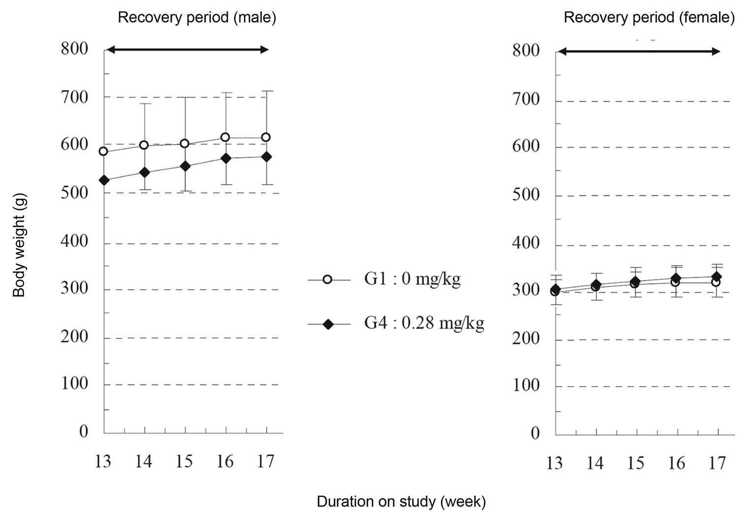
In the 13-week, repeated, dose toxicity test, the high-dosage (0.28 mg/kg) male group showed a significant decrease in body weight during the 2 to 4 week treatment period, but during the 4-week recovery period, the high-dosage group showed no significant changes in body weight compared to the control group (Fig 1, Table 2) The high-dosage and the control groups showed normal findings in the amount of food and water. In addition, compared to the control group, the high-dosage group showed no significant changes in the parameters measured during the ophthalmic examination.
In both the control and experimental groups, ketone bodies and occult blood were observed in the urine in 13-week, repeated, dose toxicity test. In the 4-week recovery period, compared to the control group the high-dosage group showed no significant changes in the urinalysis parameters.
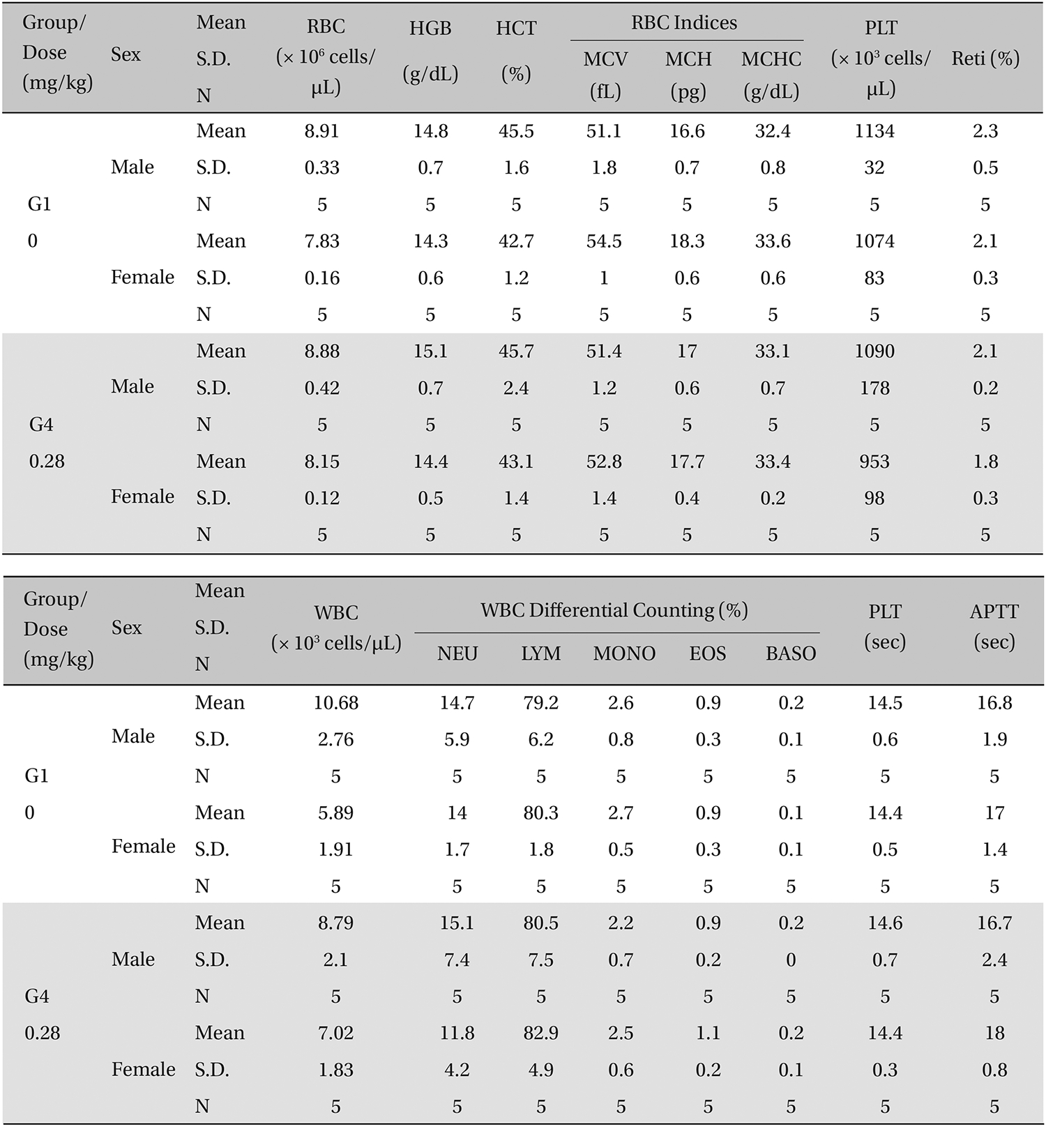
However, hematology studies revealed that the high-dosage (0.28 mg/kg) group showed significant delays in pro-thrombin time (PT) and increases in eosinophils during the 13-week toxicity test. While during 4-week recovery period, compared to the control group, the high-dosage male group showed no significant changes, and the high-dosage female group showed a significant increase in RBCs and a decrease in MCV, but these data were not considered to be due to toxic effect (Table 1).
[Table. 1] Hematological parameters for the SD rats during the 4-week recovery period study

Hematological parameters for the SD rats during the 4-week recovery period study
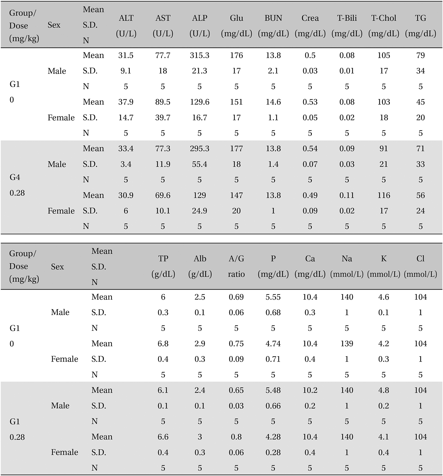
Serum biochemistry revealed that the medium (0.14 mg/ kg)- and the high-dosage male groups showed some significant changes during the 13-week toxicity test. While during the 4-week recovery period, compared to the control group, the high dosage female group showed a significant increase in total bilirubin, but these data were not considered to be due to toxic effects (Table 2).
In the measurement of organ weights during the 13- week treatment period, compared to the control group the high-dosage group showed a significant decrease in liver weight and increase in heart and lung weights and the medium- dosage female group showed significant changes in kidney and adrenal gland weights. However, during the 4-week recovery period, compared to the control group the high-dosage group did not show a significant change in the weight of any organ.
[Table. 2] Blood chemistry values for the SD rats during the 4-week recovery period study

Blood chemistry values for the SD rats during the 4-week recovery period study
During the 13-week treatment period, the medium- and the high-dosage groups showed some fatty liver changes in the periportal area. These changes were not detected during the recovery period. During the 13-week toxicity test, examination of the thigh muscle into which the SBV had been injected showed cell infiltration, inflammation, degeneration, necrosis of muscle fiber, and fibrosis in all experimental groups. But these histological changes were not observed during the recovery period. These results indicate that the abnormalities observed in the organs and tissues are temporary and reversible. No abnormalities were observed in any other organs (Fig 2, Table 3).
Apitherapy (bee sting therapy or bee acupuncture therapy) has been a folk remedy in many cultures, and BVP is a modern and scientific improvement on traditional method. As with other pharmacopuncture techniques, BVP combines physical stimulation of acupuncture with the chemical stimulation of the bee venom. BVP therapy is the first attempt to inject animal poison from nature into the human body for treatment purposes and has opened a new area of treating disorders with a potent poison. However, because BVP can cause allergic reactions, allergic responses, especially anaphylactic shock, put practitioners and patients at great risk [19].
To solve this problem, many trials have been performed over a long time and finally SBV was developed [14, 15, 20, 21]. SBV is a type of pharmacopuncture using melittin that has been isolated and refined from bee venom. Melittin is a well-known water-soluble toxic peptide present in the venom of apismellifera, comprising about 50% of its dry weight. This peptide is able to disrupt membranes, producing many effects on living cells. This small peptide has a molecular weight of 2836 and is made of 26 amino acids. Three-dimensional analysis has revealed that the four subunits of melittin combine in a helical structure [22, 23].
Grade- ±: minimal, +: mild
[Table. 3] Histopathological findings for the SD rats during the 4-week recovery period study
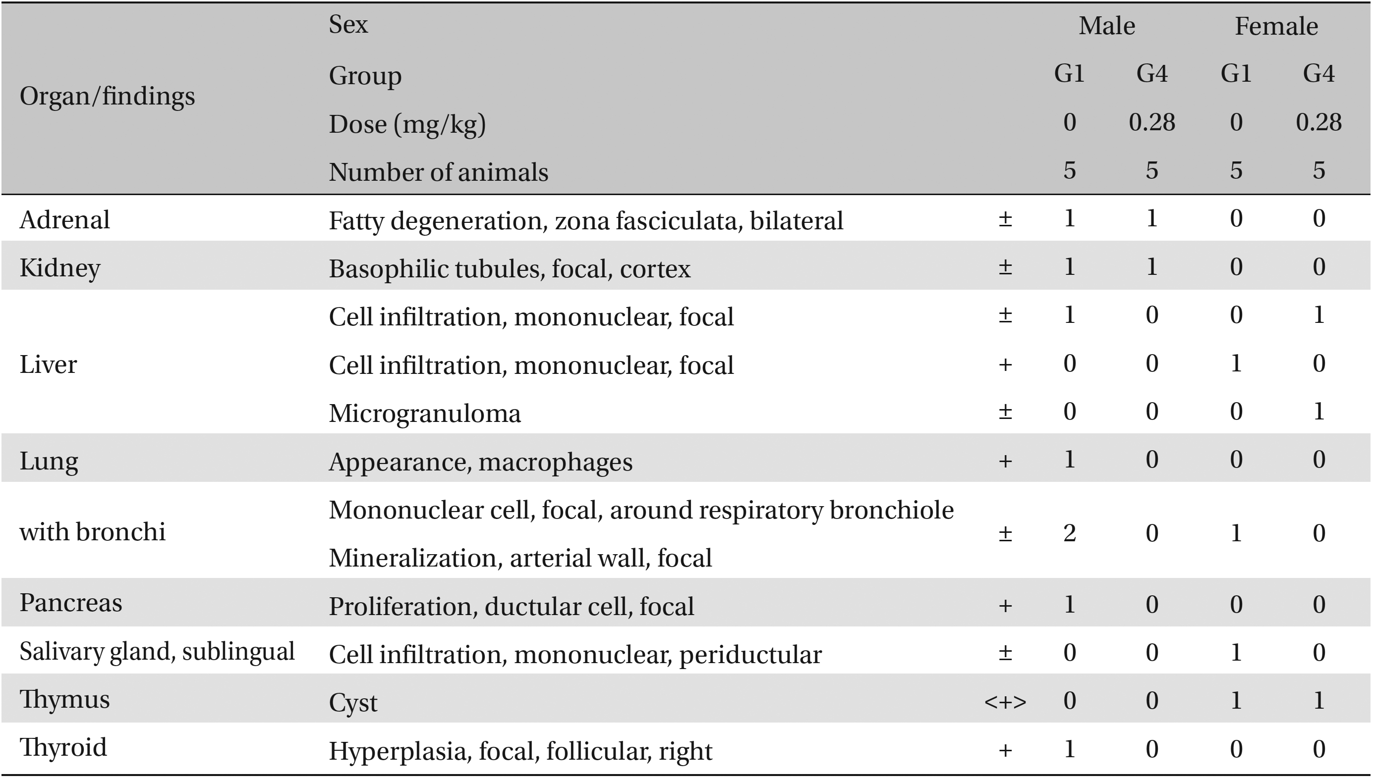
Histopathological findings for the SD rats during the 4-week recovery period study
The principle behind refining melittin from bee venom is a protein separation technique using gel filtration. When bee venom is passed into a gel where only substances with less than a 10,000 molecular weight can enter, then enzymes with weights exceeding 10,000 are passed first followed by smaller peptides. SBV is pure melittin obtained through this process. SBV’s analgesic, anti-inflammatory, and anti-bacterial functions and its effect on decreasing abdominal fat accumulation have been proven through numerous experiments [24-27]. SBV regulates the activation of nuclear factor-κB (NF-κB), a genetic modulator of immunity and inflammation. NF-κB inhibits lipopolysaccharide LPS, tumor necrosis factor-α (TNF-α), and sodium nitroprusside (SNP) to suppress inflammation. In the presence of cancer cells, NF-κB p50 expression, which increases during the process of oncogenesis in colorectal cancer, is suppressed to inhibit inflammatory disorders such as rheumatoid arthritis [28, 29].
We should confirm the acute or chronic adverse effects that one material can cause, as well as the dose-response correlations, to evaluate the toxicity. Animal experimentation is the most indispensable way to obtain this information. In other words, a toxicity test is an important safety assessment of a test substance [30], and the purpose of toxicity studies is to ensure the safety of using a new drug [
Toxicity studies of SBV have reported 50% lethal dose, LD50, for SBV in SD rats of over 30 mg/kg [16], and the max imum reported dose of SBV in beagle dogs in a single-dose toxicity test was over 9 mg/kg [17]. No significant changes were observed for doses below 0.14 mg/kg in SD rats, and no direct side effects of SBV were observed for doses of 0.56 mg/kg in beagle dogs suggesting that a high dosage of SBV of 0.28 mg/kg should be used for a 13-week, repeated, dose toxicity test with a 4-week recovery period in beagle dogs or SD rats [13, 18].
As some significant changes had been observed during the 13-week repeated dose test, we conducted a 4-week recovery test, during which we checked the reversibility of the changes that occurred during the toxicity test. During the 13-week repeated dose test, some experimental groups showed gait disturbance, particularly at higher doses of SBV. However, this symptom lasted for only 10 minutes after the injection and was not observed during the 4-week recovery period. A significant decrease in body weight was observed in the high dosage (0.28 mg/kg) group during the 13-week treatment period. Compared to the control group, the high-dosage group did not show a significant change in weight during the 4-week recovery period. Some groups showed significant changes in organ weights compared to the control group during the 13-week treatment period; however, compared to the control group, the high-dosage group did not show any significant change in organ weight during the 4-week recovery period. Thus, these changes are likely temporary and reversible.
Histopathological examination revealed some fatty liver changes in the periportal area in the medium- and the high-dosage groups during the 13-week treatment period. These changes were not observed during the recovery period. Examination of the thigh muscle into which the SBV had been injected revealed cell infiltration, inflammation, degeneration, and necrosis of muscle fiber, as well as fibrosis, in all experimental groups during the treatment period, but these histological changes were not observed during the recovery period, indicating that the abnormalities of organs and tissues were temporary and reversible.
In conclusion, the present study corroborated the beneficial effects of PEMEs of


![Thigh muscle tissue obtained from 4-week recovery test of Sweet Bee Venom after a 13-week, repeated, intramuscular dose toxicity test
in Sprague-Dawley rats. (A) In the treatment group (0.28 mg/kg every day for 13 weeks), degeneration, fibrosis, inflammation, panniculitis, and necrosis of muscle fiber
were observed. These changes were shown to be severe, depending on the dose. (B) These results show that Sweet Bee Venom has influenced the
muscle fiber, but these changes disappeared during 4-week recovery period. Pathological changes were detected by hematoxylin & eosin (H&E)
staining (× 100 [B], ×200 [A]). In (A) the stars show degeneration of muscle fiber, the small arrow shows regeneration of muscle fiber, the three big
arrows show inflammatory cell infiltration, and the cross shows fibrosis.](http://oak.go.kr/repository/journal/13091/DHOCBS_2014_v17n2_18_g002.jpg)


