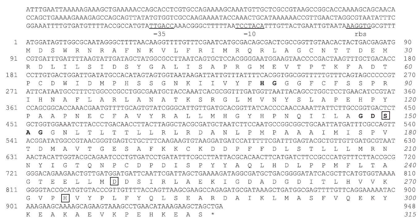
검색 전체 메뉴
PDF
맨 위로
OA 학술지
Molecular Cloning, Purification, and Characterization of a Cold-Adapted Esterase from Photobacterium sp. MA1-3
  • 비영리 CC BY-NC
  • 비영리 CC BY-NC
ABSTRACT
Molecular Cloning, Purification, and Characterization of a Cold-Adapted Esterase from Photobacterium sp. MA1-3
KEYWORD
Photobacterium sp. , Cold-adapted esterase , Gene expression , Substrate specificity
  • Introduction

    Lipases and esterases (glycerol ester hydrolases, E.C. 3.1.1.) are hydrolases that act on the carboxyl ester bonds present in acylglycerols to liberate organic acids and glycerol. Esterases (E.C. 3.1.1.1) differ from lipases (E.C. 3.1.1.3) mainly based on their substrate specificity and interfacial activation (Long, 1971). Lipases, which have a hydrophobic domain covering the active site, show a preference for triglycerides of long chain fatty acids, and thus have different properties to esterases, which have an acyl binding pocket (Pleiss et al., 1998). Lipases and esterases have been recognized as useful biocatalysts because of their versatility in a wide range of industrial applications, including their use in detergents or as additives in the food industry (Harwood 1989; Jaeger and Reetz 1998). Due to their wide diversity enzymatic properties, large numbers of lipases/esterases isolated from bacteria, fungi, plants, and higher animals have been reported (Jaeger et al., 1999; Schmidt and Verger, 1998; Villeneuve et al., 2000). In particular, lipases/esterases of microbial origin represent the most extensively used class of these enzymes and are attracting increasing attention due to their relative ease of production and potential applications in biotechnology (Hasan et al., 2006). Microorganisms that thrive at low temperatures produce cold-adapted enzymes, which have high catalytic efficiency, generally associated with low thermal stability (Feller et al., 1996). Among these enzymes, cold-adapted lipases/esterases are useful in industrial applications as additives in laundry detergents to allow washing in cold water, the food industry, bioremediation processes, and biodiesel applications, based on their high catalytic activity at low temperature and low thermostability as well as unusual specificities (Knothe, 2005; Hasan et al., 2006; Margesin, 2007). In addition, these enzymes can potentially be used as catalysts in organic synthesis of chiral intermediates, allowing relatively unstable compounds to be produced at low temperatures (Ryu et al., 2006). Compared to other lipases, few cold-adapted lipase/esterase have been studied. These include the enzymes from Moraxella sp. strain TA144 (Feller et al., 1991), Aeromonas sp. LPB4 (Lee et al., 2003), Pseudomonas sp. strain B11-1 (Choo et al., 1998), Acinetobacter sp. No. 6 (Suzuki et al., 2001, 2002a, 2002b), Psychrobacter sp. Ant300 (Kulakova et al., 2004), Photobacterium sp. (Ryu et al., 2006), Salinisphaera sp. P7-4 (Kim et al., 2011), and Shewanella sp. Ke75 (Kim et al., 2013).

    The strain Photobacterium sp. MA1-3, previously isolated from the intestine of a blood clam, possesses a cold-adapted lipase (Kim et al., 2012). Recently, we isolated another recombinant clone bearing lipolytic activity from a gene library from this strain, and the gene was heterologously expressed in Escherichia coli cells. Here, we report the cloning, sequencing, and biochemical properties of the cloned enzyme.

    Materials and Methods

      >  Materials

    Tributyrin, p-nitrophenyl (p-NP) acetate (C2), butyrate (C4), caprylate (C8), caprate (C10), palmitate (C16), and stearate (C18) were purchased from Sigma (St. Louis, MO, USA), and p-NP myristate (C14) was from Fluka (Milwaukee, WI, USA). All other chemicals and solvents were analytical grade and are commercially available.

      >  Gene cloning and sequence analysis

    Chromosomal DNA from Photobacterium sp. MA1-3 was partially digested with Sau3AI, ligated into a pUC118-HincII vector (Takara, Kyoto, Japan), and used to transform E. coli XL1-Blue. A colony, forming a clear halo on a Luria Bertani (LB) plate containing tributyrin and ampicillin (100 μg/mL), was selected. The recombinant plasmid (pUCMA1-3) was then purified from the transformant, and the insert DNA sequence was determined. DNA sequencing was performed with an Applied Biosystems Automated DNA Sequencer model 3130 with a dye-labeled terminator sequencing kit (Applied Biosystems, Foster City, CA, USA). Sequence analysis and database similarity searches were performed using the server at the National Center for Biotechnology Information (NCBI) (http://www.ncbi.nlm.nih.gov/BLAST/). Multiple sequence alignments were performed using ClustalW (Thompson et al., 1994), and the signal peptide sequence was deduced by SignalP version 3.0.

      >  Construction of the expression vector and overexpression

    The DNA corresponding to the coding region was amplified by PCR. The putative MA1-3 esterase gene was amplified from the pUCMA1-3 plasmid using the primers: 5’-TTCATATGGATAGTTGGCGCAATAGG -3’ (NdeI adaptor restriction enzyme site is underlined) and 5’-TTCTCGAGGCTAGCTTCTTTATGTTC-3’ (XhoI). After digestion with NdeI and XhoI, the PCR product was ligated into the pET22b(+) vector (Novagen, Madison, WI, USA). The resulting recombinant plasmid, pETMA1-3, was transformed into E. coli BL21 (DE3) cells.

      >  Purification of the recombinant protein

    E. coli BL21 (DE3) cells transformed with pETMA1-3 were cultivated in LB medium containing ampicillin (100 μg/mL) at 30℃. When the optical density at 600 nm reached 0.6, 1 mM isopropylthiogalactoside (IPTG) was added, and the cultures were further incubated overnight at 18℃. E. coli cells were then harvested and ruptured by ultrasonic cell lysis. The soluble proteins were recovered from the cell extract by centrifugation (10,000 g, 20 min) and loaded onto a nickel-nitrilotriacetic (Ni-NTA) column. After washing with 60 mM imidazole, 500 mM NaCl, and 50 mM Tris-HCl buffer (pH 7.9), the bound esterase was then eluted using 1000 mM imidazole, 500 mM NaCl, 20 mM Tris-HCl buffer (pH 7.9) and dialyzed against a 50 mM Tris-HCl buffer (pH 8.0) to characterize its biochemical properties. The protein concentration was determined by the BCA method with bovine serum albumin as the standard (Sigma).

      >  Esterase assay

    Esterase activity was measured using p-nitrophenyl (p-NP) esters with fatty-acid chain lengths of C2-C18 (Ryu et al., 2006). The standard assay mixture (1.0 ml) contained 10 mM p-NP butyrate (C4) in ethanol, 50 mM Tris-HCl buffer, pH 8.0, and 10 μl of purified enzyme. Blank reactions were also run with a composition identical to the assay mixture without the enzyme. The mixture was incubated at 30℃ for 5 min and the absorbance of p-NP librated was then measured at 405 nm. For long-chain p-nitrophenyl esters (C12-C18), 20 μl of esterase solution was added to 880 μL of reaction buffer containing 50 mM Tris-HCl (pH 8.0), 0.1% gum Arabic, and 0.2% deoxycholate. After 5-min incubation at 30℃, the reaction was initiated by adding 100 μl of 8 mM substrate in isopropanol. The reaction was stopped by addition of 0.5 ml of 3 M HCl. After centrifugation, 333 μl of supernatant was mixed with 1 ml of 2 M NaOH, and the absorbance at 405 nm was measured. One unit of enzyme activity was defined as the release of 1 μmol of p-nitrophenol per min from p-NP ester.

      >  Biochemical properties of recombinant esterase

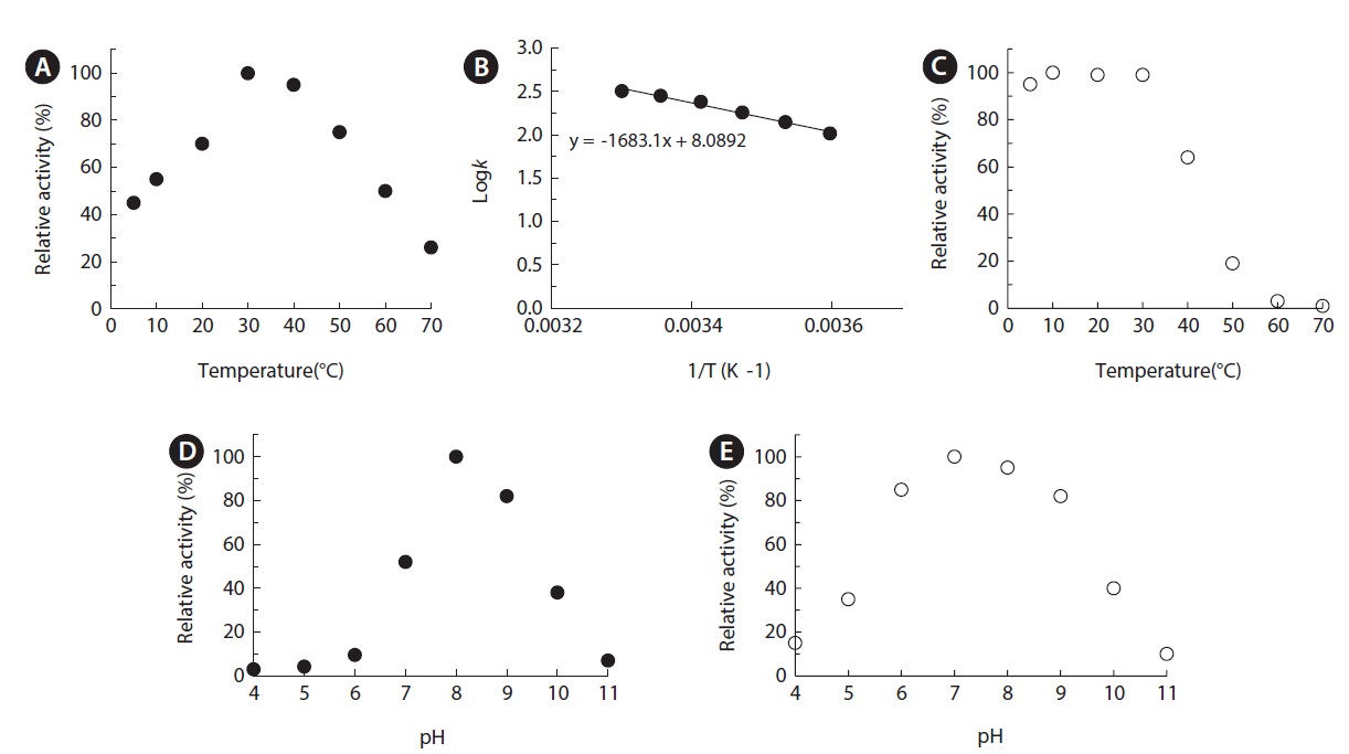
    The optimum temperature was assayed at various temperatures in the range of 5-80℃ at 50 mM Tris-HCl (pH 8.0). For thermostability, the enzyme was preincubated at various temperatures for 30 min in 50 mM Tris-HCl buffer (pH 8.0). After rapid centrifugation, they were removed and the residual enzyme activity of the supernatant was measured as standard assay. Various buffers were used to study the effects of pH: sodium acetate/acetic acid (pH 4-6), Tris/acetate (pH 6-7), Tris/HCl (pH 7-9), and sodium tetraborate/NaOH (pH 9-11). To determine pH stability, the enzyme was preincubated at 25℃ in buffers of various pH for 1 h and the remaining activity was measured by standard assay. The effects of various metal ions and inhibitors on enzyme activity were assessed after preincubation in 50 mM Tris-HCl buffer (pH 8.0) at 25℃ for 30 min. Blank reactions were performed with each measurement under different conditions and the values for nonenzymatic hydrolysis of substrates were subtracted from the results.

      >  GenBank accession number

    The nucleotide sequence of the Photobacterium sp. MA1-3 esterase gene has been deposited in the GenBank database under accession number KF431955.

    Results and Discussion

      >  Gene cloning and sequence analysis

    A DNA library of Photobacterium sp. MA1-3 was constructed with the Sau3A1 enzyme and used to transform E. coli XL1-Blue. E. coli transformant that showed a clear zone on TBN-LB plates after 48 h of incubation were selected. The recombinant plasmid from a clone was shown to contain a DNA insert of 3.2 kb, with one complete open reading frame (ORF) corresponding to a putative esterase/lipase gene based on the results of a homology search. The complete DNA sequence of the MA1-3 esterase was determined (Fig. 1). It contained a ORF of 948 bp, encoding a polypeptide chain of 315 amino acids with a predicted molecular weight of 35,020 and a pI value of 6.06. No signal sequence was found, as determined by SignalP version 3.0, suggesting that the MA1-3 esterase is a cell-bound protein. A putative ribosomal binding-site (RBS) was located 5 bp upstream of the MA1-3 esterase. In addition, a putative transcriptional initiation site with a -10 region was located 32 bp upstream of the MA1-3 esterase start codon and separated from the –35 region by 15 bp. The predicted amino acid sequence of the cloned MA1-3 esterase was compared with other protein sequences deposited in GenBank using the basic local alignment search tool (BLAST) program (Fig. 2A). The esterase showed 74%, 68%, 52%, and 36% identities with

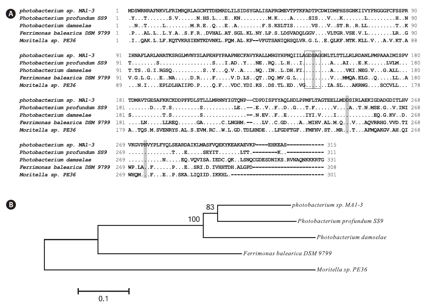
    the putative esterase/lipases from Photobacterium profundum SS9, Photobacterium damselae, Ferrimonas balearica, and Moritella sp. PE36, respectively. The MA1-3 esterase was only 44% identical to the lipases of the same strain Photobacterium sp. MA1-3 (Kim et al., 2012).

    The MA1-3 esterase primary structure contained a -G-D-S-A-G- sequence (positions 148-152), which corresponds well with the pentapeptide -G-x-S-x-G- signature motif that is generally conserved in many esterase enzymes. Based on sequence comparisons with other esterases, it was concluded that Ser 150 (in the motif GDSAG), Asp 248, and His 274 comprise the catalytic triad. Finally, an HG sequence (His80, Gly81), which constitutes an oxyanion hole in the three-dimensional protein structure, was found in the esterase (Grochulski et al., 1993; Martinez et al., 1994). Sequence analysis suggested that MA1-3 esterase may be a functional esterase with a novel amino acid sequence.

    Fig. 2B shows the phylogenetic tree, indicating the evolutionary relationship with other bacterial esterases based on the amino acid sequence. The phylogram generated using Phylip showed that Photobacterium sp. MA1-3 esterase was more closely related to a putative esterase from Photobacterium profundum SS9 than to other lipases and esterases identified to date.

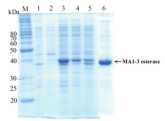
      >  Expression and identification of the recombinant esterase

    E. coli BL21 (DE3) was transformed with the plasmid, pETMA1-3, and was induced to express the recombinant protein using 1 mM IPTG. When cultivated and incubated at 37℃, the resulting protein was insoluble; however, at the lower culture temperature of 18℃, the resulting protein was both soluble and insoluble (Fig. 3). Approximately 20% of the

    recombinant esterase protein was produced in soluble form in E. coli cells. The recombinant enzyme was then purified to homogeneity by His-Bind resin affinity chromatography with a six-histidine tag at the C-terminus. Sodium dodecyl sulfate-polyacrylamide gel electrophoresis analysis of the eluted fraction showed a distinctly expressed protein band of 39 kDa (Fig. 3). This mass is in agreement with the mass of the esterase (35 kDa) with an additional 36 amino acid residues corresponding to the N-terminus 6× histidine-tag and the linker region. The specific activity of the purified enzyme was 20.2 U/mg toward p-NP butyrate at 30℃. Its activity was 3.8- and 2.9-fold higher than those of PWTSB and PWTSC, which had specific activities of 5.3 and 6.9 U/mg toward PNPB, respectively (Wei et al., 2009). In contrast, Ke75 esterase showed a lightly higher specific activity of 22.75 U/mg (Kim et al., 2013).

      >  Substrate specificity

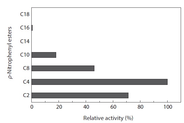
    The substrate specificity of MA1-3 esterase was examined with various p-NP esters with aliphatic acyl-chains of different lengths from 2 to 18 carbon atoms (Fig. 4). Analysis of the enzyme hydrolytic activity against these p-NP esters showed a strong preference toward short acyl chains of p-NP-2, p-NP-4 and p-NP-8 esters, with p-NP-C4 being the most easily hydrolyzed substrate. The reactivities of these substrates to the enzyme were dependent on their acyl-chain length, and the substrates with C14, C16, and C18 acyl groups were virtually inert as substrates for the enzyme. Based on the substrate preference profile, MA1-3 esterase was classified as a true carboxylesterase (Jaeger et al., 1993, 1999; Bornscheuer, 2002).

      >  Effects of pH and temperature on enzyme activity and stability

    The temperature activity profile of MA1-3 esterase was examined over the temperature range of 5–80℃ under assay conditions with p-NP butyrate as the substrate (Fig. 5A). Enzyme activity peaked at 30℃. The activation energy of the enzymes derived from cold-active organisms is usually lower than those from their mesophilic counterparts (Feller et al., 1996). As expected, the activation energy of MA1-3 esterase was 7.69 kcal/mol in the range of 5℃ to 30℃, which is lower than those of other cold-adapted esterases: 11.2 kcal/mol for the esterase of Pseudomonas sp.B11-1 (Suzuki et al., 2003); 9.0 kcal/mol for the esterase of Acinetobacter sp. No. 6 (Suzuki et al., 2002a); and 11.25 kcal/mol for the esterase of Acinetobacter lwoffii 16C-1 (Kim and Park, 2002). These observations suggest that the catalytic efficiency of this esterase is high over this temperature range. In fact, this esterase showed as much as 45% of the maximum activity at 5℃. The enzyme was stable within the temperature range 5~40℃. However, it was thermally unstable and lost its activity at temperatures above 50℃ (Fig. 5C). Taken together, these observations indicated that MA1-3 esterase is a typical cold-adapted enzyme

    The optimal pH of MA1-3 esterase was determined to be 8.0, and it retained at least 80% of its maximum activity between pH 8.0 and 9.0, indicating that it is an alkaline enzyme (Fig. 5D). Although its esterase activity was somewhat different depending on the various incubation buffers used, the MA1-3 esterase was fairly stable after incubation in buffers ranging in pH from 7.0 to 10.0 (Fig. 5E). The maximum activity at alkaline pH is a useful characteristic for detergent applications.

      >  Effects of metal ions and inhibitors on esterase activity

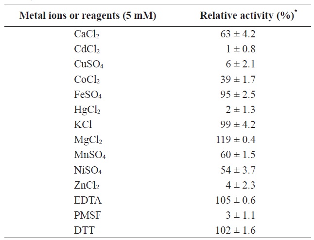
    The effects of various metal ions and inhibitors on enzyme activity were determined (Table 1). Divalent salts, such as MgCl2, simulated the esterase activity to 119% compared to

    control, whereas the activity was decreased by 50~60% in the presence of MnSO4 NiSO4, and CoCl2 Moreover, its activity was strongly inhibited by 5 mM metal ions such as CdCl2, CuCl2, ZnCl2, and HgCl2.

    To confirm that the enzyme was a serine hydrolase, the activity of MA1-3 esterase was determined in the presence of 1 mM phenylmethylsulfonyl fluoride (PMSF), a catalytic serine enzyme inhibitor. Similar concentrations of a metal-chelating

    [Table 1.] Effects of various metal ions and inhibitors on MA1-3esterase activity

    label

    Effects of various metal ions and inhibitors on MA1-3esterase activity

    agent, ethylenediaminetetraacetic acid (EDTA), and a reducing agent, dithiothreitol (DTT), were also investigated to eliminate the possible involvement of metal cations or cysteine in the enzyme mechanism. MA1-3 esterase was significantly inhibited (98%) by PMSF, while the other two additives (DTT and EDTA) had little effect on its activity. The inhibitory effect of PMSF on MA1-3 esterase indicated the involvement of serine-mediated catalytic activity in this enzyme.

    In this study, a novel esterase produced by Photobacterium sp. MA1-3 exhibited high activity at low temperatures and alkaline pH. Its low activation energy in the range of 5-30℃ indicated that it is a cold-adapted enzyme. These results suggest that this enzyme may be useful as a biocatalyst and detergent additive for low temperature applications.

참고문헌
  • 1. Bornscheuer UT 2002 Microbial carboxylesterases: classification, properties and applications in biocatalysis. [FEMS Microbiol Rev] Vol.26 P.73-81 google cross ref
  • 2. Choo DW, Kurihara T, Suzuki T, Soda K, Esaki N. 1998 A cold-adapted lipase of an alaskan psychrotroph, Pseudomonas sp. strain B11-1: gene cloning and enzyme purification and characterization. [Appl Environ Microbiol] Vol.64 P.486-491 google
  • 3. Feller G, Thiry M, Gerday C. 1991 Nucleotide sequence of the lipase gene Lip2 from the antarctic psychrotroph Moraxella TA144 and site-specific mutagenesis of the conserved serine and histidine residues. [DNA Cell Biol] Vol.10 P.381-388 google cross ref
  • 4. Feller G, Narinx E, Arpigny JL, Aittaleb M, Baise E, Geniot S, Gerday C 1996 Enzymes from psychrophilic organisms. [FEMS Microbiol Rev] Vol.18 P.189-202 google cross ref
  • 5. Grochulski P, Li Y, Schrag JD, Bouthillier F, Smith P, Harrison D, Rubin B, Cygler M. 1993 Insights into interfacial activation from an open structure of Candida rugosa lipase. [J Biol Chem] Vol.268 P.12843-12847 google
  • 6. Harwood J. 1989 The versatility of lipases for industrial uses. [Trends Biochem Sci] Vol.14 P.125-126 google
  • 7. Hasan F, Shah AA, Hameed A. 2006 Industrial applications of microbial lipases. [Enzyme Microb Technol] Vol.39 P.235-251 google cross ref
  • 8. Jaeger KE, Reetz MT. 1998 Microbial lipases from versatile tools for biotechnology [Trends Biotechnol] Vol.16 P.396-403 google cross ref
  • 9. Jaeger KE, Ransac S, Koch HB, Ferrato F, Dijkstra BW. 1993 Topological characterization and modeling of the 3D structure of lipase from Pseudomonas aeruginosa. [FEBS Lett] Vol.332 P.143-149 google cross ref
  • 10. Jaeger KE, Dijkstra BW, Reetz MT. 1999 Bacterial biocatalysts: molecular biology, three-dimensional structures, and biotechnological applications of lipases. [Annu Rev Microbiol] Vol.53 P.315-351 google cross ref
  • 11. Kim HE, Park KR 2002 Purification and characterization of an esterase from Acinetobacter lwoffii 16C-1. [Curr Microbiol] Vol.44 P.401-405 google cross ref
  • 12. Kim YO, Park IS, Kim HK, Nam BH, Kong HJ, Kim WJ, Kim DG, Kim KK, Lee SJ. 2011 A novel cold-adapted esterase from Salinisphaera sp. P7-4: gene cloning, overexpression, and characterization. [J Gen Appl Microbiol] Vol.57 P.357-364 google cross ref
  • 13. Kim YO, Khosasih V, Nam BH, Lee SJ, Suwanto A, Kim HK 2012 Gene cloning and catalytic characterization of cold-adapted lipase of Photobacterium sp. MA1-3 isolated from blood clam. [J Biosci Bioeng] Vol.114 P.589-595 google cross ref
  • 14. Kim YO, Park IS, Kim HK, Nam BH, Kong HJ, Kim WJ, Kim DG, Kim BS, Jee YJ, Song JH, Lee SJ. 2013 Shewanella sp. Ke75 esterase with specificity for p-nitrophenyl butyrate: gene cloning and characterization. Vol.55 google cross ref
  • 15. Knothe G 2005 Dependence of biodiesel fuel properties on the structure of fatty acid alkyl esters. [Fuel Process Technol] Vol.86 P.1059-1070 google cross ref
  • 16. Kulakova L, Galkin A, Nakayama T, Esaki N. 2004 Cold-active esterase form Psychrobacter sp. Ant300: gene cloning, characterization, and the effects of Gly→Pro substitution near the active site on its catalytic activity and stability. [Biochim Biophys Acta] Vol.1696 P.59-65 google cross ref
  • 17. Lee HK, Ahn MJ, Kwak SH, Song WH, Jeong BC. 2003 Purification and characterization of cold active lipase from psychrotrophic Aeromonas sp. LPB4. [J Microbiol] Vol.41 P.22-27 google
  • 18. Long C. 1971 Biochemists Handbook. P.273-274 google
  • 19. Margesin R 2007 Alpine microorganisms: useful tools for low-temperature bioremediation. [J Microbiol] Vol.45 P.281-285 google
  • 20. Martinez C, Nicolas A, van Tilbeurgh H, Egloff MP, Cudrey C, Verger R, Cambillau C 1994 Cutinase, a lipolytic enzyme with a preformed oxyanion hole. [Biochemistry] Vol.33 P.83-89 google cross ref
  • 21. Pleiss J, Fischer M, Schmid RD. 1998 Anatomy of lipase binding sites: the scissile fatty acid binding site. [Chem Phys Lipids] Vol.93 P.67-80 google cross ref
  • 22. Ryu HS, Kim HK, Choi WC, Kim MH, Park SY, Han NS, Oh TK, Lee JK 2006 New cold-adapted lipase from Photobacterium lipolyticum sp. nov. that is closely related to filamentous fungal lipases. [Appl Microbiol Biotechnol] Vol.70 P.321-326 google cross ref
  • 23. Schmid RD, Verger R. 1998 Lipases; interfacial enzymes with attractive applications. [Angew Chem Int Ed] Vol.37 P.1608-1633 google cross ref
  • 24. Suzuki T, Nakayama T, Kurihara T, Nishino T, Esaki N. 2001 Cold-active lipolytic activity of psychrotrophic Acinetobacter sp. [strain no] Vol.92 P.144-148 google cross ref
  • 25. Suzuki T, Nakayama T, Kurihara T, Nishino T, Esaki N. 2002a A cold-active esterase with a substrate preference for vinyl esters from a psychrotroph, Acinetobacter sp. strain no. 6: gene cloning, purification, and characterization. [J Mol Catal B Enzym] Vol.6 P.255-263 google cross ref
  • 26. Suzuki T, Nakayama T, Kurihara T, Nishino T, Esaki N. 2002b Primary structure and catalytic properties of a cold-active esterase from a psychrotroph, Acinetobacter sp. strain no. 6 isolated from Siberian soil. [Biosci Biotechnol Biochem] Vol.66 P.1682-1690 google cross ref
  • 27. Suzuki T, Nakayama T, Choo DW, Hirano Y, Hirano Y, Kurihara T, Nishino T, Esaki N. 2003 Cloning, heterologous expression, renaturation, and characterization of a cold-adapted esterase with unique primary structure from a psychrotroph Pseudomonas sp. strain B11-1. [Protein Expr Purif] Vol.30 P.171-178 google cross ref
  • 28. Thompson JD, Higgins DG, Gibson TJ. 1994 CLUSTAL W: improving the sensitivity of progressive multiple sequence alignment through sequence weighting, positions-specific gap penalties and weight matrix choice. [Nucleic Acids Res] Vol.22 P.4673-4680 google cross ref
  • 29. Villeneuve P, Muderhwa JM, Graille J, Haas MJ. 2000 Customizing lipases for biocatalysis: a survey of chemical, physical and molecular biological approaches. [J. Mol. Catal B: Enzym] Vol.9 P.113-148 google cross ref
  • 30. Wei P, Bai L, Song W, Hao G. 2009 Characterization of two soil metagenome-derived lipases with high specificity for p-nitrophenyl palmitate. [Arch Microbiol] Vol.191 P.233-240 google cross ref
이미지 / 테이블
  • [ Fig. 1. ]  Nucleotide sequence of the MA1-3 esterase gene and its deduced amino acid sequence. Potential promoter regions (-10, -35) and ribosomal binding-site (RBS) are labeled and underlined. The conserved Ser, Asp, and His residues that comprise a putative catalytic triad are boxed. Both conserved pentapeptide sequence (Gly-X-Ser-X-Gly) and HG sequence (oxyanion hole) are indicated in bold. The sequence has been submitted to GenBank under accession number KF431955.
    Nucleotide sequence of the MA1-3 esterase gene and its deduced amino acid sequence. Potential promoter regions (-10, -35) and ribosomal
binding-site (RBS) are labeled and underlined. The conserved Ser, Asp, and His residues that comprise a putative catalytic triad are boxed. Both conserved
pentapeptide sequence (Gly-X-Ser-X-Gly) and HG sequence (oxyanion hole) are indicated in bold. The sequence has been submitted to GenBank under
accession number KF431955.
  • [ Fig. 2. ]  Protein sequence alignment (A) and phylogenetic tree (B) of MA1-3 esterase and five similar enzymes. (A) Identity sequences are displayed by dots (...), and deletions of amino acid residues are indicated by dashes (---). Residues involved in the catalytic triad are shaded. Conserved pentapeptide containing the catalytic serine is shown in box. The accession numbers for each sequence are as follows: Photobacterium sp. MA1-3 (KF431955), Photobacterium profundum SS9 (YP_130084), Photobacterium damselae (WP_005299527), Ferrimonas balearica (YP_003912034), Moritella sp. PE36 (WP_006032431). (B) A phylogenetic tree of the aligned sequences was constructed using the Neighbor-Joining algorithm in MEGA (version 4.0). The degree of confidence for each branch point was determined by bootstrap analysis (1,000 repetitions).
    Protein sequence alignment (A) and phylogenetic tree (B) of MA1-3 esterase and five similar enzymes. (A) Identity sequences are displayed by dots (...), and deletions of amino acid residues are indicated by dashes (---). Residues involved in the catalytic triad are shaded. Conserved pentapeptide containing the catalytic serine is shown in box. The accession numbers for each sequence are as follows: Photobacterium sp. MA1-3 (KF431955), Photobacterium profundum SS9 (YP_130084), Photobacterium damselae (WP_005299527), Ferrimonas balearica (YP_003912034), Moritella sp. PE36 (WP_006032431). (B) A phylogenetic tree of the aligned sequences was constructed using the Neighbor-Joining algorithm in MEGA (version 4.0). The degree of confidence for each branch point was determined by bootstrap analysis (1,000 repetitions).
  • [ Fig. 3. ]  Sodium dodecyl sulfate (SDS)-polyacrylamide gel electrophoresis (PAGE) of MA1-3 esterase. Lane M, Standard protein molecular weight markers; lane 1, lysate supernatants of uninduced transformant; lane 2 and 3, soluble and insoluble protein induced by isopropylthiogalactoside (IPTG) at 37℃ ; lane 4 and 5, soluble and insoluble protein induced by IPTG at 18℃ lane 6, purified esterase by nickel-nitrilotriacetic (Ni-NTA) affinity column chromatography. Arrow indicates MA1-3 esterase.
    Sodium dodecyl sulfate (SDS)-polyacrylamide gel electrophoresis
(PAGE) of MA1-3 esterase. Lane M, Standard protein molecular weight
markers; lane 1, lysate supernatants of uninduced transformant; lane 2
and 3, soluble and insoluble protein induced by isopropylthiogalactoside
(IPTG) at 37℃ ; lane 4 and 5, soluble and insoluble protein induced by
IPTG at 18℃ lane 6, purified esterase by nickel-nitrilotriacetic (Ni-NTA)
affinity column chromatography. Arrow indicates MA1-3 esterase.
  • [ Fig. 4. ]  Substrate specificity of MA1-3 esterase. Hydrolysis activity was measured toward various p-nitrophenyl esters.
    Substrate specificity of MA1-3 esterase. Hydrolysis activity was
measured toward various p-nitrophenyl esters.
  • [ Fig. 5. ]  Effects of temperature and pH on the activity and stability of MA1-3 esterase. (A) Hydrolytic activity was assayed at various temperatures (B) The logarithm of the enzyme turnover rate (k) (s-1) was plotted against the reciprocal of absolute temperature (T). The values shown are activation energy calculated from the linear part of the Arrhenius plot. (C) Residual hydrolytic activity was measured after treatment for 30 min at a range temperatures (D) Hydrolytic activity was assayed at various pHs (E) Residual hydrolytic activity was measured after treatment for 30 min at a range pH values.
    Effects of temperature and pH on the activity and stability of MA1-3 esterase. (A) Hydrolytic activity was assayed at various temperatures (B) The logarithm of the enzyme turnover rate (k) (s-1) was plotted against the reciprocal of absolute temperature (T). The values shown are activation energy calculated from the linear part of the Arrhenius plot. (C) Residual hydrolytic activity was measured after treatment for 30 min at a range temperatures (D) Hydrolytic activity was assayed at various pHs (E) Residual hydrolytic activity was measured after treatment for 30 min at a range pH values.
  • [ Table 1. ]  Effects of various metal ions and inhibitors on MA1-3esterase activity
    Effects of various metal ions and inhibitors on MA1-3esterase activity
(우)06579 서울시 서초구 반포대로 201(반포동)
Tel. 02-537-6389 | Fax. 02-590-0571 | 문의 : oak2014@korea.kr
Copyright(c) National Library of Korea. All rights reserved.