검색 전체 메뉴
PDF
맨 위로
OA 학술지
Expression of bovine lactoferrin N-lobe by the green alga, Chlorella vulgaris
  • 비영리 CC BY-NC
  • 비영리 CC BY-NC
ABSTRACT
Expression of bovine lactoferrin N-lobe by the green alga, Chlorella vulgaris
KEYWORD
Chlorella vulgaris , electrotransfomation , expression , hygromycin , lactoferrin
  • INTRODUCTION

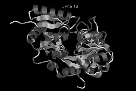
    Lactoferrin (also called lactotransferrin) is a member of the transferrin family of proteins (Aisen and Listowski 1980, Brock 1985, Rose et al. 1986) and is found in milk, saliva, and tears. Lactoferrin is secreted from mammary glands, mucous, and secondary granules of neutrophils, which function in the nonimmunological defense system. Moreover, bovine colostrum contains 5 mg mL-1 lactoferrin, while normal milk contains only about 0.02 mg mL-1 lactoferrin. Human lactoferrin consists of 691 amino acids, which associate with 2 glycan chains of glycoproteins (Metz-Boutigue et al. 1984), and bovine lactoferrin contains 689 amino acids (Picerce et al. 1991), with a predicted molecular weight of about 76 kDa, or 83 kDa when accounting for its glycoconjugates. The lactoferrin glycoprotein consists of 2 lobes, with each lobe having an Fe3+ binding site. The antibacterial core of lactoferrin, called lactoferricin, can be produced through pepsin-mediated cleavage. This peptide has antimicrobial properties and is therefore a potential therapeutic agent in various clinical settings.

    Currently, most commercially obtainable recombinant protein is produced using bacteria, yeast fungi, or bioreactors designed for animal cell culture. Bacterial protein expression systems lack critical post-transcriptional and post-translational regulatory mechanisms, such as splicing, glycosylation, and protein assembly. Moreover, there is a high possibility that purified proteins may contain bacterial endotoxin and protease contaminants. Similarly, yeast expression systems, while eukaryotic in nature, do not have machinery for the glycosylation. Currently, most proteins generated for medical use are cultured and produced in animal cells. However, it is difficult to mass produce such proteins, the cost of culture medium is high, and cultures are easily contaminated. Moreover, low yields have high costs, making this type of expression system unsustainable for clinical applications. Additionally, while plant-based expression systems have many advantages for mass production of stable, functional proteins, challenges to these methods include environmental pollution by genetically modified plants, allergic reactions to plant components, contamination of protein, and regulation of medical protein permission (Choi and Sim 2009).

    Genetic engineering technology has been under development for more than ten years now, and eukaryotic microalga represent an alternative expression system to overcome issues of other expression systems based in animals, fungi, bacteria, and plants. Recently, many studies have investigated the production of antibodies, medical proteins, and vaccines in microalgae (Yang et al. 2006, Tran et al. 2009, Dreesen et al. 2010). Moreover, microalgae have the potential for mass culture in growth conditions similar to that required for general crops. For these reasons, microalgae have become the focus of novel gene manipulation methods to increase the productivity of natural components and novel compound. However, while studies are currently being conducting to investigate the potential for genetic transformation in microalga models, such as Chlamydomonas reinhardtii, gene manipulation technology in microalgae is still in its infancy, and recombinant products from microalgae have not been commercialized.

    In the present study, to the best of our knowledge, we report the first successful expression of bovine lactoferrin N-lobe gene in Chlorella vulgaris via electrotransformation.

    MATERIALS AND METHODS

      >  Codon optimization and the expression vector

    In this study, bovine lactoferrin N-lobe (including a lactoferricin) was used for codon optimization and determined to be Lfb-N. In order to optimize the Lfb-N, Chlorella-codon usage database was chosen to reflect online at http://www.kazusa.or.jp/codon/ (Nakamura et al. 2000). Chlorella-codon optimized Lfb-N DNA sequence (including an SpeI restriction site on the 5′ end and a 6× His-tag sequence and BstEII restriction site on the 3′ end) was synthesized (Bioneer, Daejeon, Korea), and the correct sequence was confirmed by DNA sequence analysis (Bioneer). The codon-optimized Lfb-N gene was inserted into the pGEM T easy vector, and the obtained construct (pLfb-N) was transformed in Escherichia coli XL1-blue 380strain using the heat shock method. The algal expression vector pCAMBIA1304 containing a GUS-mgfp5 fusion reporter and a selectable marker for hygromycin driven by the CaMV35s promoter was used for transformation.

      >  Construction of the expression vector

    The vector construction was carried out as described by Ebrahimi et al. (2012), with some modifications. For construction of the expression vector, the purified pLfb-N vector and the pCAMBIA1304 vector were digested with SpeI and BstEII (Fermentas, Pittsburgh, PA, USA). The digested pLfb-N and pCAMBIA1304 vector were electrophoresed on a 1.0% agarose gel and purified from the gel using a gel extraction kit (Qiagen, Valencia, CA, USA). The purified Lfb-N gene and the pCAMBIA1304 vector were ligated using T4 DNA ligase (Takara Bio, Shiga, Japan) at 16℃ overnight, and the obtained construct (pCAMLfb-N) was transformed in Escherichia coli strain XL1-blue using the heat shock method (Fig. 1). Transformed colonies were selected on LB agar plates containing 50 mg L-1 kanamycin. The pCAMLfb-N recombinant plasmid was verified by colony polymerase chain reaction (PCR), digestion, and sequencing. PCR analysis was performed with the primer pair1-F(5′-CTAGTGTTCGATGGTGCACCATTTCCC-3′) and 1-R (5′-CTCACACGTGGTGGTGGTGGTG-3′) specific to the Lfb-N gene to amplify a 1.0 kb fragment. A thermocycler (Takara Bio) was programmed for 1 cycle at 95℃ for 2 min; 30 cycles of 1 min at 94℃ for denaturation, 2 min at 58℃ for primer template annealing, 3 min at 72℃ for extension; and 1 cycle at 72℃ for 7 min as a final extension. The Lfb-N gene was then inserted into the reporter gene region of pCAMBIA1304 after the CaMV35s promoter and before the NOS terminator by ligation.

      >  Electrotransformation

    Electrotransformation was carried out as described by Wang et al. (2007a), with some modifications. Fifteen mililiters of C. vulgaris culture grown to a cell density of OD600 = 0.3-0.5 was harvested by centrifugation at 6,000 rpm for 10 min at room temperature, then treated by osmosis (0.2 M mannitol and 0.2 M sorbitol) for 1 h, followed again by centrifugation. The resulting pellets were re-suspended in 1 mL of electroporation buffer (500 mM NaCl, 5 mM KCl, 5 mM CaCl2, 20 mM Hepes, 200 mM mannitol, and 200 mM sorbitol; pH 7.2). The 5 μg of pCAMLfb-N was added into a 1 mL cell suspension and kept on ice for 5-10 min. The 1 mL of DNA-cell mixture was transferred into 0.2 cm electroporation cuvette, then

    electrotransformation was performed by electroporator ECM 2001 (BTX, Holliston, MA, USA) under voltage 1,000 V at 25 μF and 200 ohm to generate the transgenic C. vulgaris. After electroporation, C. vulgaris cells were transferred to 6-well plate containing 5 mL of BG-11 medium and cultured in the dark at 25℃ for 24 h. The cultured cells were harvested by centrifugation at 6,000 rpm for 10 min at room temperature and discard the supernatant. The cells were resuspended in 200 μL of BG-11 medium at room temperature and plated onto selection agar plate (BG-11 agar plates containing 20 mg L-1 hygromycin). The selection agar plate cultures were kept in the incubation under continuous cool fluorescent light with 50 μmol m-2 s-1 at 25℃. Transformed colonies appeared in a 5 days, and independent colonies were maintained on selection medium. Hygromycin-resistant colonies were grown in BG-11 medium containing hygromycin and used for molecular analysis.

      >  Genomic DNA extraction and screening of putative transgenic Chlorella vulgaris DNA by PCR

    Extraction of C. vulgaris genomic DNA was performed as described by Dawson et al. (1997), with some modifications. Microalgae were harvested from 5 mL culture medium (approximately 1 × 10⁷ cells mL-1) and resuspended in 500 μL buffer solution (54 mM hexadecyltrimethylammonium bromide, 0.25 mM Tris [pH8.0], 1.4 M NaCl, 10 mM ethylenediaminetetraacetic acid [EDTA], and 2% β-mercaptoethanol). The mixture was incubated at 65℃ for 2 h and shaken every 15 min. After incubation, an equal volume of phenol-chloroform was added, and the aqueous phase was recovered after 5 min of centrifugation at 8,000 ×g and 25℃ for 10 min. We extracted DNA several times until the aqueous layer was no longer cloudy. Genomic DNA was then precipitated with 2 volumes of 100% ethanol, centrifuged at 8,000 ×g for 15 min, washed with 70% ethanol, dried, and resuspended in 30 μL Tris hydrochloride (TE) buffer.

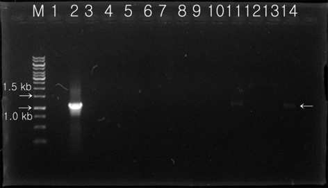
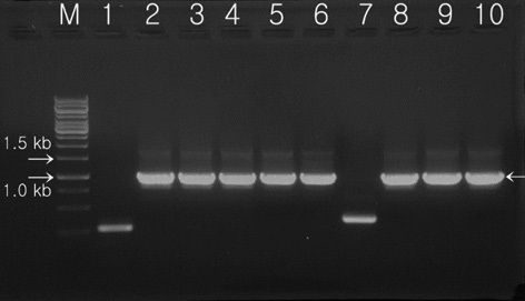
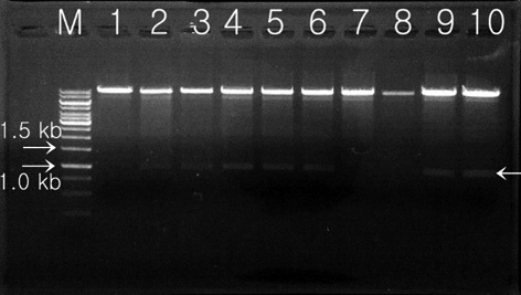
    Transformed colonies was randomly selected and cultured for 7 days in 5 mL BG-11 broth (containing 20 mg L-1 hygromycin) at 25℃, with shaking at 180 rpm. Light conditions were set at 50 μmol m-2 s-1 using a fluorescent light for 14 h, with a 10 h night cycles. Cultured transformants were recovered after processing with a centrifugal separator, and extracted genomic DNA was used as a temple for PCR. Two oligonucleotide primers were synthesized for detection of the transferred Lfb-N DNA fragment by PCR analysis. PCR analysis was performed with the primer pair 2-F (5′-GAGAACACGGGGGACTCTTG-3′) and 2-R (5′-GGGGAAATTCGAGCTGGTCA-3′) specific to the Lfb-N gene to amplify a 1.1 kb fragment containing 3′ end of CaMV35s and 5′ end of NOS terminator. A thermocycler (Takara Bio) was programmed for 1 cycle at 95℃ for 2 min; 30 cycles of 1 min at 94℃ for denaturation, 2 min at 60℃ for primer template annealing, and 3 min at 72℃ for extension; and 1 cycle at 72℃ for 7 min as a final extension.

      >  Protein extraction and western blot analysis

    A 50-mL sample of cell suspension (108 cells mL-1) was centrifuged for 5 min at 2,000 ×g and resuspended in 2 mL lysis buffer (50 mM Hepes, pH 7.9, 250 mM KCl, 0.1 mM EDTA, 1 mM dithiothreitol, 0.5 mM phenylmethylsulfonyl fluoride). The cells were disrupted by sonicating 3 times for 10 s with a micro tip sonicator using a power setting of 30 W (Cannons and Shiflett 2001). The lysate was centrifuged at 12,000 ×g for 5 min, and the supernatant was concentrated using a 10 kDa filter membrane. The total protein concentration was determined with Bradford reagent (Bio-Rad, Hercules, CA, USA) (Bradford 1976) using bovine serum albumin (BSA) as a standard. Equal amounts of total soluble protein were subjected vulto sodium dodecyl sulfate polyacrylamide gel electrophoresis (SDS-PAGE). After the separation, proteins were electroblotted on to nitrocellulose membranes, blocked with BSA, incubated with mouse anti-6× HIS monoclonal antibodies (Abcam, Cambrige, MA, USA) raised in goats, and detected by an anti-mouse IgG-alkaline phosphatase conjugate (Sigma, St. Louis, MO, USA) following standard procedures (Lorimer et al. 2009). In addition, we also predicted the 3D structure of recombinant bovine lactoferrin N-lobe using ESyPred3D.

    RESULTS

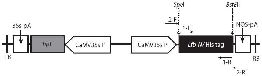
      >  Codon optimization and construction of the expression vector pCAMLfb-N

    In this study, we optimized the Lfb-N gene to resemble Chlorella nuclear gene codons, i.e., to have a GC content of 61% and a gene size of 1,014 bps. In order to express heterologous genes in microalgae, a codon bias that is similar to the GC-bias of nuclear genes and the AT-bias of chromosomal genes should be expressed or optimized for codon usage (Choi and Sim 2009). In order to optimize for codon usage, we used the codon adaptation index (CAI), a quantitative tool to predict heterologous gene expression levels based on their codon usage. Since the chloroplast, mitochondrial, and nuclear genomes of microalgae may exhibit different codon biases, as is the case in C. vulgaris, genome-specific CAI values should be used for optimal translation.

    The digested Lfb-N was cloned between 3′ end of CaMV35s and 5′ end of the NOS terminator in the pCAMBIA1304 vector between BstEII and SpeI restriction sites (Fig. 1). The constructed plasmid was named pCAMLfb-N. Transformed colonies were confirmed by colony PCR (Fig. 2) and restriction enzyme digestion with BstEII and SpeI (Fig. 3). Colony containing of the codon-optimized Lfb-N gene was confirmed in transformants T2, T3, T4, T5, T6, T9, and T10. The plasmid sequence of each transformant was confirmed by DNA sequencing, and T10 was finally selected from sequencing analysis.

      >  Electrotransformation and screening of putative Chlorella vulgaris by PCR analysis

    The pCAMLfb-N was used to transform C. vulgaris by electrotransformation. Transformed cells were selected on BG-11 plates containing 20 mg L-1 hygromycin. A total of 153 transformants were screened. A total of 12 putative

    hygromycin-resistant single colonies, confirmed within 7 days on selection media, were randomly selected, grown in liquid media before the DNA was extracted, and used in PCR analysis. The primers 2-F and 2-R were used to carry out the PCR amplification for screening of C. vulgaris harboring the Lfb-N gene (Fig. 4). Nontransformed C. vulgaris was used as a negative control, and pCAMLfb-N was used as a positive control. Amplification with Lfb-N-specific primers (2-F and 2-R) detected the 1.1 kb DNA fragment from 2 putative transgenic lines (CT-9 and CT-12), but not from an additional 10 putative transgenic lines. The 1.1 kb DNA fragment was detected in the positive control but not in the negative control. Finally, results showed that the Lfb-N genes were integrated into the C. vulgaris genome.

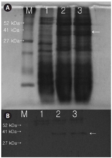
      >  Protein expression and western blot analysis

    Expression of the protein product from the introduced Lfb-N gene was tested by SDS-PAGE and western blot analysis. Results showed that a protein band with a molecular weight of about 35 kDa was expressed in transformed C. vulgaris; this band was absent in the proteins extracted from nontransformed C. vulgaris (Fig. 5A). Western blot analysis of protein extracts from the transformed lines revealed the presence of the Lfb-N protein where the protein was not detected in untransformed cells, indicating the stability of the transgene expression (Fig. 5B). The sequence of this protein was 99% identical to that of lactoferrin from 2 different species (Bos taurus seq. ID: gb|ABF69106.1| and Bos grunniens seq. ID: gb|ACB29795.1|). This 3D structure demonstrated the presence of lactoferricin, the antibacterial core, within Phe18 to Phe42 and the existence of a α-helix and β-sheet (Fig. 6). Our experimental data is probably the first documentation of expression of the bovine lactoferrin N-lobe, in viral promoter CaMV35s in an algal system using electrotransformation.

    DISCUSSION

      >  Algal transformation

    In microalgae, the expression of heterologous proteins presents several difficulties. First, microalgae have an unusual GC-rich codon bias in their nuclear genes; therefore, codon optimization must be performed on any gene for which high levels of protein expression are desired (Heitzer et al. 2007). Additionally, expression levels of optimized foreign genes may vary considerably due to position effect that is driven by random integration of the gene of interest and strong silencing mechanism that drives by epigenetic phenomena similar to those in land plants (Schroda 2006). In C. vulgaris and other algae,

    as in land plants, silenced multiple-copy transgenes exhibit high levels of DNA methylation (Cerutti et al. 1997, Babinger et al. 2001). In contrast, single-copy transgenes are subject to transgene silencing without detectable cytosine methylation (Cerutti et al. 1997).

    The nuclear genome and the plastid genome have highly divergent codon usage, with the chloroplast preferring an A or T in the wobble position, while the nuclear genome prefers a G or C (Nakamura et al. 2000). Using GFP, early work showed that codon optimization to reflect the genome bias could increase transgene protein accumulation 5-fold in the nucleus (Fuhrmann et al. 1999) and up to 80-fold in the chloroplast (Franklin et al. 2002). Today, recombinant genes are universally codon optimized for improved protein expression in almost every system (Puigbo et al. 2007, Xia 2007, Puigbo et al. 2008b). For codon optimization, several free software and web applications have recently been developed to estimate CAI values and optimize the codon usage of sequences. E-CAI (http://genomes.urv.es/CAIcal/E-CAI) determines whether differences in CAIs between sequences are significant or arise from biases in G+C or amino-acid composition (Puigbo et al. 2008b). The online OPTIMIZER application (http://genomes.urv.es/OPTIMIZER) optimizes the codon usage of provided sequences using preconstructed usage tables based on a ‘one amino acid-one codon’ basis, Monte-Carlo algorithms, or a novel algorithm for optimization with minimal changes (Puigbo et al. 2007). CAI-cal (http://genomes.urv.es/CAIcal) provides an integrated set of tools for the optimization of codon usage (Puigbo et al. 2008a). Some gene design software packages such as Gene Composer include gene optimization functionalities (Lorimer et al. 2009). A database of CAI and codon usage indices for most sequenced species is available online at http://www.kazusa.or.jp/codon/ (Nakamura et al. 2000).

    Various genetic transformation methods have been used to express recombinant proteins through introduction of genes into microalgae like Chlorella sp. The applied methods include particle bombardment, glass beads method, Agrobacterium tumefaciens-mediated transformation, and electrotransformation. Bombardment of target cells with DNA-coated metallic particles is a widespread, simple, effective and highly reproducible transformation method. This method has been successfully employed for the transformation of most standard cellular expression systems, and it is therefore not surprising that it is also useful for the study of microalgae. The main drawback of the particle bombardment method is the cost of the required specialized equipment. Although the number of transformants recovered following particle bombardment can be low, it remains the most effective method for the transformation of chloroplasts, as it allows for the delivery of multiple copies of recombinant DNA through both the cellular and chloroplast membranes, increasing the chance for a successful integration event to occur (Boynton and Gillham 1993). This method has been shown to be effective for the stable nuclear (Mayfield and Kindle 1990) and chloroplast (Boynton et al. 1988, El-Sheekh 2000) transformation of C. reinhardtii. A simple and effective transformation method consists of agitating cell wall-deficient microalgal cells with recombinant DNA, polyethylene glycol, which greatly increases transformation efficiency, and glass beads. Despite the drop in cell viability to 25% following agitation with the beads, a nuclear transformation efficiency of 103 transformants μg-1 DNA was achieved using this method (Kindle 1990) and an efficiency of 50 transformants μg-1 DNA was achieved for the transformation of C. reinhardtii chloroplasts (Kindle et al. 1991). Compared to the particle bombardment method, the glass beads method is simpler, more efficient for nuclear transformations, and much less expensive as it does not require specialized equipment. A recent study showed that the glass beads method is also more efficient than particle bombardment for the transformation of Dunaliella salina (Feng et al. 2009). Transformation by the tumour-inducing A. tumefaciens is another efficient means of delivering genetic material, although this method has so far been mainly used to modify plant cells. Transformation results from the stimulation of cell division by products encoded by T-DNA transferred from Agrobacterium to the target cell. The T-DNA and virulence (vir) regions are located on the tumour inducing plasmid (pTi). The vir system processes and transfers any DNA between the short flanking repeats that delimit the T-DNA, making Agrobacterium an efficient DNA delivery system (Akhond and Machray 2009). Using the Agrobacterium-mediated transformation method, C. reinhardtii was successfully transformed with uidA (β-glucuronidase), gfp (green fluorescent protein) and hpt (hygromycin phosphotransferase) reporter genes, with a fifty-fold increase in resulting transformants compared to the glass beads method (Kumar et al. 2004).

    In this study, we used electrotransformation to move recombinant DNA into microalgae, allowing the growth of hygromycin-resistant colonies. The effectiveness of microalgal electroporation, or the induction of macromolecular uptake by exposing cell walls to high intensity electrical field pulses, was first reported by Brown et al. (1991). Electroporation specifically disrupts lipid bilayers, leading to efficient molecular transport across the plasma membrane (Azencott et al. 2007). Efficient electrotransformation was achieved in both wild-type and cell wall-deficient strains (Brown et al. 1991). The transformation efficiency of electroporation is two orders of magnitude higher than the glass beads method, and only requires relatively simple equipment (Shimogawara et al. 1998). Important parameters affecting the effectiveness of electroporation include field strength, pulse length, medium composition, temperature and membrane characteristics (Brown et al. 1991) as well as the concentration of DNA (Wang et al. 2007a). Electroporation was successfully used for the transformation of C. reinhardtii (Tang et al. 1995, Shimogawara et al. 1998, Kovar et al. 2002, Ladygin 2003, 2004), Chlorella sp. (Chow and Tung 1999, Wang et al. 2007a), D. salina (Geng et al. 2004, Sun et al. 2005, 2008, Wang et al. 2007b), Dunaliella viridis (Sun et al. 2006) and Dunaliella tertiolecta (Walker et al. 2005) species, and Nannochloropsis oculata (Chen et al. 2008).

      >  Expression of recombinant lactoferrin

    Studies of recombinant lactoferrin expression have been conducted in several hosts, including E. coli (Weickert et al. 1996). However, lactoferrin expressed in E. coli has been shown to easily form inclusion bodies. Therefore, many researchers have attempted to use insect or animal cells as hosts, but these hosts are more time consuming to work with and do not produce high yields. Another alternative method to produce recombinant lactoferrin was to adopt plant cells as hosts. In a previous study, human lactoferrin was inserted into the dP35SCaMV vector (enhanced 35S promoter), transformed into Nicotiana tabacum xanthi NC through agro-infiltration, and expressed as an 80-kDa recombinant protein (Salmon et al. 1998). Additionally, the potato has been used to express plant microsomal retention signal peptide (SEK-DEL) linked to full-length recombinant human lactoferrin under control of the masP2 and CaMV35s promoters (Chong and Langridge 2000). The antibiotic core of lactoferrin, located in the N-lobe, has also been expressed in Nicotiana benthmiana via agro-infection, resulting in the production of a 40 kDa protein (Li et al. 2004), and in edible rice under control of the CaMV35s promoter, resulting in the production of a 45 kDa protein (Takase et al. 2005).

    Thus, genetic transformation in a plant-based expression system has some benefits, enabling the production of stable, functional protein. However, mass culture via such plant-based systems is limited due to spatial restrictions and risks of exposure of genetically transformed organisms into the ecosystem. However, as we have shown, recombinant protein production using microalgae can be performed using micro-organism culture within a bioreactor, the volume of the culture is adjustable, and the risk of exposure to the ecosystem is relatively low.

    In conclusion, we confirmed that recombinant bovine lactoferrin N-lobe could be expressed from the microalga C. vulgaris. This system allowed for more stable expression of recombinant lactoferrin N-lobe than in other expression systems. The bovine lactoferrin N-lobe, which exhibits a 400-fold increase in antibacterial activities compared to full-length lactoferrin, is difficult to separate from the full-length protein using proteases. However, this important peptide, known as lactoferricin, shows antibacterial activities against gram-positive and -negative bacteria, but does not show antibacterial activities against bifidobacteria, a bacterium found in the intestines. Therefore, the production of recombinant bovine lactoferrin N-lobe protein is expected to be beneficial in a variety of clinical applications and fields.

참고문헌
  • 1. Aisen P., Listowsky I. 1980 Iron transport and storage proteins. [Annu. Rev. Biochem.] Vol.49 P.357-393 google cross ref
  • 2. Akhond M. A. Y., Machray G. C. 2009 Biotech crops: technologies, achievements and prospects. [Euphytica] Vol.166 P.47-59 google cross ref
  • 3. Azencott H. R., Peter G. F., Prausnitz M. R. 2007 Influence of the cell wall on intracellular delivery to algal cells by electroporation and sonication. [Ultrasound Med. Biol.] Vol.33 P.1805-1817 google cross ref
  • 4. Babinger P., Kobl I., Mages W., Schmitt R. 2001 A link between DNA methylation and epigenetic silencing in transgenic Volvox carteri. [Nucleic Acids Res.] Vol.29 P.1261-1271 google cross ref
  • 5. Boynton J. E., Gillham N. W. 1993 Chloroplast transformation in Chlamydomonas. [Methods Enzymol.] Vol.217 P.510-536 google
  • 6. Boynton J. E., Gillham N. W., Harris E. H., Hosler J. P., Johson A. M., Jones A. R., Randolph-Anderson B. L., Robertson D., Klein T. M., Shark K. B., Sanford J. C. 1988 Chloroplast transformation in Chlamydomonas with high velocity microprojectiles. [Science] Vol.240 P.1534-1538 google cross ref
  • 7. Bradford M. M. 1976 A rapid and sensitive method for the quantitation of microgram quantities of protein utilizing the principle of protein-dye binding. [Anal. Biochem.] Vol.72 P.248-254 google cross ref
  • 8. Brock J. H. 1985 Metal proteins with non-redox roles (Part II). Topics in molecular and structural biology. In Harrison, P. (Ed.) P.183-262 google
  • 9. Brown L. E., Sprecher S. L., Keller L. R. 1991 Introduction of exogenous DNA into Chlamydomonas reinhardtii by electroporation. [Mol. Cell Biol.] Vol.11 P.2328-2332 google
  • 10. Cannons A. C., Shiflett S. D. 2001 Transcriptional regulation of the nitrate reductase gene in Chlorella vulgaris: indentification of regulatory elements controlling expression. [Curr. Genet.] Vol.40 P.128-135 google cross ref
  • 11. Cerutti H., Johnson A. M., Gillham N. W., Boynton J. E. 1997 Epigenetic silencing of a foreign gene in nuclear transformants of Chlamydomonas. [Plant Cell] Vol.9 P.925-945 google cross ref
  • 12. Chen H. L., Li S. S., Huang R., Tsai H. -J. 2008 Conditional production of a functional fish growth hormone in the transgenic line of Nannochloropsis oculata (Eustigmatophyceae). [J. Phycol.] Vol.44 P.768-776 google cross ref
  • 13. Choi S. P., Sim S. J. 2009 Production of CO2 by biological process using microalgae. [KIC News] Vol.12 P.1-11 google
  • 14. Chon D. K. X., Langridge W. H. R. 2000 Expression of full-length bioactive antimicrobial human lactoferrin in potato plants. [Transgenic Res.] Vol.9 P.71-78 google cross ref
  • 15. Chow K. -C., Tung W. L. 1999 Electrotransformation of Chlorella vulgaris. [Plant Cell Rep.] Vol.18 P.778-780 google cross ref
  • 16. Dawson H. N., Burlingame R., Cannons A. C. 1997 Stable transformation of Chlorella: rescue of nitrate reductase-deficient mutants with the nitrate reductase gene. [Curr. Microbiol.] Vol.35 P.356-362 google cross ref
  • 17. Dreesen I. A. J., Charpin-El Hamri G., Fussenegger M. 2010 Heat-stable oral alga-based vaccine protects mice from Staphylococcus aureus infection. [J. Biotechnol.] Vol.145 P.273-280 google cross ref
  • 18. Ebrahimi N., Memari H. R., Ebrahimi M. A., Ardakani M. R. 2012 Cloning, transformation and expression of human gamma interferon gene in tomato (Lycopersicon esculentum Mill.). [Biotechnol. Biotechnol. Equip.] Vol.26 P.2925-2929 google cross ref
  • 19. El-Sheekh M. M. 2000 Stable chloroplast transformation in Chlamydomonas reinhardtii using microprojectile bombardment. [Folia Microbiol.] Vol.45 P.496-504 google cross ref
  • 20. Feng S., Xue L., Liu H., Lu P. 2009 Improvement of efficiency of genetic transformation for Dunaliella salina by glass beads method. [Mol. Biol. Rep.] Vol.36 P.1433-1439 google cross ref
  • 21. Franklin S., Ngo B., Efuet E., Mayfield S. P. 2002 Development of a GFP reporter gene for Chlamydomonas reinhardtii chloroplast. [Plant J.] Vol.30 P.733-744 google cross ref
  • 22. Fuhrmann M., Oertel W., Hegemann P. 1999 A synthetic gene coding for the green fluorescent protein (GFP) is a versatile reporter in Chlamydomonas reinhardtii. [Plant J.] Vol.19 P.353-361 google cross ref
  • 23. Geng D. -G., Han Y., Wang Y. -Q., Wang P., Zhang L. -M., Li W. -B., Sun Y. -R. 2004 Construction of a system for the stable expression of foreign genes in Dunaliella salina. [Acta. Bot. Sin.] Vol.46 P.342-346 google
  • 24. Heitzer M., Eckert A., Fuhrmann M., Griesbeck C. 2007 Influence of codon bias on the expression of foreign genes in microalgae. [Adv. Exp. Med. Biol.] Vol.616 P.46-53 google
  • 25. Kindle K. L. 1990 High-frequency nuclear transformation of Chlamydomonas reinhardtii. [Proc. Natl. Acad. Sci. U. S. A.] Vol.87 P.1228-1232 google cross ref
  • 26. Kindle K. L., Richards K. L., Stern D. B. 1991 Engineering the chloroplast genome: techniques and capabilities for chloroplast transformation in Chlamydomonas reinhardtii. [Proc. Natl. Acad. Sci. U. S. A.] Vol.88 P.1721-1725 google cross ref
  • 27. Kovar J. L., Zhang J., Funke R. P., Weeks D. P. 2002 Molecular analysis of the acetolactate synthase gene of Chlamydomonas reinhardtii and development of a genetically engineered gene as a dominant selectable marker for genetic transformation. [Plant J.] Vol.29 P.109-117 google cross ref
  • 28. Kumar S. V., Misquitta R. W., Reddy V. S., Rao B. J., Rajam M. V. 2004 Genetic transformation of the green alga-Chlamydomonas reinhardtii by Agrobacterium tumefaciens. [Plant Sci.] Vol.166 P.731-738 google cross ref
  • 29. Ladygin V. G. 2003 The transformation of the unicellular alga Chlamydomonas reinhardtii by electroporation. [Microbiology] Vol.72 P.585-591 google cross ref
  • 30. Ladygin V. G. 2004 Efficient transformation of mutant cells of Chlamydomonas reinhardtii by electroporation. [Process Biochem.] Vol.39 P.1685-1691 google cross ref
  • 31. Li Y., Geng Y., Song H., Zheng G., Huan L., Qiu B. 2004 Expression of a human lactoferrin N-lobe in Nicotiana benthmiana with potato virus X-based agroinfection. [Biotechnol. Lett.] Vol.26 P.953-957 google cross ref
  • 32. Lorimer D., Raymond A., Walchli J., Mixon M., Barrow A., Wallace E., Grice R., Burgin A., Stewart L. 2009 Gene composer: database software for protein construct design, codon engineering, and gene synthesis. [BMC Biotechnol.] Vol.9 P.36 google cross ref
  • 33. Mayfield S. P., Kindle K. L. 1990 Stable nuclear transformation of Chlamydomonas reinhardtii by using a C. reinhardtii gene as the selectable marker. [Proc. Natl. Acad. Sci. U. S. A.] Vol.87 P.2087-2091 google cross ref
  • 34. Metz-Boutigue M. -H., Jollees J., Mazurier J., Schoentgen F., Legrand D., Spik G., Montreuil J., Jollees P. 1984 Human lactotransferrin: amino acid sequence and structural comparisons with other transferrins. [Eur. J. Biochem.] Vol.145 P.659-676 google cross ref
  • 35. Nakamura Y., Gojobori T., Ikemura T. 2000 Codon usage tabulated from international DNA sequence databases: status of the year 2000. [Nucleic Acids Res.] Vol.28 P.292 google cross ref
  • 36. Picerce A., Colavizza D., Benaissa M., Maes P., Tartar A., Montreuil J., Spik G. 1991 Molecular cloning and sequence analysis of bovine lactotransferrin. [Eur. J. Biochem.] Vol.196 P.177-184 google cross ref
  • 37. Puigboo P., Bravo I. G., Garcia-Vallvee S. 2008a CAI-cal: a combined set of tools to assess codon usage adaptation. [Biol. Direct] Vol.3 P.38 google cross ref
  • 38. Puigboo P., Bravo I. G., Garcia-Vallvee S. 2008b E-CAI: a novel server to estimate an expected value of Codon Adaptation Index (eCAI). [BMC Bioinformatics] Vol.9 P.65 google cross ref
  • 39. Puigboo P., Guzmaan E., Romeu A., Garcia-Vallvee S. 2007 OPTIMIZER: a web server for optimizing the codon usage of DNA sequences. [Nucleic Acids Res.] Vol.35S P.W126-W131 google cross ref
  • 40. Rose T. M., Plowman G. D., Teplow D. B., Dreyer W. J., Hellstroom K. E., Brown J. P. 1986 Primary structure of the human melanoma-associated antigen p97 (melanotransferrin) deduced from the mRNA sequence. [Proc. Natl. Acad. Sci. U. S. A.] Vol.83 P.1261-1265 google cross ref
  • 41. Salmon V., Legrand M., Slominny M. -C., El Yazide I., Spick G., Gruber V., Bournat P., Olagnier D., Mison M., Theisen B., Meerot B. 1998 Production of human lactoferrin in transgenic tobacco plants. [Protein Expr. Purif.] Vol.13 P.127-135 google cross ref
  • 42. Schroda M. 2006 RNA silencing in Chlamydomonas: mechanisms and tools. [Curr. Genet.] Vol.49 P.69-84 google cross ref
  • 43. Shimogawara K., Fujiwara S., Grossman A., Usuda H. 1998 High-efficiency transformation of Chlamydomonas reinhardtii by electroporation. [Genetics] Vol.148 P.1821-1828 google
  • 44. Sun G., Zhang X., Sui Z., Mao Y. 2008 Inhibition of pds gene expression via the RNA interference approach in Dunaliella salina (Chlorophyta). [Mar. Biotechnol.] Vol.10 P.219-226 google cross ref
  • 45. Sun Y., Gao X., Li Q., Zhang Q., Xu Z. 2006 Functional complementation of a nitrate reductase defective mutant of a green alga Dunaliella viridis by introducing the nitrate reductase gene. [Gene] Vol.377 P.140-149 google cross ref
  • 46. Sun Y., Yang Z., Gao X., Li Q., Zhang Q., Xu Z. 2005 Expression of foreign genes in Dunaliella by electroporation. [Mol. Biotechnol.] Vol.30 P.185-192 google cross ref
  • 47. Takase K., Hagiwara K., Onodera H., Nishizawa Y., Ugaki M., Omura T., Numata S., Akutsu K., Kumura H., Shimazaki K. 2005 Constitutive expression of human lactoferrin and its N-lobe in rice plants to confer disease resistance. [Biochem. Cell Biol.] Vol.83 P.239-249 google cross ref
  • 48. Tang D. K. H., Qiao S. Y., Wu M. 1995 Insertion mutagenesis of Chlamydomonas reinhardtii by electroporation and heterologous DNA. [Biochem. Mol. Biol. Int.] Vol.36 P.1025-1035 google
  • 49. Tran M., Zhou B., Pettersson P. L., Gonzalez M. J., Mayfield S. P. 2009 Synthesis and assembly of a full-length human monoclonal antibody in algal chloroplasts. [Biotechnol. Bioeng.] Vol.104 P.663-673 google
  • 50. Xia X. 2007 An improved implementation of codon adaptation index. [Evol. Bioinform. Online] Vol.3 P.53-58 google
  • 51. Walker T. L., Becker D. K., Dale J. L., Collet C. 2005 Towards the development of a nuclear transformation system for Dunaliella tertiolecta. [J. Appl. Phycol.] Vol.17 P.363-368 google cross ref
  • 52. Wang C., Wang Y., Su Q., Gao X. 2007a Transient expression of the GUS gene in a unicellular marine green alga, Chlorella sp. MACC/C95, via electroporation. [Biotechnol. Bioprocess Eng.] Vol.12 P.180-183 google cross ref
  • 53. Wang T., Xue L., Hou W., Yang B., Chai Y., Ji X., Wang Y. 2007b Increased expression of transgene in stably transformed cells of Dunaliella salina by matrix attachment regions. [Appl. Microbiol. Biotechnol.] Vol.76 P.651-657 google cross ref
  • 54. Weickert M. J., Doherty D. H., Best E. A., Olins P. O. 1996 Optimization of heterologous protein production in Escherichia coli. [Curr. Opin. Biotechnol.] Vol.7 P.494-499 google cross ref
  • 55. Yang Z., Li Y., Chen F., Li D., Zhang Z., Liu Y., Zheng D., Wang Y., Shen G. 2006 Expression of human soluble TRAIL in Chlamydomonas reinhardtii chloroplast. [Chin. Sci. Bull.] Vol.51 P.1703-1709 google cross ref
OAK XML 통계
이미지 / 테이블
  • [ Fig. 1. ]  T-DNA region of pCAMLfb-N and primers designed for detecting the transgene. Primers 1-F and 1-R were used to amplify a 1.0-kb DNA fragment from the transcript of recombinant bovine lactoferrin N-lobe, whereas primers 2-F and 2-R were used to amplify a 1.1-kb polymerase chain reaction fragment from the contaminated plasmid DNA. LB and RB, left and right borders; 35s-pA, CaMV35s-poly A terminator; hpt, hygromycin-selectable marker; CaMV35s P, cauliflower mosaic virus promoter; SpeI and BstEII, restriction sites; NOS-pA, NOS-poly A terminator.
    T-DNA region of pCAMLfb-N and primers designed for detecting the transgene. Primers 1-F and 1-R were used to amplify a 1.0-kb DNA
fragment from the transcript of recombinant bovine lactoferrin N-lobe, whereas primers 2-F and 2-R were used to amplify a 1.1-kb polymerase
chain reaction fragment from the contaminated plasmid DNA. LB and RB, left and right borders; 35s-pA, CaMV35s-poly A terminator; hpt,
hygromycin-selectable marker; CaMV35s P, cauliflower mosaic virus promoter; SpeI and BstEII, restriction sites; NOS-pA, NOS-poly A terminator.
  • [ Fig. 2. ]  Proof of the presence of the Lfb-N gene in pCAMLfb-N by colony polymerase chain reaction. The Lfb-N gene was confirmed in all selected colonies except T1 and T7. M, 1-kb DNA ladder; lanes 1-10, randomly selected colonies (T1-T10).
    Proof of the presence of the Lfb-N gene in pCAMLfb-N by colony polymerase chain reaction. The Lfb-N gene was confirmed in all selected colonies except T1 and T7. M, 1-kb DNA ladder; lanes 1-10, randomly selected colonies (T1-T10).
  • [ Fig. 3. ]  Proof of the presence of the Lfb-N gene in pCAMLfb-N by digestion reaction. The Lfb-N gene was confirmed in all selected colonies except T1, T7, and T8. M, 1-kb DNA ladder; lanes 1-10, randomly selected colonies (T1-T10).
    Proof of the presence of the Lfb-N gene in pCAMLfb-N by digestion reaction. The Lfb-N gene was confirmed in all selected colonies except T1, T7, and T8. M, 1-kb DNA ladder; lanes 1-10, randomly selected colonies (T1-T10).
  • [ Fig. 4. ]  Polymerase chain reaction analysis of Lfb-N gene expression in Chlorella vulgaris. Expression of the Lfb-N gene was confirmed in CT-9 and CT-12. M, 1-kb DNA ladder; lane 1, C. vulgaris (nontransformant); lane 2, pCAMLfb-N; lanes 3-14, transformant colonies (CT-1 to CT-12).
    Polymerase chain reaction analysis of Lfb-N gene expression in Chlorella vulgaris. Expression of the Lfb-N gene was confirmed in CT-9 and CT-12. M, 1-kb DNA ladder; lane 1, C. vulgaris (nontransformant); lane 2, pCAMLfb-N; lanes 3-14, transformant colonies (CT-1 to CT-12).
  • [ Fig. 5. ]  Sodium dodecyl sulfate polyacrylamide gel electrophoresis (SDS-PAGE) and western blot analysis of the total soluble protein extract from transformed Chlorella vulgaris. (A) Total soluble proteins were extracted and subjected to SDS-PAGE (50 μg per lane). (B) Western blot analysis confirmed the expression of this 35-kDa protein in transformed but not nontransformed C. vulgaris. Lane 1, nontransformed C. vulgaris; lanes 2-4, transformed C. vulgaris (lane 2, CT-9; lane 3, CT-12).
    Sodium dodecyl sulfate polyacrylamide gel electrophoresis
(SDS-PAGE) and western blot analysis of the total soluble protein
extract from transformed Chlorella vulgaris. (A) Total soluble proteins
were extracted and subjected to SDS-PAGE (50 μg per lane). (B)
Western blot analysis confirmed the expression of this 35-kDa
protein in transformed but not nontransformed C. vulgaris. Lane 1,
nontransformed C. vulgaris; lanes 2-4, transformed C. vulgaris (lane 2,
CT-9; lane 3, CT-12).
  • [ Fig. 6. ]  3-D structure of recombinant bovine lactoferrin N-lobe protein. The 3-D structure of recombinant bovine lactoferrin N-lobe protein was determined. The existence of 14 α-helix and 16 β-sheet structures was confirmed.
    3-D structure of recombinant bovine lactoferrin N-lobe
protein. The 3-D structure of recombinant bovine lactoferrin N-lobe
protein was determined. The existence of 14 α-helix and 16 β-sheet
structures was confirmed.
(우)06579 서울시 서초구 반포대로 201(반포동)
Tel. 02-537-6389 | Fax. 02-590-0571 | 문의 : oak2014@korea.kr
Copyright(c) National Library of Korea. All rights reserved.