The taxonomy of Cryptophyceae has been based on differences in external morphology, with an emphasis on the use of ultrastructural features (Santore 1982, 1984, Hill and Wetherbee 1986, Hill 1991, Novarino 1991, 2003, Novarino and Lucas 1993, Clay et al. 1999, Laza-Martinez 2012, Laza-Martinez et al. 2012). In addition, electron microscopy has revealed a multitude of previously unknown cellular features that have been used to describe genera and species and to amend diagnoses, providing many ultrastructural features of taxonomic usefulness (Santore 1984, Novarino and Lucas 1993) and showing that significant diversity exists among cryptomonads. The most useful structures for determining taxonomic and phylogenetic affinities among cryptomonad groups are flagella and flagellar-associated rootlets. The flagellar apparatus, consisting of microtubular roots and other fibrous components, has also been used to understand taxonomy and phylogenetic affinities in other algal groups due to its universality and diversity at higher taxonomic level than intraspecies (Stewart and Mattox 1978, Moestrup 1982, Mattox and Stewart 1984, O’Kelly and Floyd 1984, Grain et al. 1988, Shin et al. 2002).
Since the genus Rhinomonas was established by Hill and Wetherbee (1988), ultrastructural studies have rarely been undertaken. The type species Rhinomonas pauca was characterized by a very small size (6-10 μm long), a chloroplast with an inner projecting pyrenoid, a nucleomorph located in an invagination of the periplastidial compartment of the pyrenoid and rosette scales on flagella (Hill and Wetherbee 1988). In the description of the type species, general ultrastructural features were described, but the flagellar apparatus was not. The general ultrastructures of the genus Rhinomonas were similar to those of the genera Rhodomonas (= Pyrenomonas) and Storeatula (Oakley and Dodge 1976, Klaveness 1981, Santore 1982, Hill and Wetherbee 1988, Hill 1991, Novarino 1991, Kugrens et al. 1999). Therefore, here we describe the ultrastructure of the flagellar apparatus of R. reticulata var. atrorosea and compare it with those of closely related cryptomonads.
Cultures of R. reticulata var. atrorosea were obtained from the Culture Collection of Marine Phytoplankton, Bigelow Laboratory for Ocean Sciences, West Boothbay Harbor, Maine 04575, USA (CCMP No. 978/6A) and maintained in f/2 medium under a 14 : 10 h light-dark cycle at 20-22℃.
For TEM, a clonal culture of R. reticulata var. atrorosea was fixed, dehydrated and embedded following the methods of Nam et al. (2012). Polymerized blocks were thin-sectioned at a thickness of 70 nm. Serial sections were collected on formvar-coated slot copper grids, stained with 3% (w/v) uranyl acetate and Reynold’s lead citrate (Reynolds 1963) and then examined and photographed with a JEM-1010 transmission electron microscope operated at 80 kV (JEOL, Tokyo, Japan). Images of the sections were recorded on Kodak EM Film 4489 (Eastman Kodak Co., Rochester, NY, USA) and scanned to tagged image file (TIF) format using Epson Perfection V700 Photo (Epson Korea Co., Ltd., Seoul, Korea). Three-dimensional reconstructions were generated via Catia V5R16 (Dassault-Aviation, Argenteuil, France).
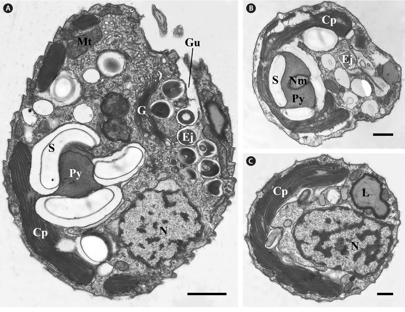
The majority of the organelles of R. reticulata var. atrorosea CCMP 987/6A can be observed in Fig. 1. R. reticulata var. atrorosea had a single chloroplast (Fig. 1). The pyrenoid was slightly dorsal to the center, and the thylakoid membrane did not traverse the pyrenoidal matrix (Fig. 1A & B). The nucleomorph was located in an invagination of the periplastidial compartment into the pyrenoid (Fig. 1B). A Golgi body was positioned ventrally and anteriorly near the gullet (Fig. 1A). Several ejectosomes were observed in two rows near the gullet (Fig. 1A & B). The nucleus was positioned in the posterior of the cell (Fig. 1A & C).
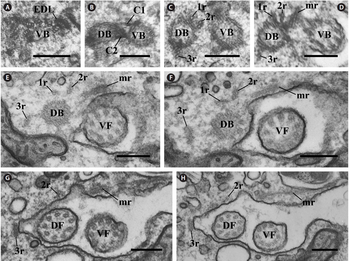
The flagellar apparatus of R. reticulata var. atrorosea was asymmetrical and very complex (Fig. 2A-D). Two flagella protruded on the left and dorsal side of the gullet. The flagellar apparatus consisted of the following eight major components: a one-stranded microtubular root (1r), a two-stranded microtubular root (2r), a three-stranded microtubular root (3r), another microtubular root (mr), a rhizostyle (Rhs), a mitochondrion-associated lamella (ML), a striated fibrous root (SR), and a striated fiber-associated microtubular root (SRm). Additionally, an electron-dense layer and two connecting structures were observed. The electron-dense layer was connected to a triplet of the ventral basal body (Fig. 2A). Two fibrous connections (C1 and C2) were observed between the two basal bodies (Fig. 2B).
In the flagellar apparatus of R. reticulata var. atrorosea, the microtubular roots were well-developed and composed of the following six types: 1r, 2r, 3r, mr (Figs 2C-H & 3A-D), Rhs, and SRm. The 1r and 2r originated from the
[Fig. 1.] Transmission electron micrographs of general structure. (A) Longitudinal section of Rhinomonas reticulata var. atrorosea showing the
chloroplast (Cp), Golgi body (G), gullet (Gu), nucleus (N), starch (S), pyrenoid (Py), mitochondria (Mt), and ejectosome (Ej). (B) Cross section at
the gullet level showing the Cp, Ej, Py, and nucleomorph (Nm). (C) Cross section at the nuclear level showing the Cp and N. L, lipid. Scale bars
represent: A, 1 μm; B & C, 0.5 μm.
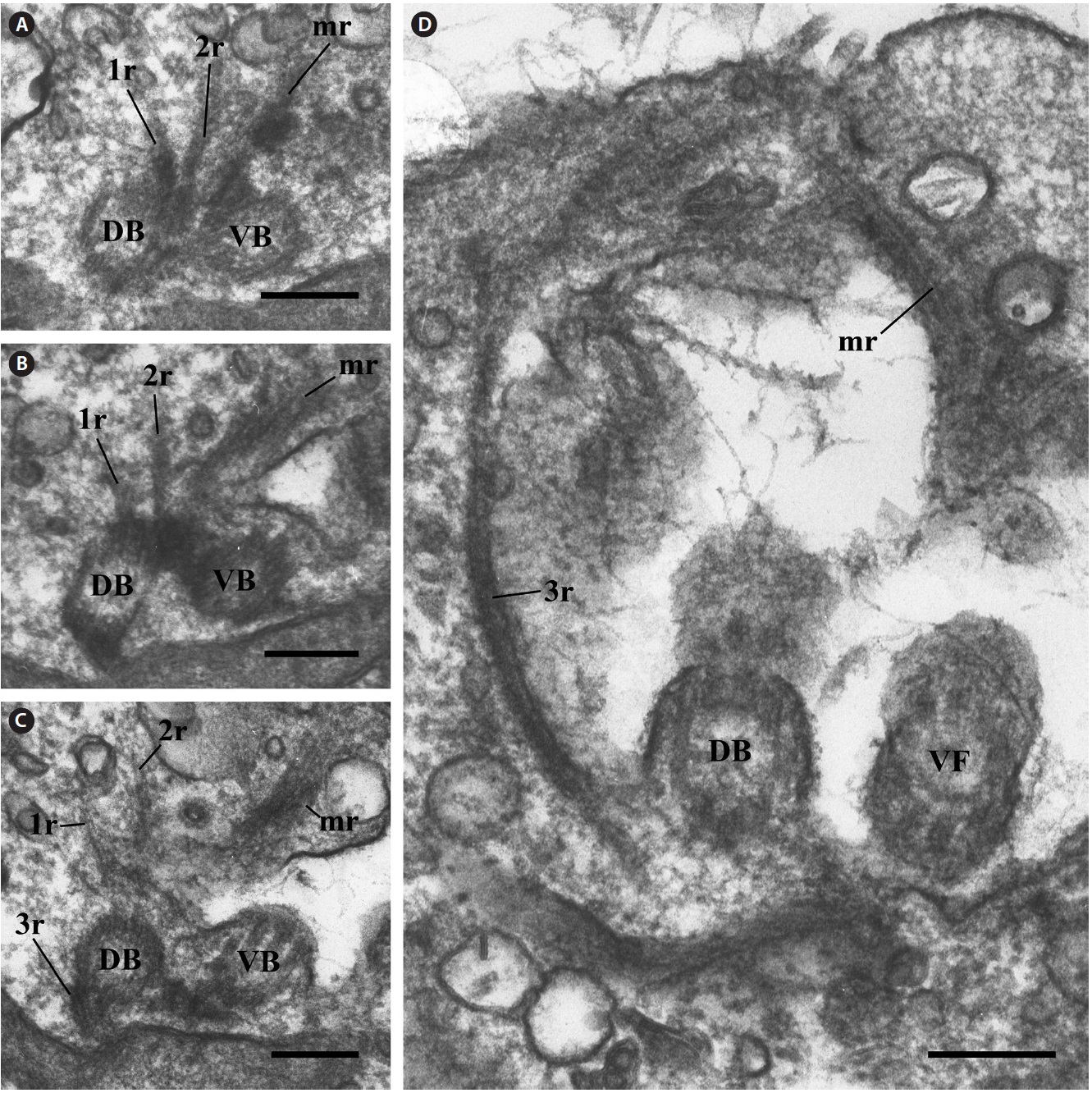
left side of the dorsal basal body (Fig. 2C). They extended more or less parallel with the dorsal basal body (Fig. 2E & F) and the dorsal-left side under the vestibulum together (Fig. 3A-D). Serial sections revealed that the 2r was located more ventrally than the 1r at the origin point (Fig. 2C & D) and extended through a larger radius of curvature than the 1r (Fig. 3A-C). The 3r originated from the right side of the proximal dorsal basal body (Fig. 2C) and extended along the dorsal side of the vestibulum toward the left side of the cell (Figs 2E-H & 3D). The mr originated between two basal bodies, near the dorsal basal body (Fig. 2C). It extended from the ventral side of the cell (Fig. 2E-H) to the anterior left side under the vestibulum similar to the 1r and 2r (Fig. 3A-D). The 1r, 2r, and mr turned counterclockwise, whereas the 3r turned clockwise (Fig. 3A-D). The 3r and mr were also longer than the 1r and 2r (Fig. 3A-D).
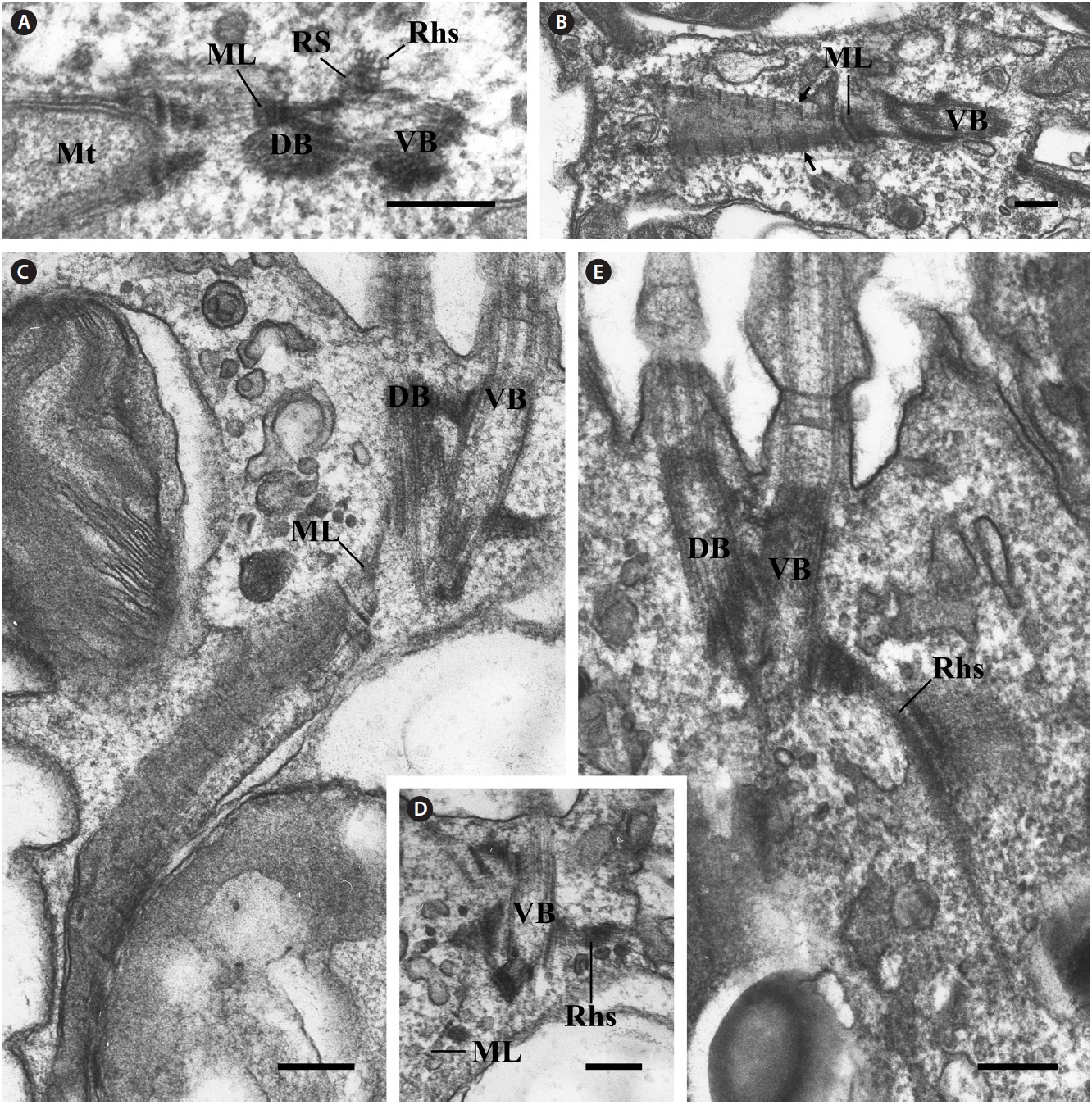
The most prominent and conspicuous component of the flagellar apparatus was the Rhs. The Rhs originated from a rhizostyle-associated striated fiber (RS) between two basal bodies (Fig. 4A). In the cross section, the Rhs comprised three microtubules without a wing-like lamellar projection (Fig. 4A), and it passed near the ventral basal body and extended to the posteriad of the cell through the ventral side (Fig. 4D & E).
An unusual component of the flagellar apparatus was the ML. The ML originated from the RS (Fig. 4A). However, the ML extended toward the dorsal side of the basal body to a mitochondrion (Fig. 3C) in the opposite direction from the Rhs (Fig. 4D) and divided into two bands
[Fig. 2.] Transmission electron micrographs of the connecting structures and microtubular roots at the basal body level. Serial cross sections of the two basal bodies from the posterior to the anterior direction showing the spatial relationships among the microtubular roots of the flagellar apparatus. (A) Cross section of the proximal ventral basal body (VB) showing that the electron dense layer (EDL) is connected to a triplet of ventral basal bodies. (B) Cross section of the two basal bodies showing the connecting structures. C1 and C2 connect with the two basal bodies. (C & D) Serial cross sections of the two basal bodies showing the origin points of the one-stranded microtubular root (1r), two-stranded microtubular root (2r), three-stranded microtubular root (3r), and another microtubular root (mr). (E-H) Serial cross sections of the two basal bodies showing the four root types. Immediately after genesis, the 1r, 2r, and 3r extended in parallel with the dorsal basal body, but the mr extended toward the ventral side of the cell. DB, dorsal basal body; DF, dorsal flagellum; VF, ventral flagellum. Scale bars represent: A-H, 0.2 μm.
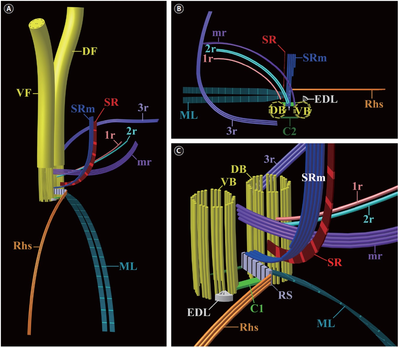
[Fig. 6.] Diagrammatic reconstructions of the flagellar apparatus in Rhinomonas reticulata var. atrorosea. Not to scale. (A) Diagram showing the overall flagellar apparatus. (B) Diagram showing a plane view from above. (C) Diagram showing a magnified view from the oblique left side. C1, connecting fiber 1; C2, connecting fiber 2; DB, dorsal basal body; DF, dorsal flagellum; EDL, electron dense layer; mr, microtubule root; ML, mitochondrion-associated lamella; Rhs, rhizostyle; RS, rhizostyle-associated striated fiber; SR, striated fibrous root; SRm, striated fiber-associated microtubular root; VB, ventral basal body; VF, ventral flagellum; 1r, one-stranded microtubular root; 2r, two-stranded microtubular root; 3r, three-stranded microtubular root.
near the dorsal basal body (Fig. 4B). Then, the ML extended to the posterior of the cell (Fig. 4C). The ML had a regular striped pattern, and its periodicity was measured as 114-125 nm (Fig. 4B & C).
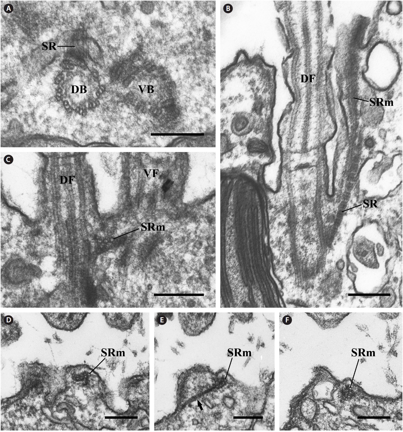
The other major components of the flagellar apparatus were the SR and the SRm. The SR had a striped pattern similar to the ML (Fig. 5B), and the SRm originated between two basal bodies (Fig. 5C). The periodicity of the SR was measured as 42.5-46.3 nm (Fig. 5B). Both structures were parallel and extended together toward the anterior of the cell along the left side of the vestibulum (Fig. 5B). In the longitudinal section of the two basal bodies, the SRm comprised three microtubules (Fig. 5C) and gradually became five (Fig. 5D-F). The diagrammatic reconstruction of R. reticulata var. atrorosea is intended to provide an accurate reconstruction of the flagellar apparatus. However, this representation is not to scale (Fig. 6A-C).
In this study, the general ultrastructures of R. reticulata var. atrorosea were found to be very similar to those of Rhodomonas salina (Santore 1982), Storeatula rhinosa (Kugrens et al. 1999), and Rhinomonas pauca (Hill and Wetherbee 1988) with respect to the chloroplast, pyrenoid and position of the nucleomorph. Therefore, general ultrastructural features may not be useful as diagnostic characteristics among closely related genera. In addition, molecular phylogeny using nuclear and nucleomorph small subunit rDNA sequence data suggested that the genus Rhinomonas was grouped with Rhodomonas (= Pyrenomonas) and Storeatula (Deane et al. 2002, Hoef-Emden et al. 2002, Von der Heyden et al. 2004, Tanifuji et al. 2010).
The overall configuration of the flagellar apparatus in R. reticulata var. atrorosea is similar to those of previous ultrastructural observations for cryptomonads (Roberts et al. 1981, Gillott and Gibbs 1983, Roberts 1984, Hill and Wetherbee 1986, Perasso et al. 1992) but is slightly more complex than those of other species. Specifically, R. reticulata var. atrorosea had one more microtubular root than previously well-studied cryptomonads (Roberts et al. 1981, Gillott and Gibbs 1983, Roberts 1984, Hill and Wetherbee 1986, Perasso et al. 1992). R. reticulata var. atrorosea had six types of microtubular roots (1r, 2r, 3r, mr, Rhs, and SRm), whereas previously reported species had three (Gillardia theta), four (Hanusia phi, Cryptomonas paramecium, and C. pyrenoidifera) or five types (diplomorphs of Proteomonas sulcata and Cryptomonas ovata) of microtubular roots. Among the microtubular roots, the mr and 2r appear to be homologous with either the lateral rootlet (lr) of the diplomorphs of Proteomonas sulcata and Hanusia phi (Cryptomonas sp. Φ in Gillott and Gibbs 1983) or the Cr root of Cryptomonas paramecium (Chilomonas paramecium in Roberts et al. 1981), C. pyrenoidifera (C. ovata var. palustris in Roberts 1984) and C. ovata (Perasso et al. 1992). The 3r is similar to the ascending microtubules of P. sulcata, H. phi, and G. theta; the twelvestranded microtubules of Cryptomonas paramecium; the four-stranded microtubules of C. pyrenoidifera; and the four-stranded microtubules of the diplomorphs of P. sulcata. This study revealed that R. reticulata var. atrorosea has an additional microtubular root.
The Rhs is the largest, most prominent microtubular root in cryptomonad cells. According to Clay et al. (1999), the Rhs is classified into three types: 1) long, keeled Rhs, 2) short, non-keeled Rhs, and 3) no Rhs. In R. reticulata var. atrorosea, the Rhs is short and non-keeled. A short, keeled Rhs was reported in C. pyrenoidifera (Roberts 1984, Fig. 28) and Urgorri complanatus (Laza-Martinez 2012), whereas Cryptomonas paramecium (Roberts et al. 1981, Fig. 1), Hanusia phi (Gillott and Gibbs 1983, Fig. 4a & b) and diplomorph cells of Proteomonas sulcata (Hill and Wetherbee 1986) have a long, keeled Rhs. A non-keeled Rhs was reported in haplomorphs of P. sulcata (Hill and Wetherbee 1986) and Guillardia theta (Gillott and Gibbs 1983). However, Hill and Wetherbee (1986) did not consider the Rhs of the P. sulcata haplomorphs to be typical due to the lack of a lamellar projection. In the case of C. pyrenoidifera, the wing structure of the Rhs was different from those of H. phi, Cryptomonas paramecium and the diplomorphs of P. sulcata in terms of its short length and origin point in an electron-dense sheet (Roberts 1984, Figs 11-14). Therefore, one of the characteristic features of R. reticulata var. atrorosea is that its Rhs originates from the RS and is short and non-keeled (see Fig. 6C).
The second distinctive structure of the flagellar system was an ML. The ML was first described in Cryptomonas paramecium by Roberts et al. (1981) and consisted of two layers. It is an unusual structure for the photosynthetic genus Cryptomonas, although little is known about other cryptomonadian genera. In R. reticulata var. atrorosea, the ML has fine striations similar to that of C. pyrenoidifera. However, the periodicity of the ML striations (approximately 120 nm) is longer than that of C. pyrenoidifera (9-15 nm). One of the most surprising features is that the ML in R. reticulata var. atrorosea divided into two directions towards a mitochondrion.
Another conspicuous component was the SR and its SRm. Most cryptomonad species, including R. reticulata var. atrorosea, have these components in the flagellar apparatus system. In previous studies and in R. reticulata var. atrorosea, the SR and SRm coexist. However, the striation periodicity of the SR ranges from 35 to 80 nm (60-80 nm in Hanusia phi, 45-50 nm in the haplomorphs of Proteomonas sulcata, 35-65 nm in Cryptomonas pyrenoidifera, and 45 nm in Cryptomonas paramecium). The SR periodicity in R. reticulata var. atrorosea was measured as 42-46 nm.
There are three microtubules in the SRm at the origin point in all previously reported species, including R. reticulata var. atrorosea. The SRm of only Cryptomonas pyrenoidifera and R. reticulata var. atrorosea are reinforced with five microtubules. The distinctive feature of the SRm found only in R. reticulata var. atrorosea is the wing-like structure (arrow in Fig. 5E). This structure is observed when the SRm comprises four microtubules. This feature has not been reported in any other cryptomonad species.
In conclusion, we revealed the structure of the flagellar apparatus in R. reticulata var. atrorosea and presented a three-dimensional reconstruction to compare its structure with those of other cryptomonads. The flagellar apparatus of R. reticulata var. atrorosea appears to be more complex than those of other cryptomonad species due to the presence of an additional microtubular root. However, drawing taxonomic or phylogenetic conclusions regarding R. reticulata var. atrorosea at this time is difficult because the available ultrastructural data of the flagellar apparatus in cryptomonads are limited.