검색 전체 메뉴
PDF
맨 위로
OA 학술지
Screening and Evaluation of Antibacterial Metabolites from Entomopathogenic Fungi
  • 비영리 CC BY-NC
  • 비영리 CC BY-NC
ABSTRACT
Screening and Evaluation of Antibacterial Metabolites from Entomopathogenic Fungi
KEYWORD
Entomopathogenic fungi , Fungal metabolites , Antibacterial activity , Escherichia coli
  • Introduction

    Entomopathogenic fungi are natural pathogens of insects and contribute to the regulation of host insect populations in the natural environment. Their mode of action against insects involves the attachment of conidia to the insect cuticle followed by germination, cuticle penetration, and internal dissemination throughout the insect (Vega et al., 2012). During this process, secreted enzymes, protein toxins, and secondary metabolites can be used by fungi to overcome the host immune system, mod host behavior, and defend host resources against competing pathogens and saprophytes (Isaka et al., 2005; Molnar et al., 2010).

    Recently, secondary metabolites isolated from entomopathogenic fungi have been reported as potential bioactive substances (Isaka et al., 2005; Molnar et al., 2010). The antimicrobial effect of entomopathogenic fungi [e.g., Beauveria bassiana (Bals.) Vuill., Metarhizium anisopliae (Metchn.) Sorokin, Isaria spp., and Lecanicillium spp.] in the suppression of plant disease caused by plant pathogens such as Alternaria solani, Botrytis cinerea, Fusarium oxysporum, Rizoctonia solani, and Pythium myriotylum have been reported by numerous studies (Goettel et al., 2008; Lee et al., 2005). Additionally, these metabolites exhibit a wide variety of insecticidal, antibacterial, antifungal, anticancer, antioxidant, and antiviral activities, and they have been suggested as potential candidates for the development of new bioactive agents (Isaka et al., 2005; Pedras et al., 2002; Wang and Xu, 2012).

    These findings confirm the potential of entomopathogenic fungi as sources of lead compounds in pharmaceutical interests. Therefore, in this study, we report our results by assessing the ability of entomopathogenic fungi isolated in Korea (Shin et al., 2013) to produce antibacterial metabolites against Escherichia coli and their biochemical characteristics.

    Materials and Methods

      >  Entomopathogenic fungal metabolites

    Entomopathogenic fungi, which exhibit virulence against the great wax moth (Galleria mellonella L. [Lepidoptera: Pyralidae]), were used in this study (Table 1) (Shin et al., 2013). For initial screening of antibacterial compounds, 1 ml of sabouraud dextrose broth containing a yeast extract medium (SDYB; 10 g Bacto Peptone, 40 g Dextrose, and 10 g yeast extract in 1000 ml distilled water; pH 6.0) was placed in a 2-ml microcentrifuge tube. The samples were then inoculated with a one-agar block (6 mm) of fungi collected from 2-week-old potato dextrose agar (PDA; Difco, USA). Samples were cultured at 25℃ in the dark and were shaken at 150 rpm for 10 days, after which the samples were centrifuged at 13,000 rpm for 10 min. The aqueous upper layer was transferred into a Plasmid DNA collection kit column (COSMO GENETECH) to filter spores and mycelia.

    For quantitative assay, all fungal conidia were obtained by scraping a 2-week-old PDA plate and suspended in a 0.05% Tween-80 solution. The conidial suspension was vigorously agitated and filtered through cotton to remove mycelial debris. After counting the number of conidia, 50 μl of the conidial suspension (2 × 10⁶ conidia/ml) was inoculated with 20 ml of SDYB medium in a 100-ml Erlenmeyer flask and cultured using the abovementioned method. After 10 days, the samples were centrifuged at 13,000 rpm for 20 min. The pellet was removed and the supernatant was filtered using a 0.45 μm membrane

    [Table 1.] Entomopathogenic fungal isolates used in this study

    label

    Entomopathogenic fungal isolates used in this study

    filter (ADVACTEC No. 2) to separate the crude extract from the mycelium and spore mass. All fungal cell-free culture filtrates were stored at -70℃ until antibacterial activity could be detected.

      >  Bacterial strain

    For the preparation of bacteria, E. coli XL1Blue was cultured using a LB media (10 g tryptone, 5 g yeast extract, and 5 g sodium chloride in 1000 ml distilled water; pH 7.0). One ml of E. coli culture suspension incubated overnight was inoculated into 100 ml of LB media and grown to the mid-exponential phase (O.D.650 nm = ~0.5).

      >  Antibacterial activity assay

    Antibacterial activity against E. coli was determined using a broth dilution method (du Toit and Rautenbach, 2000). The bacterial cells of the mid-exponential phase were diluted with LB media to 1 × 10³ CFU/ml (for the screening assay) and 5 × 10⁵ CFU/ml (for the quantitative assay), respectively. For the screening assay, 100 μl of bacterial suspensions were placed in 96 wells with 100 μl of the fungal culture filtrate and incubated at 37℃ for 16 h. To determine bacterial growth inhibition, the absorbance of the each well was measured at 650 nm by using a microplate reader (Molecular Devices, UK). A bacterial culture well grown in SDYB or LB medium without the fungal culture filtrate was used as the positive control well.

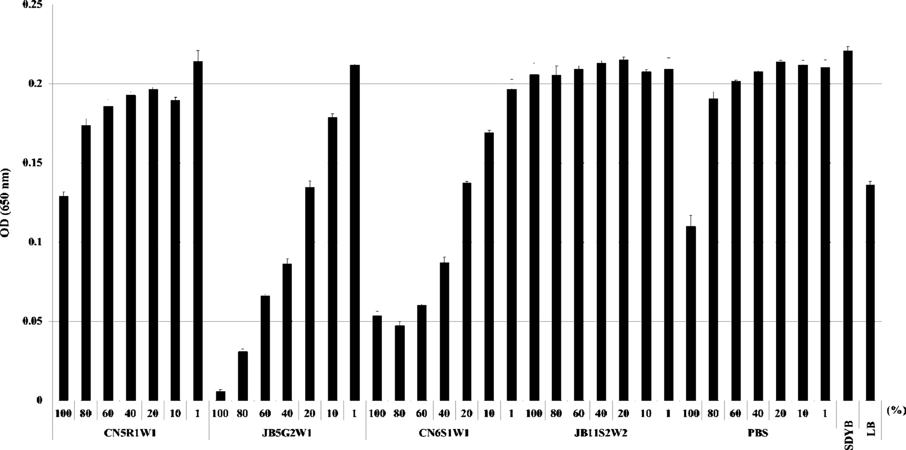
    For the quantitative assay, 100 μl of bacterial suspensions were placed in 96 wells and then mixed with 100 μl of the fungal culture filtrate at different concentrations (i.e., 1%, 10%, 20%, 40%, 60%, 80%, and 100%). The fungal culture filtrate was diluted with SDYB media. For the control, bacterial suspensions were cultured in SDYB media with different concentrations of PBS in place of the fungal culture filtrate. All experiments were performed in triplicate.

      >  MTT assay

    Bacterial cell viability was assessed by MTT [3-(4,5- dimethylthiazol-2-yl)-2,5-diphenyltetrazolium bromide] assay based on the reduction of MTT to formazan dye by active mitochondria. Briefly, 20 μl of the MTT solution (5 mg/ml in PBS) was added to each well and incubated for 4 h. Then, the MTT-spiked media was carefully removed by centrifugation at 13,000 rpm for 10 min, and formazan formed in the cells was dissolved in 200 μl of dimethyl sulfoxide. Absorbance at 540 nm was read using a micro plate reader (Molecular Devices, UK).

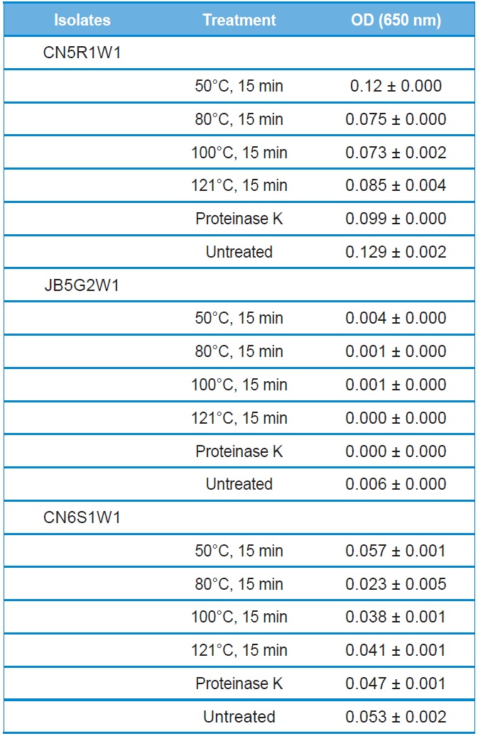
      >  Stability of antibacterial activity of culture filtrate

    For determining the heat stability, the culture filtrate was treated at various temperatures (50℃, 80℃, 100℃, and 121℃) for 15 min. After thermal shock, the culture filtrate was quickly cooled to 25℃, and its antibacterial activity was evaluated.

    To test its stability in the presence of a protease, the culture filtrate was treated with proteinase K (Sigma, USA) at a final concentration of 1 mg/ml under the condition of 37℃ for 2 h. Then, the culture filtrate was autoclaved at 121℃ for 15 min to inactivate the enzymes before the antibacterial activity assay was conducted.

    Results

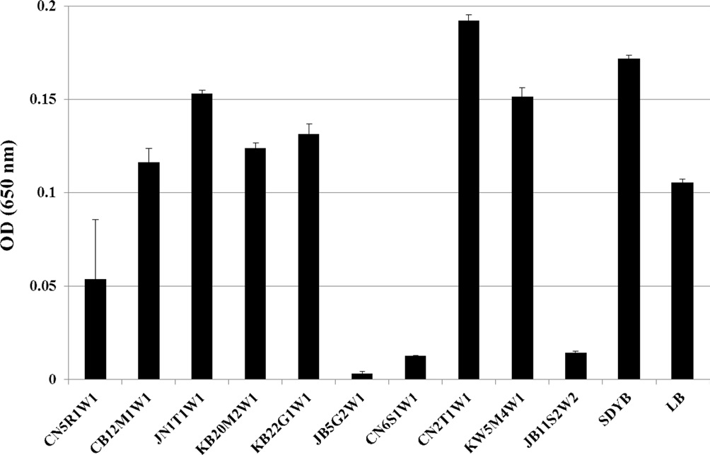
      >  Screening of fungi showing antibacterial activity

    Primary screening for the antibacterial activity of entomopathogenic

    fungal isolates was carried out using a broth dilution assay. Among the 10 isolates tested, the culture filtrate of 4 isolates (i.e., B. bassiana CN5R1W1, M. anisopliae var. anisopliae JB5G2W1, CN6S1W1, and Myrothecium sp. JB11S2W2) exhibited high antibacterial activity (Fig. 1). Bacterial growth was reduced to approximately 2?20 and 3?30 times that in the LB and SDYB media, respectively. These fungal isolates were used for further studies to clarify the production of antibacterial compounds.

      >  Quantitative assay of antibacterial activity

    Fungi were selected for scale-up culture with the same concentration of conidia and they were them subjected to a quantitative assay. The culture filtrate of M. anisopliae var. anisopliae JB5G2W1 showed the highest antibacterial activity, followed by that of M. anisopliae var. anisopliae CN6S1W1 and B. bassiana CN5R1W1 (Fig. 2). Antibacterial activity increased with increasing concentrations of the culture filtrate. However, the culture filtrate of Myrothecium sp. JB11S2W2, which showed a high activity rate at the initial screening stage, did not show any significant antibacterial activity in the quantitative assay.

      >  Cell viability assay

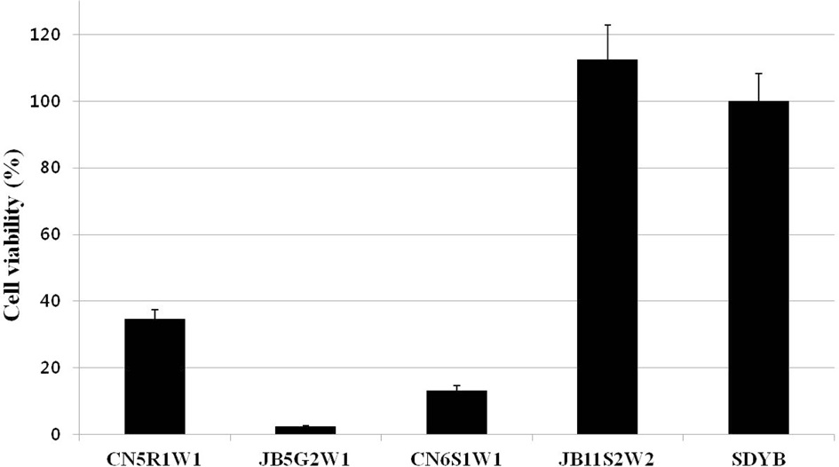
    Surviving bacterial cell numbers in each well treated with 100% of the culture filtrate were determined indirectly by MTT

    assay. As a result, for all fungal culture filtrates, no differences were detected between the O.D. (Fig. 2) and cell viability values (Fig. 3) obtained by both assays.

      >  Stability of antibacterial activity

    The stability of culture filtrates to heat and protease treatments were evaluated for antibacterial activity. Heat treatment did not influence the antibacterial activity of the culture filtrate for all tested isolates (Table 2). Furthermore, the culture filtrate retained its antibacterial activity even after treatment at 121℃ for 15 min. The hydrolytic enzyme, proteinase K, also had no effect on the antibacterial activity of all culture filtrates (Table 2).

    Discussion

    Beauveria bassiana and M. anisopliae are the most commonly isolated species in the world (Vega et al., 2012), thus making them the most widely known among entomopathogenic fungi and fostering their use as biocontrol agents against a variety of pests in various countries (de Faria and Wraight, 2007). These 2 species are also the most commonly used in the study of fungal metabolites (Pedras et al., 2002; Wang and Xu, 2012). In the present study, B. bassiana and M. anisopliae (including Isaria farinosa and Myrothecium sp.) culture filtrates were tested for their antibacterial activity against E. coli. Initial screening results showed that 4 of 10 isolates exhibited high antibacterial activity against bacterial cells (1 × 10² CFU). These results corresponded with previous reports that entomopathogenic fungi can exhibit antibacterial activity (Isaka et al., 2005; Pedras et al., 2002; Wang and Xu, 2012). However, Myrothecium sp. JB11S2W2 showing high antibacterial activity in the initial screening assay did not show any antibacterial activity against a high concentration of bacterial cells (i.e., 5 × 10⁴ CFU; Fig. 2). This result may be explained by 2 lines of reasoning. First, the data obtained may be because of differences in the antibacterial compound yield between initial screening and quantitative assays. In the initial screening assay, the concentration of conidia for inoculation

    [Table 2.] Effect of temperature and enzymatic shock on the antibacterial activity of culture filtrates of entomopathogenic fungi. Data represent mean values of 3 replicates with standard error

    label

    Effect of temperature and enzymatic shock on the antibacterial activity of culture filtrates of entomopathogenic fungi. Data represent mean values of 3 replicates with standard error

    was not determined, but it was determined and inoculated in the quantitative assay. The conidial concentration for inoculation may have been insufficient to produce a substantial yield of antibacterial compounds from Myrothecium sp. JB11S2W2. Second, it may be related to differences in the tested bacterial concentrations. Although antibacterial compound(s) from Myrothecium sp. JB11S2W2 was sufficient to reduce the growth of low concentrations of bacteria (i.e., 1 × 10² CFU), it may not have affected all bacterial cells at high concentrations (i.e., 5 × 10⁴ CFU). These reasons should be clarified through further studies.

    To determine whether the antibacterial activity of the fungal culture filtrate showed cytotoxic or cytostatic effects against E. coli, an MTT assay was performed. The results showed that the antibacterial compound did not completely suppress bacterial growth (Fig. 3). The data presented could not confirm the reason for the antibacterial activity of the fungal culture filtrate. Further studies need to be performed to elucidate this finding.

    Antibacterial compound(s) produced from selected fungal isolates remained stable in response to heat and proteolytic enzyme treatments (Table 2). These results suggest that the antibacterial compound(s) is not a protein. Heat-stable antimicrobial compounds are not commonly produced. Some Class II bacteriocins have been reported to be heat stable (Bharti et al., 2012; Drider et al., 2006). Therefore, the antibacterial compound produced from our fungal isolates may have useful applications in various fields of study.

    Fungi produce a wide range of compounds with biological activities against other organisms; most of these are products of secondary metabolites (Vey et al., 2001). In recent years, beauvericin and destruxins have become well-known secondary metabolites produced from many entomopathogenic fungi such as B. bassiana and M. anisopliae. These metabolites exhibit a wide variety of insecticidal, antibacterial, antifungal, anticancer, and antiviral activities (Pedras et al., 2002; Wang and Xu, 2012). These findings confirm the potential of entomopathogenic fungi as a source for lead structures of pharmaceutical interest. In our study, the antibacterial activity against E. coli was tested and demonstrated by using only entomopathogenic fungi that showed insecticidal activity against the great wax moth. Therefore, other biological activities should also be evaluated, and further investigation into the antibacterial activity compound(s) from our fungal isolates is also required. These studies will increase the usefulness of entomopathogenic fungi as biological control agents in addition to other biological applications.

참고문헌
  • 1. Bharti P, Anand V, Chander J, Singh IP, Singh TV, Tewari R (2012) Heat stable antimicrobial activity of Burkholderia gladioli OR1 against clinical drug resistant isolates. [Indian J Med Res] Vol.135 P.666-671 google
  • 2. de Faria MR, Wraight SP (2007) Mycoinsecticides and mycoacaricides: a comprehensive list with worldwide coverage and international classification of formulation types. [Biol Control] Vol.43 P.237-256 google cross ref
  • 3. Drider D, Fimland G, Hechard Y, McMullen LM, Prevost H (2006) The continuing story of class IIa bacteriocins. [Microbiol Mol Biol Rev] Vol.70 P.564-582 google cross ref
  • 4. du Toit EA, Rautenbach M (2000) A sensitive standardised micro-gel well diffusion assay for the determination of antimicrobial activity. [J Microbiol Methods] Vol.42 P.159-165 google cross ref
  • 5. Goettel MS, Koike M, Kim JJ, Aiuchi D, Shinya R, Brodeur J (2008) Potential of Lecanicillium spp. for management of insects, nematodes and plant diseases. [J Invertebr Pathol] Vol.98 P.256-261 google cross ref
  • 6. Isaka M, Kittakoop P, Kirtikara K, Hywel-Jones NL, Thebtaranonth Y (2005) Bioactive substances from insect pathogenic fungi. [Acc Chem Res] Vol.38 P.813-823 google cross ref
  • 7. Lee SY, Nakajima I, Ihara F, Kinoshita H, Nihira T (2005) Cultivation of entomopathogenic fungi for the search of antibacterial compounds. [Mycopathologia] Vol.160 P.321-325 google cross ref
  • 8. Molnar I, Gibson DM, Krasnoff SB (2010) Secondary metabolites from entomopathogenic Hypocrealean fungi. [Nat Prod Rep] Vol.27 P.1241-1275 google cross ref
  • 9. Pedras MS, Irina Zaharia L, Ward DE (2002) The destruxins: synthesis, biosynthesis, biotransformation, and biological activity. [Phytochemistry] Vol.59 P.579-596 google cross ref
  • 10. Shin TY, Lee WW, Ko SH, Choi JB, Bae SM, Choi JY, Lee KS, Je YH, Jin BR, Woo SD (2013) Distribution and characterization of entomopathogenic fungi from Korean soils. [Biocontrol Sci Techn] Vol.23 P.288-304 google cross ref
  • 11. Vega FE, Meyling NV, Luangsa-ard JJ, Blackwell M (2012) Fungal Entomopathogens; In Insect Pathology: Second Edition. Vega FE, Kaya HK (eds) P.171-220 google
  • 12. Vey A, Hoagland RE, Butt TM (2001) Toxic metabolites of fungal biocontrol agents; In Fungi as Biocontrol Agents . Butt TM, Jackson C, Magan N (eds) P.311-346 google
  • 13. Wang Q, Xu L (2012) Beauvericin, a bioactive compound produced by fungi: a short review. [Molecules] Vol.17 P.2367-2377 google cross ref
OAK XML 통계
이미지 / 테이블
  • [ Table 1. ]  Entomopathogenic fungal isolates used in this study
    Entomopathogenic fungal isolates used in this study
  • [ Fig. 1. ]  Growth of E. coli in LB media with culture filtrates of entomopathogenic fungi for 16 h. Vertical bars correspond to standard error.
    Growth of E. coli in LB media with culture filtrates of entomopathogenic fungi for 16 h. Vertical bars correspond to standard error.
  • [ Fig. 2. ]  Antibacterial activity of culture filtrates of entomopathogenic fungi at different concentrations (1%, 10%, 20%, 40%, 60%, 80%, and 100%) against E. coli for 16 h. Vertical bars correspond to standard error.
    Antibacterial activity of culture filtrates of entomopathogenic fungi at different concentrations (1%, 10%, 20%, 40%, 60%, 80%, and 100%) against E. coli for 16 h. Vertical bars correspond to standard error.
  • [ Fig. 3. ]  Bacterial cell viability for fungal culture filtrates. Cell viability was assessed by MTT assay. Vertical bars correspond to standard error.
    Bacterial cell viability for fungal culture filtrates. Cell viability was assessed by MTT assay. Vertical bars correspond to standard error.
  • [ Table 2. ]  Effect of temperature and enzymatic shock on the antibacterial activity of culture filtrates of entomopathogenic fungi. Data represent mean values of 3 replicates with standard error
    Effect of temperature and enzymatic shock on the antibacterial activity of culture filtrates of entomopathogenic fungi. Data represent mean values of 3 replicates with standard error
(우)06579 서울시 서초구 반포대로 201(반포동)
Tel. 02-537-6389 | Fax. 02-590-0571 | 문의 : oak2014@korea.kr
Copyright(c) National Library of Korea. All rights reserved.