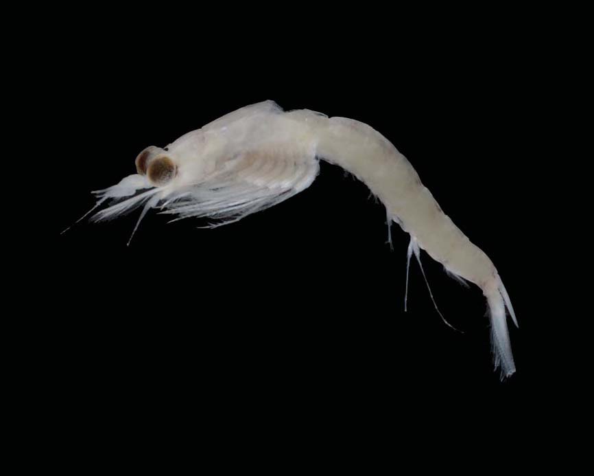
A mysid species, Parastilomysis paradoxa Ii, 1936 belonging to the subfamily Mysinae Hansen, 1910 which comprises approximately 90% of all mysid species, is newly reported from Korean waters. The genus Parastilomysis Ii, 1936 which is also new to Korea, is distinctly different from other genera by having biramous third and fourth pleopods of the male, and telson with an apical cleft. In the present paper, authors provide detailed descriptions and illustrations of P. paradoxa based on the specimens collected from the southern coast of the Korean peninsula, and also discuss on the zoogeographical distribution herein. Parastilomysis paradoxa belongs to Mysidae Haworth, 1825, and is the 50th species in Korea.
The subfamily Mysinae Hansen, 1910 is the most speciose group in family Mysidae Haworth, 1825 and more than 90 percent of the mysid species are distributed worldwide (Tattersall and Tattersall, 1951). The mysinae species is defined by a combination of characters: 1) pleopods of the male variously modified, usually rudimentary in the female, 2) uropod segment undivided, and 3) endopod shorter than exopod.
Up to date 32 species of 14 genera in this subfamily have been reported from Korean waters. During the investigation of the mysid fauna from the southern part of Korea,
All Specimens were collected with a light trap equipment. Specimens were preserved in 70% ethyl alcohol and illustrations have been prepared using a drawing tube on a compound microscope equipped with differential interference contrast (Model BX-60; Olympus, Tokyo, Japan). Images were recorded using a digital camera (Model D7000; Nikon, Tokyo, Japan), and produced with Helicon Focus software (Model Helicon Focus; Helicon Soft Ltd., Kharkov, Ukraine). Body length was measured from the tip of the rostrum to the distal apex of the telson excluding the spine. The simple setae and plumage of the plumose setae on the margin of antennules, antennae, mouthparts, and uropods are omitted from the figures. Terminology for the dissection and measurement is adopted from Tattersall and Tattersall (1951). All specimens have been deposited in the Marine Arthropod Depository Bank of Korea (MADBK), Seoul National University.
Order Mysida Haworth, 1825
Family Mysidae Haworth, 1825
Subfamily Mysinae Haworth, 1825
1*Genus Parastilomysis Ii, 1936
1*Parastilomysis paradoxa Ii, 1936 (Figs. 1-4)
Parastilomysis paradoxa Ii, 1936: 3, figs. 1-14; 1964: 418, fig. 106; Murano, 1970a: 264; 1970b: 146; Mauchline and Murano, 1977: 70; Muller, 1993: 211; Fukuoka et al., 2005: 31, figs. 1, 2.
Material examined. 1♂, Korea: Gyeongsangnam-do, Geoje- si, Nambu-myeon, Dae-po Port, 20 May 2011, Kim M; 3♂♂, Jeollanam-do, Yeosu-si, Samsan-myeon, Geomun-ri, Geomundo Isl., Seo-do port, 28 Jul 2011, Kim M.
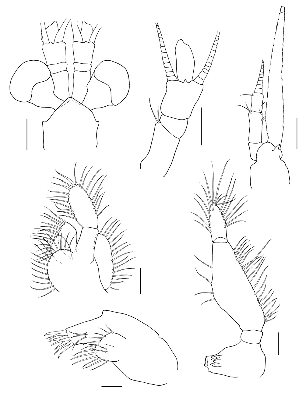
Description. Carapace (Fig. 2A) with anterior margin produced into rounded rostral plate with blunt tip, reaching base of first segment of antennular peduncle; pseudo-rostral process overlapped below carapace, triangular shape in dorsal view; antero-lateral corners rounded.
Eye (Figs. 1,2A) large, reaching 1/2 of third segment of antennular peduncle; cornea distinctly reniform, occupying 1/2 of whole eye.
Antennal scale (Fig. 2C) narrow lanceolate with round apex, about 8 times as long as broad; distal suture occupying 1/24 of whole length; all margins armed with setae. Antennal peduncle (Fig. 2C) 3-segmented; second one longest, 1.3 times as long as first and third one.
Antennular peduncle (Fig. 2B) 3-segmented; first segment twice as long as second one, armed with 2 plumose setae on proximal margin; third segments about 1.5 times as long as second one, with developed processus masculinus.
Maxilla and maxillule (Fig. 2D, E) shown no marked difference from those in other species.
Mandibular palp (Fig. 2F) 3-segmented; second segment about 8.5 times as long as first one, and about 2.4 times as long as third one.
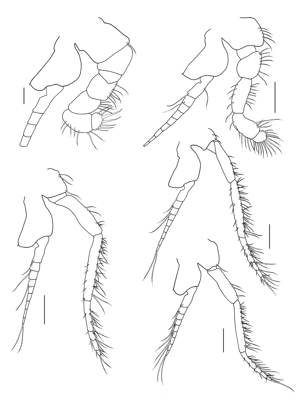
Endopod of first and second thoracopods (Fig. 3A, B) showing no marked difference from those in other species.
Endopod of third to eighth thoracopods (Fig. 3C-E) with 3-subsegmented carpopropodus; basal plate of exopod without spinules on outer distal corner except first, second, and eighth thoracopods which armed with one spinule.
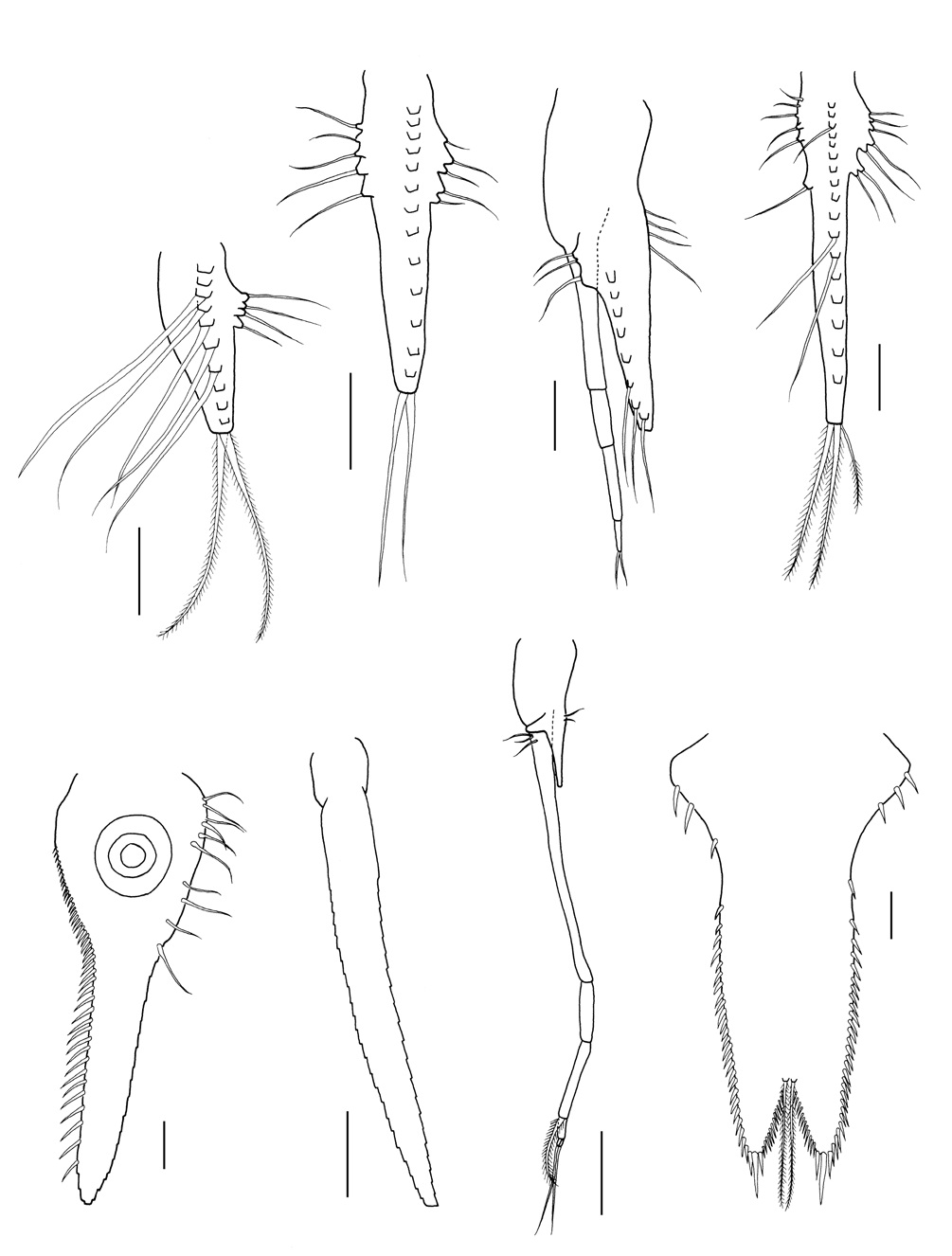
First, second and fifth pleopod of male (Fig. 4A, B, D) uniramous, rudimentary and unsegmented.
Third pleopod of male (Fig. 4C) biramous; endopod thick and unsegmented, about 1.6 times as long as exopod; exopod 4-segmented, first one longest and subequal endopod in length, second one about 2/5 times as long as first one, third
one twice as long as distal one, distal segment terminated by 2 short setae.
Fourth pleopod of male (Fig. 4G) also biramous; endopod small and unsegmented, about 4.2 times as long as exopod; exopod 4-segmented, first one about 2.6 times as long as endopod, second one 1/4 times as long as first one, third one about 1.3 times as long as second one and bearing inwardly curved one strong spinous seta on distal segment, and fourth one 1/4 times as long as third one and terminated by two plumose setae.
Inner uropod (Fig. 4E) bearing about 50 spines in ventral statocyst region, gradually increased in length to distal part; statocyst large and well developed.
Outer uropod (Fig. 4F) about 1.5 times longer than inner one, all margins armed with setae.
Telson (Fig. 4H) broadly linguiform with apical cleft, about 1.7 times as long as basal part; lateral margin of proximal 1/3 armed with 3-4 strong spines, subsequent 1/6 margin naked, remaining region with 25-27 spines tightly; cleft 1/6 times as long as whole length, bearing 15-20 slender spines on entire margin and with one pair of plumose setae in front of anterior end of cleft; each apex truncated and armed with 3 stout spines, medial one longest and robust, and spines on both sides 1/2 times as long as medial one.
Distribution. Korea (the South Sea), Japan (Honshu and Kyushu), China (the East China Sea and Yellow Sea).
Remarks. The examined specimens agree morphologically with the original description of Ii (1936)’s original description in the main characteristics except for the following points: 1) Ii (1936, 1964) noted that the basal plate in the exopod of the first to seventh thoracopods has a spinule on the outer distal corner, and the eighth thoracopod has no spinule. On the other hand, the specimens collected from Geomundo Island of Korea do not have any spinules on the exopod of all thoracopods, and another specimen the from same site has a spinule only in first, second, and eighth thoracopods, respectively. 2) there is a variation in the arrangement of spines at the proximal part of the telson. Three individuals have a naked region at the 1/6 part from the base of the telson, while one of the specimens collected from Geomundo Island has no naked region, which is composed of several stout spines subsequently and increased in length distally. This characteristic is consitent with the description of Fukuoka et al. (2005). 3) As to the third pleopod of the male, the first segment of the exopod is slightly shorter than half of the endopod in the previous record (Ii, 1936, 1964). Whereas in the present specimens, the first segment is longer than the half of the endopod, and the endopod is extended beyond the first segment of the exopod.
Korean name: 1*두갈래꼬리곤쟁이속 (신칭) Korean name: 1*두갈래꼬리곤쟁이 (신칭)