It has been estimated that there are as many as 15,000 species of chironomids worldwide, many of which remain undescribed or undiscovered. Such estimates need to be treated with caution, but there is good evidence to support an estimate in excess of 10,000 species (Cranston, 1995). Sasa and Kikuchi (1995) reported a total of 810 species of chironomids in Japan during the period 1936-1995, and 176 species were added 3 years thereafter to the fauna of chironomids in Japan (Sasa, 1998). In Korea, 32 species of chironomidae including four new species were reported for the first time (Ree and Kim, 1981). Since then, 14 new and 27 unreported species were reported in 11 scientic papers during the period of 1982-2009. In the past three years (2010-2012), 32 new and 47 unreported species were reported (Na, 2004; Ree and Jeong, 2010; Ree et al., 2010, 2011, 2012; Kang, 2012; Ree, 2012). In this report, the fauna of Korean Chironomidae is extended 157 species (including 50 new species), 57 genera, and five subfamilies.
Chironomid adults were collected by sweeping grasses around breeding sites with an insect net during the daytime. Swarming males were collected by sweeping with an insect net in the evenings. At night, a light trap was operated, and light-attracted adults resting on the walls and windows of stores, restaurants and official buildings were aspirated using a sucking tube. The collected specimens were preserved in 75% ethanol. The antennae, head, wings, abdomen, and hypopygium of each specimen were dissected using two fine dissecting needles under a stereomicroscope, and mounted using either phenol balsam or Hoyer’s solution. When Hoyer’s mounting media was used, the cover glasses were ringed with lacquer. Terminology follows Saether (1980).
Measurements of the slide-mounted specimens were obtained from observing the specimens under a compound microscope equipped with a micrometer. Values were transformed to mm or μm using the scale of the micrometer. Abbreviations used are as follows: wing length (WL), antennal ratio (AR), fore leg ratio (LR), radius-medial cross vein (RM), medius-cubital cross-vein (MC), and forked Cu (fCu).
Type specimens were deposited in the collection of Arthro-pods
of Medical Importance Resource Bank, Department of Environmental Medical Biology, Yonsei University.
Order Diptera
Family Chironomidae Holiday
Subfamily Orthocladiinae Edwards
Genus Orthocladius v.d. Wulp
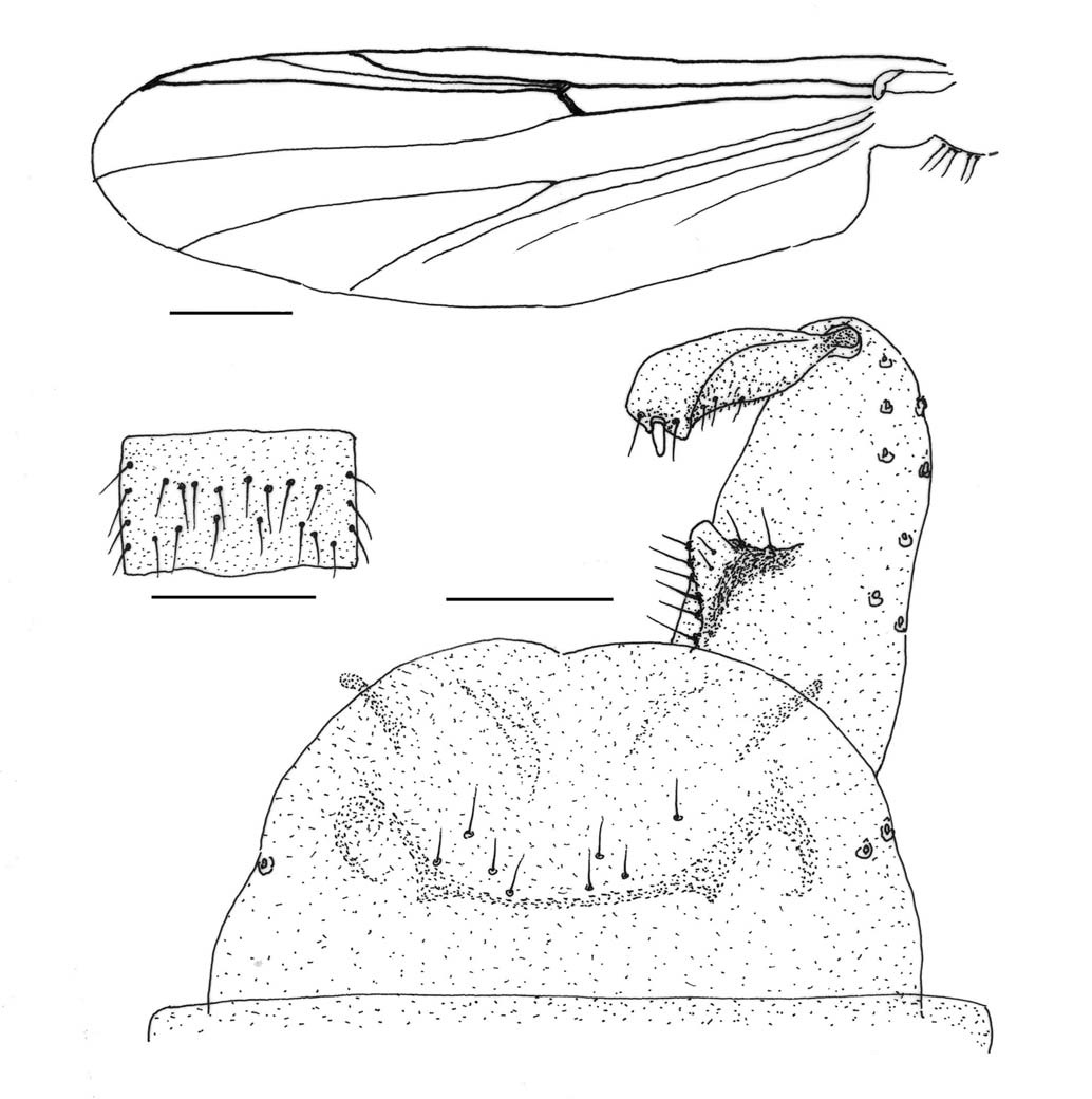
1*Ortholadius manhaei n. sp. (Fig. 1)
Material examined. Holotype: ♂ (RCH-3460), Korea, Gangwon-do, Inje-gun, Manhae village, 11 Nov 2005, Ree HI. Paratypes: 10♂♂, same as holotype.
Diagnosis. Dark brown, medium to large species (WL 3.0 mm). Eye bare. Wing membrane bare. All veins bare, except R. Costa slightly extended. Squama densely fringed. Anal point relatively short (48 μm long), slightly tapered distally, with 4-5 lateral setae. Gonocoxal inner lobe double. AR 1.72. LR 0.85.
Description (male). Head: Eye bare, not produced dorsomedially. Antenna dark brown, 13 segmented, with 5 subapical sensory setae (Fig. 1B). AR 1.72. Palp dark brown, with 5 segments: 25, 79, 139, 137, 203 μm(1 : 3.2 : 5.6 : 5.5 : 8.1). Clypeus with 23 setae. Thorax: Yellowish brown in ground color. Antepronotum brown, well developed, with 9 setae ventrally. Scutum dark brown, vittae inconspicuous, 7-10 dorsocentrals arising from large pale pit, 6 prealars. Scutellum dark brown, with 18 setae. Postnotum dark brown. Wing (Fig. 1A): WL 3.0 mm. Membrane bare. All veins bare, except R. R2+3 ending at 2/5 between R1 and R4+5. Costa slightly extended from R4+5. FCu slightly distal to RM. M3+4 ending under end of R4+5. Cu1 almost straight. An far distal to FCu. Anal lobe well developed. Squama fringed. Arculus and brachiolum dark brown, bare. Legs: All segments uniformly dark brown. Fore tibia with a long narrow spur apically; mid tibia, mid tarsus I, mid tarsus II with 2 short apical spurs, respectively. Pulvillus absent. LR 0.85. Abdomen: All segments dark brown, with irregularly arranged numerous setae. Hypopygium (Fig. 1C): Anal tergite bilobed distally, with 5 short median setae, anal tergal band absent. Anal point relatively short (48 μm long, 20 μm wide at base), slightly tapered distally, with 4-5 lateral setae and round tip. Gonocoxite rather long; gonocoxal inner lobe double: dorsal lobe (Fig. 1D) semi-square with 1 apical, 2 inner lateral, and 4-5 ventral setae, ventral lobe slightly larger, triangular, covered with microtrichia. Gonostylus roughly parallel-sided, with developed crista dorsalis and megaseta.
Female. Unknown.
Etymology. This species is named after late Manhae, who is a famous poet in Korea. The specimens were collected at Manhae village.
Distribution. Korea.
Remarks. This species is somewhat similar to
Genus Paratrichocladius Santos Abreu
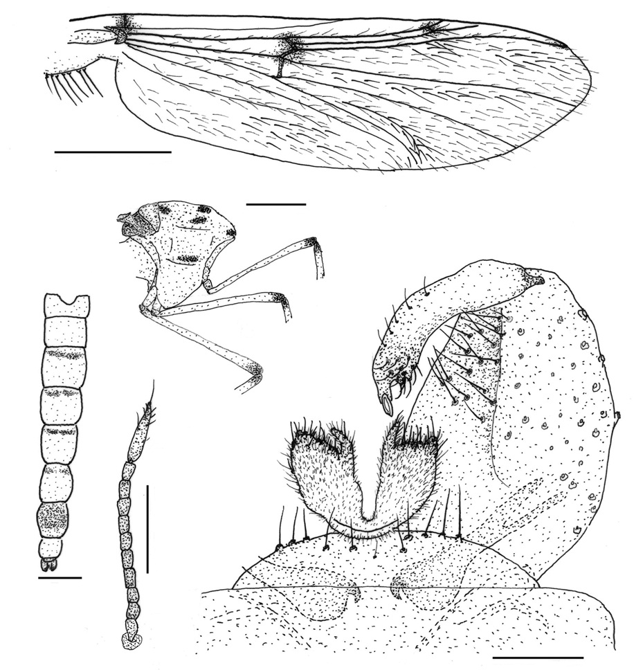
1*Paratrichocladius tamaator Sasa, 1981 (Fig. 2)
Material examined. 2♂♂: Korea, Gyeonggi-do, Munsan, 20 May 2003, Ree HI; 6♂♂: Jeju-do, Seoguipo-si, 2 Sep 1988, Ree HI.
Diagnosis. Brownish, small to medium species (WL 1.4 mm). Eye hairy. Costa extended from R4+5. Cu1 almost straight. Squama with setae. Anal point absent. Anal tergite band absent. Gonocoxal inner lobe triangular, notched dorsally on middle, with rounded tip. AR 1.13. LR 0.59.
Description (Male). Head: Eye hairy, shortly extended dorsomedially. Antenna pale dark brown, 13 segmented, with 7-9 subapical sensory setae. AR 1.13. Palp pale dark brown, with 5 segments: 36, 43, 89, 102, 189 μm(1 : 1.2 : 2.5 : 2.8 : 5.3). Clypeus brown, with 10 setae. Thorax: Brown in ground color. Antepronotum brown, sharply narrowed dorsally, bare. Scutum brown, vittae inconspicuous; 7 small, decumbent acrosticals; 13-14 dorsocentrals, arising from pale pit; 4-5 prealars. Scutellum dark brown, with 8-10 setae. Postnotum brown. Wing (Fig. 2A): WL 1.4 mm. Membrane bare. Costa slightly produced from R4+5. R2+3 ending middle of R1 and R4+5. R4+5 distal to M3+4. FCu slightly distal to RM. Cu1 almost straight. Anal lobe moderately developed. Squama with setae. Legs: Femur brown, lighter basally. Tibia and tarsi I-V light brown. Fore tibia with a narrow, long spur; mid tibia with a very short spur; hind tibia with a long spur and 12-14 comb spurs. Pulvillus absent. LR 0.59. Abdomen: All segments brown; 3-5 lateral setae on tergites III-IV (Fig. 2B). Hypopygium (Fig. 2C): Anal tergite roundly produced distally, with 8 median setae, anal tergal band absent. Gonocoxal inner lobe triangular, notched dorsally on middle, with round tip, with 7-9 inner-lateral setae and 2 small dorsal setae. Gonostylus expanded distally with pale megaseta.
Distribution. Japan, Korea.
Remarks.
2*Genus Rheocricotopus Thienemannn and Harnisch
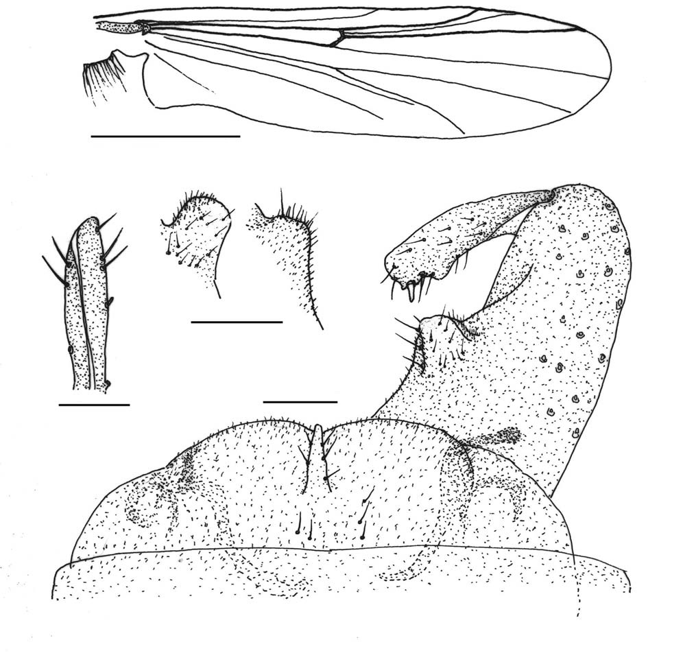
3*Rheocricotopus chalybeatus (Edwards, 1929) (Fig. 3)
Material examined. 5♂♂ (RCH 4040, 4041, 4050, 4106, 4122), Korea, Jeju-do, Seoguipo-si, 2 Sep 1988, Ree HI.
Diagnosis. Small to medium sized, dark brown species (WL 1.4 mm). Eye hairy, reniform. Scutum with large, oval humeral pit. Costa not produced. Anal point triangular, with round tip and several lateral setae. Gonocoxite with two inner lobes. Gonostylus parallel-sided, abruptly bent inward at tip, with distinct crista dorsalis. AR 1.15. LR 0.63.
Description (male). Head (Fig. 3B): Eye reniform, hairy. Antenna brown, with 13 segments. AR 1.15. Palp brown, with 5 segments: 29, 54, 93, 107, 171 μm(1 : 1.9 : 3.2 : 3.7 : 5.9). Clypeus brown, with 13-15 setae. Thorax: Brown in ground color. Antepronotum dark brown, well developed, with 1-3 ventral setae. Scutum brown, vittae dark brown, with large oval humeral pit (Fig. 3C): Acrosticals minute (not able to count); 15 erect dorsocentrals, arising from distinct pale pit; 3-4 prealars. Scutellum dark brown, with 8 setae. Postnotum dark brown. Haltere yellowish brown, elliptic. Wing (Fig. 3A): WL 1.4 mm. Membrane bare. Costa not produced from R4+5. R2+3 ending on middle of R1 and
R4+5. R4+5 distal to M1+2. Cu1 slightly bent in middle. An reaching FCu. FCu slightly distal to RM. Anal lobe moderately developed. Squama with setae. Legs: All segments brown, Tarsi I-V of mid and hind legs slightly lighter (brownish yellow). Fore tibia with a long, narrow apical spur; mid tibia with 2 short spurs; hind tibia with 1 short, 1 long and 10-12 comb spurs. Pulvillus moderately developed. LR 0.63. Abdomen: All tergites dark brown; all sternites brownish yellow. Tergal setae arise as shown in Fig. 3D. Hypopygium (Fig. 3E): Anal tergite with large, triangular bilobes distally, without any setae. Anal point wide at base, tapered apically, with round tip and several lateral setae. Gonocoxite rather long, slightly narrowed distally, with 2 gonocoxal inner lobes: dorsal lobe triangular with round tip, bare; ventral lobe wider with many setae and microtrichia. Gonostylus parallel-sided, abruptly bent inward at tip, with distince cristatdorsalis and megaseta.
Distribution. Europe, Japan, Mongolia, Korea.
Remarks. The morphological key characters of the Korean specimens are consistent with those described for European and Japanese specimens (Edwards, 1929; Lehmann, 1969; Sasa and Kawai, 1987). This species showed a wide range of the geographical variation in body size, i.e. the WL of England specimen was 2.5mm(Edward, 1929), that of Germany’s 1.6-2.0 mm (Lehmann, 1969), that of Japanese’s 1.3 mm (Sasa and Kawai, 1987), and that of Korean’s 1.4 mm. In China, nine species of
Subfamily Tanypodinae Thienemann and Zavrel
Genus Ablabesmyia Johannsen
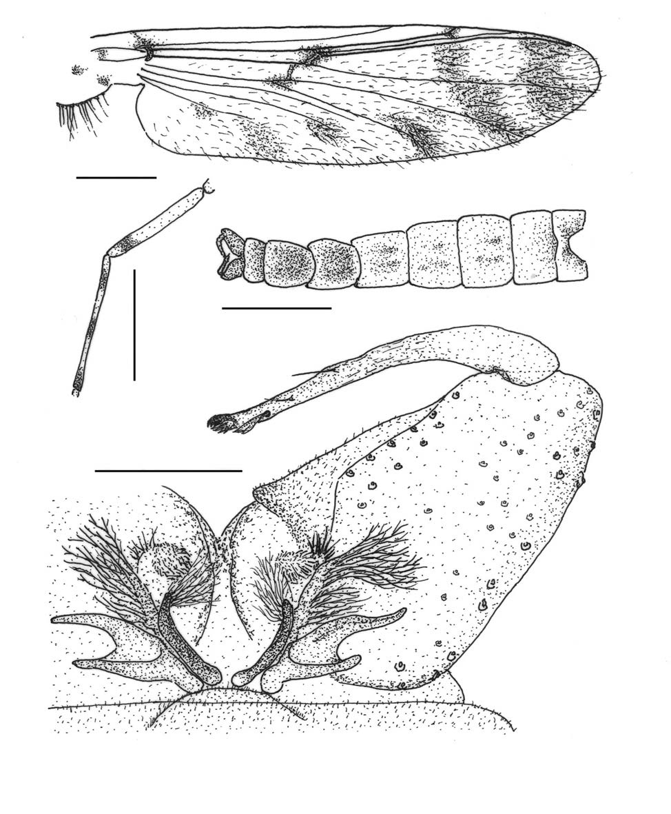
1*Ablabesmyia jeongi n. sp. (Fig. 4)
Material examined. Holotype: ♂ (RCH-5876), Korea, Gangweon-do, Chuncheon-si, Wudu-dong, 1 May 2009, Jeong KY. Paratypes: 3♂♂ (RCH-5881, 5888, 5891), same as holotype.
Diagnosis. Yellowish brown, medium-sized species (WL 2.9 mm). Wing membrane covered with macrotrichia, 3 cloudy patches on anterior and middle of cell r4+5, RM, and FCu. MC distal to FCu and proximal to RM. Gonocoxite large; gonocoxal basal lobe complicated in structure, basal blade and dorsal blade directed outward. AR 1.90.
Description (Male). Head: Eye bare, dorsomedially produced. Antenna dark brown, 14 segmented, 14th segment short with an apical seta. AR 1.90. Palp dark brown, with 5 segments: 65, 137, 209, 144, 335 μm(1 : 2.1 : 3.2 : 2.2 : 5.2). Clypeus dark brown, roughly square, with 35 setae. Thorax: Yellowish brown in ground color. Antepronotum yellowish brown. Scutum brown, with inconspicuous dark brown vittae; 30-34 short dorsocentrals and 28-30 prealars. Scutellum yellowish brown, with 29 short setae. Postnotum dark brown. Wing (Fig. 4A): WL 2.9 mm. Membrane with macrotrichia, faint cloudy patches on anterior part and middle of r4+5, on middle of m1+2, on anterior end of Cu1, on middle of an, and on RM and FCu. MC distal to FCu and proximal to RM. R4+5 distal to M1+2 and proximal to M3+4. Anal lobe moderately developed. Squama fringed. Legs: Femur pale yellow with a subapical dark ring. Tibia pale yellow with subbasal, middle and apical dark rings (Fig. 4B). Tarsus I pale with middle and apical dark rings, tarsi II-III pale with an apical dark ring, tarsi IV-V brownish yellow. Pulvillus absent. Abdomen (Fig. 4C): Tergites I-V pale with faint shade on middle; tergites VI-VII yellowish brown. Hypopygium (Fig. 4D): Anal tergite extremely small. Gonocoxite large; basal lobe of gonocoxite complicated in structure: basal blade and dorsal blade directed outward, lateral filaments long, branched in number. Gonostylus narrow, long, with several teeth and an apical filament.
Female. Unknown.
Etymology. This species is named after Dr. K.Y. Jeong, who collected the type specimens.
Distribution. Korea.
Remarks. This new species has a gonocoxal basal lobe with very unique structures, particularly the outward direction of the long dorsal blade and the short basal blade, which have not previously been observed in other congeners.
2*Genus Hayesomyia Murray and Fittkau
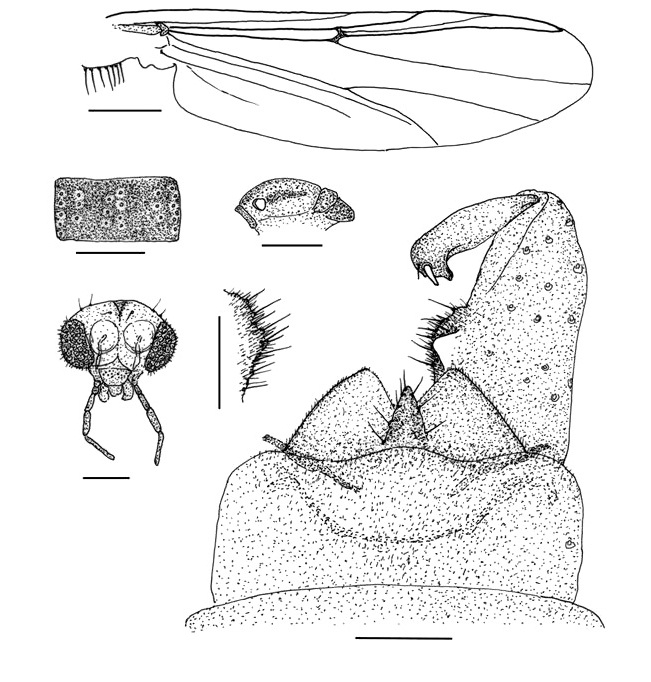
3*Hayesomyia tripunctata (Goetghebuer, 1922) (Fig. 5)
Material examined. 1♂ 2♀♀, Korea, Jeollanam-do, Gureeub, 2 Oct 1977, Ree HI.
Diagnosis. Reddish pale yellow, medium sized species (WL 2.1 mm). Eye bare. Scutum with 4 pairs of dark spots. Wing membrane thickly covered with hairs, 3 dark spots on R2+3, RM, and humeral cross-vein. R2+3 present. Gonocoxal basal lobe large, belobed (inner branch narrow and outer branch broad). Gonostylus abruptly narrowed at tip. AR 1.96. LR 1.14.
Description (male). Head: Eye bare, with narrow dorsomedial extension. Antenna pale dark brown, with 14 segments. AR 1.96. Palp pale yellow, with 4 segments: 82, 168, 193, 250 μm(1 : 2.1 : 2.4 : 3.1). Clypeus brownish yellow, with 28 setae. Thorax (Fig. 5B): Reddish yellow in ground color. Antepronotum yellow, notched medially. Scutum brownish yellow with 4 pairs of dark spots: 1 pair on anterior margin of central vittae, 1 pair on posterior margin of central vittae, 1 pair on anterior margin of lateral vittae, and 1 pair on posterior margin of lateral vittae. One dark spot on anterior anepisternum II. 19 long, erect acrosticals; 17 dorsocentrals; 9 prealars. Scutellum yellowish brown with 17 setae. Postnotum dark brown. Wing (Fig. 5A): WL 2.1 mm. Membrane thickly covered with macrotrichia. 3 dark spots on R2+3, RM, and humeral cross-vein. Costa not produced. R4+5 slightly beyond M3+4. R2+3 present. MCu slightly distal to FCu. An well beyond FCu. Anal lobe developed. Squama fringed.
Arculus dark. Legs (Fig. 5B): All segments pale yellow, with dark spots on apical end of femur and on basal end of tibia. Apical spur of fore tibia with 2 lateral teeth each side; 2 subequal spurs of mid tibia with several lateral teeth, respectively; hind tibia with 1 long spur with 1-2 lateral teeth, 1 short spur with several lateral teeth, and 7-8 comb spines. Tarsus III of mid leg without prominently developed apical setae. Pulvillus absent. LR 1.14. Abdomen (Fig. 5C): Tergites I, II, VIII pale; tergites III-VI pale with basal dark markings; tergite VII brown (distal 1/3 yellowish brown). Hypopygium (Fig. 5D): Anal tergite small, roundly produced distally, with 12 setae; phallapodeme pale, broad ante-riorly, .
hooked at tip, deeply forked posteriorly. Gonocoxite relatively short (164 μm long×129 μm wide); gonocoxal basal lobe large, bilobed, inner branch narrow and outer branch broad, covered with small setae distally and with microtrichia. Gonostylus parallel-sided, slightly bent inward, abruptly narrowed at tip, with several subapical setae ventrally
Female. General morphology same as in male, except for usual sexual differences. Antenna with 11 segments (Fig. 5E).
Distribution. Europe, Korea.
Remarks. The genus
Korean name: 1*만해깃깔따구 (신칭) Korean name: 1*타마깃깔따구 (신칭), 2*유아기깔따구속 (신칭), 3*삼각유아기깔따구 (신칭) Korean name: 1*정알락깔따구 (신칭), 2*집게깔따구속 (신칭), 3*열점집게깔따구 (신칭)