검색 전체 메뉴
PDF
맨 위로
OA 학술지
Embryonic Development of Siberian Sturgeon Acipenser baerii under Hatchery Conditions: An Image Guide with Embryological Descriptions
  • 비영리 CC BY-NC
  • 비영리 CC BY-NC
ABSTRACT
Embryonic Development of Siberian Sturgeon Acipenser baerii under Hatchery Conditions: An Image Guide with Embryological Descriptions
KEYWORD
Acipenser baerii , Siberian sturgeon , Embryonic development , Uneven holoblastic cleavage
  • Introduction

    The Acipenseriformes (Chondrostei) represent a group of primitive, ray-finned fish, comprising Acipenseridae (sturgeons) and Polyodontidae (paddlefishes) (Bemis et al., 1997; Birstein et al., 1997). They are often referred to as ‘living fossils’ of the actinopterygian lineage and have distinctive morphologic and/or developmental attributes (Billard and Lecointre, 2001). Furthermore, some sturgeon species exhibit repeated rounds of genome duplication, resulting in diversification into different ploidy levels among sturgeon species (Blacklidge and Bidwell, 1993; Kim et al., 2005). Based on these unique and interesting characteristics, this extant primitive fish group is regarded as a useful model for evolutionary genetics and genomic studies (Cho et al., 2007; Kim et al., 2009; Akbarzadeh et al., 2011).

    In addition to the interest in their evolutionary aspects, sturgeons have long been recognized as very valuable natural resources for fisheries in Northern Hemisphere countries, particularly with regard to caviar production. However, over the last few decades, wild sturgeon stocks have faced considerable threats to their natural habitats as a result of various anthropogenic and industrial activities (Pikitch et al., 2005). Currently, all extant sturgeon species are considered to be ‘critically endangered’ and are included in the Convention on International Trade in Endangered Species (CITES) list, which applies to global trading. Thus, sustainable, aquaculture-based production has become mandatory for the commercial exploitation of sturgeons and their products (Karpinsky, 2010; Webb and Doroshov, 2011).

    In Korea, the introduction of sturgeon species into the aquaculture domain was first noted in 1999 for the Siberian sturgeon Acipenser baerii (Seong and Baik, 1999). Early successful production of the Siberian sturgeon fingerlings using adult brooders on Korean fish farms was achieved by both private and nonprofit organizations in the early 2000s (personal communication). These pioneering works have encouraged the progressive expansion of Siberian sturgeon culturing in Korea over the past decade, although the efficiency and capacity of farming practices for Siberian sturgeon remain to be improved. The establishment of effective guidelines for embryonic and larval development is one of the most important prerequisites for developing an optimal protocol for reproductive control in hatchery management. Despite its importance, comprehensive information on the embryonic development of the Siberian sturgeon, guided by a complete set of relevant image data, has not become available to date, with the exceptions of brief and general descriptions in the literature. The objective of the present study was to provide a comprehensive image-based guide to the embryologic development of Siberian sturgeons that are artificially propagated under hatchery conditions.

    Materials and Methods

      >  Broodfish and hormonal artificial spawning

    The first experiment for the morphologic staging of embryonic development was performed in March 2011, and this experiment was replicated in March 2012. The broodfish used for artificial spawning and gamete collection were individuals that were produced in 2003. The broodfish were maintained at ambient temperature (range, 12 to 20℃) at Dinoville Aquafarm Inc. (Hamyang, Korea). Fish were sexed by external gonad biopsy at 6 years of age. Out of six (in 2011) or four (in 2012) female candidates, four (average body weight [BW], 14 ± 2.6 kg) and three individuals (13 ± 4.6 kg BW) were selected for hormonal induction of spawning in 2011 and 2012, respectively. When necessary, polarization index (PI) observations using catheterized oocytes were conducted (PI scores < 0.1) (Van Eenennaam et al., 1996). In addition, four males each for 2011 (7 ± 2.7 kg BW) and 2012 (7 ± 3.1 kg BW) were selected based on the presence of milt, which was obtained by either palpation of the abdomen or catheterization using a silicon tube-connected syringe into the genital duct. The selected females were administered the primary intramuscular injection of the luteinizing hormone-releasing hormone analogue des- Gly10, [D-Ala6] LH-RH ethylamide (LHRHa; Syndel Laboratories Ltd., Qualicum Beach, BC, Canada) at 10-15 μg/kg BW. At 12 h after the primary injection, each female was injected again with LHRHa at 90-135 μg/kg BW as a resolving dose. The male broodfish were injected with LHRHa at 100 μg/kg BW. The temperature of the water during the induced spawning was adjusted to 15.0 ± 0.5℃ in both years. After injection, the fish were incubated in individual spawning tanks until a low number of ovulated eggs was released, which usually occurred around 36 h after the second injection of hormone.

      >  Artificial insemination and egg incubation

    When ovulation followed by spawning was identified, milt was collected from the males using a silicon tube-connected aspirator and stored at 2-4℃ until used. After milt collection, the eggs were removed from the spawned females by hand stripping and Caesarean section. The eggs were mixed with milt (diluted 1:200 with water) for 150 s, and then rinsed with fresh water. To remove the adhesiveness of the fertilized eggs, the eggs were mixed continuously with Fuller’s earth (Sigma- Aldrich, St. Louis, MO, USA) and washed with fresh water several times for 30 min. The fertilized eggs were placed in McDonald incubation jars until hatched. Dead embryos were removed at 6-h intervals. The water temperature was maintained at 18.0 ± 0.5℃ until hatching.

      >  Sampling of biological specimens for developmental staging

    In 2011, the development of embryos from two mating pairs was examined. Based on a modified version of the developmental stages defined for Russian sturgeon, Acipenser guldenstadtii (Dettlaff and Vassetzky, 1991), the developmental stages of the Siberian sturgeon embryos were assigned to 30 stages, ranging from just-fertilized to hatching (Table 1). After artificial insemination, the embryos were collected every hour until the beginning of gastrulation, every 2 h until the onset of heart beating, and every 4-6 h until the first occurrence of hatching. From each mating group, 30-50 embryos were sampled at each detection point and fixed in cold 4% paraformaldehyde. Under the stereomicroscope (AZ100; Nikon, Tokyo, Japan), the embryos that had reached each stage were recorded. Based on the examination in 2011, developmental progress to each stage was confirmed with embryo batches from two independent mating pairs in 2012, and the morphologic characteristics of the developing embryos at each stage were analyzed using the NIS-Elements BR image analysis software (Nikon), which was implemented in the AZ100 microscope.

    Results

      >  Early cleavage events

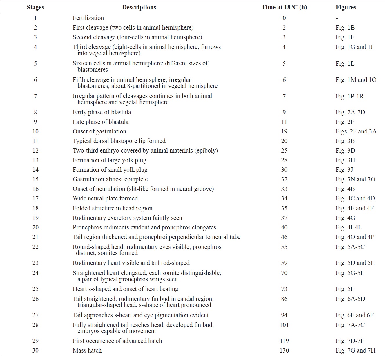
    The developmental staging results are listed in Table 1. Upon fertilization, a black pigment developed at the animal pore, and the first cleavage furrow appeared at the animal pore at 2 h post-fertilization (HPF) (Fig. 1A). This cleavage furrow was limited to the animal hemisphere (Fig. 1B and 1C). Following the first cleavage (1 h later), the second cleavage formed typical 4-cell (four equal-sized blastomeres) embryos (Fig. 1D and 1E). Although the second cleavage furrow did not divide completely the vegetal hemisphere, partial infiltration of the vegetal hemisphere was observed (Fig. 1F). At 4 HPF, 8-cell embryos (similarly sized blastomeres) were formed in the animal hemisphere through the third cleavage event (Fig. 1G and 1H). At that time-point, the cleavage infiltrated the entire vegetal hemisphere and the vegetal hemisphere

    [Table 1.] Staging of embryonic development in Siberian sturgeon Acipenser baerii

    label

    Staging of embryonic development in Siberian sturgeon Acipenser baerii

    was completely divided into four similarly sized compartments (Fig. 1I). When the 16 blastomeres were formed in the animal hemisphere (at 5 HPF; fourth cleavage), one half (newly formed) of the 16 blastomeres in the center of the animal hemisphere were much smaller than the remaining half (earlier ones) (Fig. 1J-1L). The sizes and shapes of the blastomeres that resulted from subsequent cleavages in the animal hemisphere were noticeably dissimilar (Fig. 1M and 1N), and the furrows in the vegetal hemisphere were also formed in an irregular manner (Fig. 1O). This irregular pattern of division continued in both the animal (Fig. 1P and 1Q) and vegetal (Fig. 1R) hemispheres.

      >  Blastulation

    As cleavage continued, small blastomeres proliferated in the animal hemisphere (Fig. 2A), concomitant with irregular divisions in the vegetal hemisphere (Fig. 2B). The blastomeres in the animal hemisphere became uncountable (Fig. 2C), although large blastomeres were still formed in the vegetal

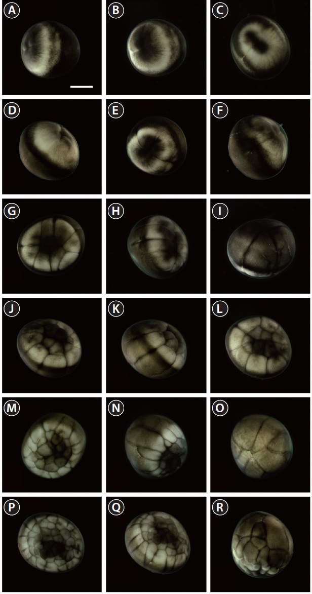
    hemisphere (Fig. 2D). Cell division meant that the blastomeres became indistinguishable from one another in the animal hemisphere under the microscope at low-power magnification, and a cleavage cavity (blastocoel) began to form at the apex of the animal hemisphere (9 HPF). Meanwhile, in the vegetal hemisphere, relatively small blastomeres were located near the marginal zone (the equator), while larger blastomeres were evident in the vegetal apex region. At 11 HPF, the primordial cleavage cavity became larger and more evident in the animal hemisphere, whereas cell division continued to generate smaller blastomeres in the vegetal hemisphere (Fig. 2E). Nearing the completion of blastulation (18-19 HPF), the smooth appearance of the animal hemisphere changed to a milky white blastula roof (Fig. 2F).

      >  Gastrulation

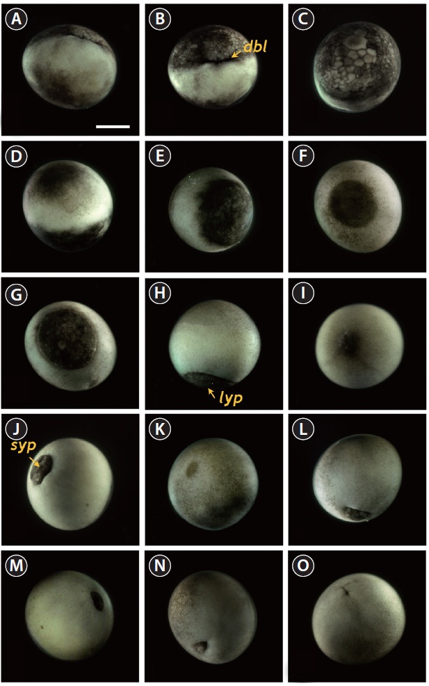
    Upon the onset of gastrulation (19-20 HPF), a band was formed close to the equator, and this was followed by the formation of a “dorsal blastopore lip” between the animal and vegetal hemispheres (Fig. 3A and 3B). At this time-point, the vegetal hemisphere contained a number of divided blastomeres of differing sizes, whereby relatively large, countable (distinguishable) blastomeres were present in the region close to the apex and smaller (often uncountable) blastomeres were detected in the region close to the marginal zone (Fig. 3C). The blastula roof of the animal hemisphere enveloped progressively the vegetal hemisphere. At 25 HPF, approximately two-thirds of the embryo were covered by blastoderm (Fig. 3D and 3E), and a darkly pigmented region was formed in a round shape at the surface of the animal pore in many, albeit not all, of the embryos (Fig. 3F). As gastrulation continued, epiboly covered more than two-thirds of the embryos, and the area of remaining vegetal material declined progressively (Fig. 3G). At 28 HPF, a large yolk plug was formed, and the darkly pigmented region at the animal pore decreased (Fig. 3H

    and 3I). About 2 h later, the size of the yolk plug was further reduced to less than one-fifth the diameter of the embryo (i.e., small yolk plug formation) (Fig. 3J). The pigmented region at the surface of the animal pore was also markedly reduced in size and coloration (Fig. 3K). The size of the yolk plug gradually decreased (Fig. 3L) until the blastopore appeared only as a small circle at the apex of the vegetal pore (Fig. 3M and 3N). As gastrulation neared completion (32 HPF), the blastopore took on a slit-like appearance, which signaled the initiation of neurulation (Fig. 3O).

      >  Neurulation

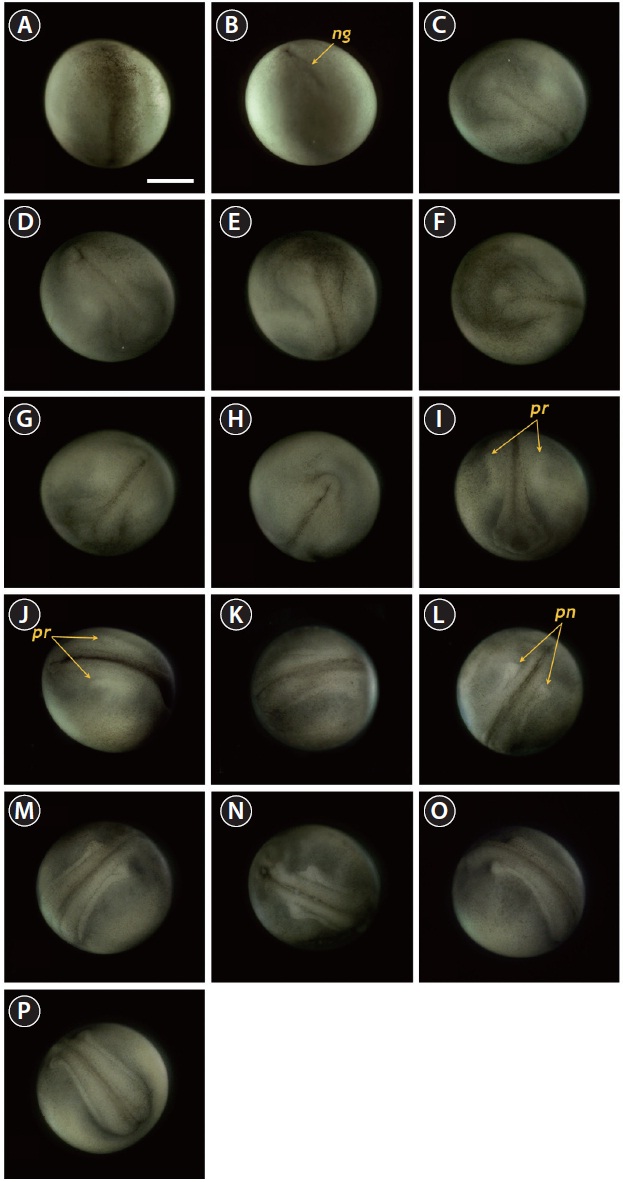
    Neurulation began with the appearance of a slit-shaped

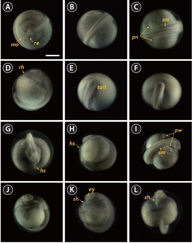
    neural groove in the blastopore (Fig. 4A and 4B), followed by the formation of the neural plate at the dorsal surface with the folded structure in the head region (33 HPF) (Fig. 4C and 4D). With the progression of development (33-35 HPF), the neural plate in the head region widened and the neural folds rose and thickened (Fig. 4E and 4F). In the dorsal region, the neural groove was more evident and the excretory system rudiments were faintly visible parallel to the neural groove (37 HPF) (Fig. 4G). At that time-point, a folded shape was seen in the tail region (Fig. 4H). As neurulation continued, the folding

    of the head region was slightly reorganized and a pair of pronephros rudiments became more evident as cords running parallel to the neural groove (40 HPF) (Fig. 4I and 4J). The neural folds rose significantly and the anterior part of the pronephros was visualized as a distinct form (Fig. 4K and 4L). Thereafter, the pronephros became elongated and thickened along with the risen neural folds (Fig. 4M and 4N). The tail region continued to thicken significantly as development progressed, although it had not yet detached from the yolk sac membrane. At this stage, the pronephroi were located almost perpendicular to the neural tube and the neural tube was mostly closed (46 HPF) (Fig. 4O and 4P).

      >  Progress to the onset of heart beating

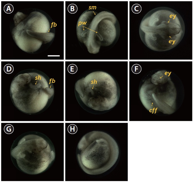
    At 55 HPF, the lateral plates were fused to the prosencephalon (forebrain), and the head region appeared as a roundshaped object in the dorsal view, in which the rudimentary

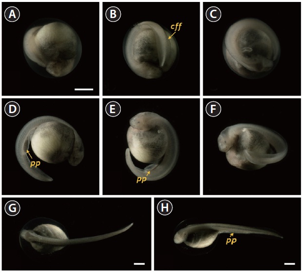
    eyes and undeveloped mouth were easily visible (Fig. 5A). At this stage, the dorsal region had a rod-shaped appearance with developed somites, and the primary pronephros-wings were visible (Fig. 5B and 5C). A rudimentary heart formed in the embryo (59 HPF), although the head had not yet separated (Fig. 5D). In the caudal region, the flattened tail was transformed to a rod-shaped structure and the tail had begun to separate from the yolk sac (Fig. 5E and 5F). As development continued (60-70 HPF), the head region thickened and began to separate from the yolk, in which the heart was seen as a short straight tube (Fig. 5G). The tail continued to lengthen and pronounced somites were visible over the entire embryonic body (Fig. 5H and 5I). At this point, a pair of highly differentiated pronephros-wings could be observed (Fig. 5I). At 73 HPF, the heart became s-shaped (the so-called s-heart) and began to beat. The eyes were more distinct in this stage (Fig. 5J-5L).

      >  Progress to hatching

    At 86 HPF, the round, rod-shaped structure of the tail straightened, and fin-bud rudiments were visible in the caudal region (Fig. 6A). Other morphologic features typical of the embryos at this stage were: separation of the anterior part of the head from the yolk sac; the triangular shape of the developed head; slightly pigmented eyes; and a more pronounced s-heart (Fig. 6B-6D). At 94 HPF, the tail reached the beating

    s-heart and fin folds appeared in the caudal region (Fig. 6E- 6H). Blood circulation was easily observable, and olfactory organs were also seen. As the tail continued to lengthen, it approached the head (101 HPF). Eye caps were evident and caudal fin folds were easily distinguishable (Fig. 7A-7C). The embryos at this stage were capable of movement. From 119 HPF, several advanced embryos began to hatch, and mass hatching was observed at 130 HPF. The average hatching success based on duplicate examinations was 80.5%. Individuals from advanced hatches were often bent in shape (Fig. 7D-7F), whereas most of the mass-hatched prolarvae (i.e., yolk-bearing) had a straight shape (Fig. 7G and 7H). At hatching, the average total length of the prolarvae was 10.5 mm. They had a pigment plug (i.e., yolk plug), which is a typical feature of sturgeon prolarvae.

    Discussion

    Unlike many teleostean embryos that show the meroblastic cleavages on the blastodisc exclusively in the animal hemisphere, sturgeon embryos display holoblastic cleavage (Conte et al., 1988). In general, the holoblastic cleavage pattern observed in the present study is in agreement with those previously defined for other Acipenseriform fishes (Dettlaff et al., 1993). In similarity with other sturgeon species, the Siberian sturgeon exhibits an uneven pattern of holoblastic cleavage in which the vegetal hemisphere is not completely divided with each cleavage furrow. This asymmetrical cleavage pattern in sturgeon embryos is different from the holoblastic cleavage seen in anuran embryos. This difference is attributable to the uneven distribution of yolk constituents between the animal and vegetal hemispheres in sturgeon embryos, a consequence of which is a reduction of the cleavage rate in the vegetal hemisphere, which contains more yolk content than the animal hemisphere (Colombo et al., 2007).

    These differential rates of cleavages result in differently sized and irregular-shaped blastomeres in the animal and vegetal hemispheres at the beginning of blastulation. With the progression of blastulation, a marginal zone of transition between the animal and vegetal hemispheres is formed with intermediate-sized blastomeres. In this zone, a “dorsal lip” appears as a marker of the initiation of gastrulation in sturgeon embryos (Bolker, 1993). Following the primary internalization by involution, germ layers arise and the overall plan for the body architecture is programmed during gastrulation. This usually involves a complex set of morphogenetic movements of cellular progenitors, syncytial yolk cells, and an extra-embryonic sheath (enveloping layers) (Cooper and Virta, 2007; Shook and Keller, 2008). During gastrulation, the cell layers (i.e., the blastula roof) in the animal hemisphere progressively cover the embryos (i.e., formation of epiboly). The covered embryo begins to show the “slit-like” neural groove from the blastopore as a sign of neurulation, and this is soon followed by the formation of the neural plate at the dorsal surface, with the neural folds in the head region. With the initial progress of neurulation, head structuring and formation of the excretory system are noticeable where the reorganized folding of the head region and a pair of pronephros rudiments running parallel to the neural groove are visible. The pronephros is the first kidney formed during development in vertebrates (so-called embryonic kidney), and it often persists in hatchlings to play essential roles in primary blood filtration and osmoregulation in fish and amphibians (Drummond et al., 1998; Ichimura et al., 2012). Although functional pronephroi are found during the early life stages of most fish types, the embryos of ganoid fishes (e.g., sturgeons) usually display pronephroses that are morphologically more distinct than those of teleost fishes. With the progression of development, sturgeon embryos acquire a pair of highly differentiated pronephros-wings. At the later phase of neurulation, one of the most notable morphogenetic changes is heart formation. A rudimentary heart is initially seen as a straight tube in Siberian sturgeon embryos, and this straightened structure becomes s-shaped (s-heart) at the onset of heart-beating. Thereafter, the embryos undergo further differentiation into their morphologic structures, particularly with respect to thickening of the head, lengthening of the caudal part, detachment of the tail from the yolk sac, appearance of the eye cap, and the formation of fin folds. Embryos at the prehatching stage are capable of movement. Hatch-out of the embryos is often signaled by the emergence of the tail from the embryonic membrane. Several larvae that show advanced hatching remain bent in shape, unlike many normal larvae that have a straight shape. Colombo et al. (2007) have reported that such larvae of advanced hatchings of shovelnose sturgeon Scaphirhynchus platorynchus typically show no pigment in the eye cups and incomplete formation of the pigment plug. However, in the present study, we found no differences in either the degree of pigmentation of the eye cups or the formation of the yolk plug, which suggests that differences in pigment accumulation patterns occur during embryonic development in the two sturgeon species.

    In the present study, a complete set of images of normal embryonic development was prepared, together with embryologic descriptions for 30 developmental stages of the Siberian sturgeon. Based on the results of this study, the effects of integral water temperature on the mitotic divisions and development of sturgeon might be examined in future studies. The results of the present study could be useful in hatchery management for the reproductive control of the Siberian sturgeon, and constitute a basis for future studies of developmental gene expression in this sturgeon species.

참고문헌
  • 1. Akbarzadeh A, Farahmand H, Mahjoubi F, Nematollahi MA, Leskinen P, Rytkonen K, Nikinmaa M 2011 The transcription of l-gulono-gamma-lactone oxidase, a key enzyme for biosynthesis of ascorbate, during development of Persian sturgeon Acipenser persicus. [Comp Biochem Physiol B Biochem Mol Biol] Vol.158 P.282-288 google cross ref
  • 2. Bemis WE, Findeis EK, Grande L 1997 An overview of Acipenseriformes. [Environ Biol Fishes] Vol.48 P.25-71 google cross ref
  • 3. Billard R, Lecointre G 2001 Biology and conservation of sturgeon and paddlefish. [Rev Fish Biol Fish] Vol.10 P.355-392 google cross ref
  • 4. Birstein VJ, Hanner R, DeSalle R 1997 Phylogeny of the Acipenseriformes: cytogenetic and molecular approaches. [Environ Biol Fishes] Vol.48 P.127-155 google cross ref
  • 5. Blacklidge KH, Bidwell CA 1993 Three ploidy levels indicated by genome quantification in Acipenseriformes of North America. [J Hered] Vol.84 P.427-430 google
  • 6. Bolker JA 1993 The mechanism of gastrulation in the white sturgeon. [J Exp Zool] Vol.266 P.132-145 google cross ref
  • 7. Cho YS, Douglas SE, Gallant JW, Kim KY, Kim DS, Nam YK 2007 Isolation and characterization of cDNA sequences of Lgulono- gamma-lactone oxidase, a key enzyme for biosynthesis of ascorbic acid, from extant primitive fish groups. [Comp Biochem Physiol B Biochem Mol Biol] Vol.147 P.178-190 google cross ref
  • 8. Colombo RE, Garvey JE, Wills PS 2007 A guide to the embryonic development of the shovelnose sturgeon (Scaphirhynchus platorynchus), reared at a constant temperature. [J Appl Ichthyol] Vol.23 P.402-410 google cross ref
  • 9. Conte FS, Doroshov SI, Lutes PB, Strange EM 1988 Hatchery Manual for the White Sturgeon (Acipencer transmontanus Richardson) with Applications to Other North American Acipenseridae. Publ. 3322. google
  • 10. Cooper MS, Virta VC 2007 Evolution of gastrulation in the rayfinned (Actinopterygian) fishes. [J Exp Zool B Mol Dev Evol] Vol.308 P.591-608 google cross ref
  • 11. Dettlaff TA, Vassetzky SG 1991 Animal Species for Developmental Studies. Vol. 2. Vertebrates. google
  • 12. Dettlaff TA, Ginsburg AS, Schmalhausen OI 1993 Sturgeon Fishes: Developmental Biology and Aquaculture. google
  • 13. Drummond IA, Majumdar A, Hentschel H, Elger M, Solnica-Krezel L, Schier AF, Neuhauss SC, Stemple DL, Zwartkruis F, Rangini Z, Driever W, Fishman MC 1998 Early development of the zebrafish pronephros and analysis of mutations affecting pronephric function. [Development] Vol.125 P.4655-4667 google
  • 14. Ichimura K, Bubenshchikova E, Powell R, Fukuyo Y, Nakamura T, Tran U, Oda S, Tanaka M, Wessely O, Kurihara H, Sakai T, Obara T 2012 A comparative analysis of glomerulus development in the pronephros of medaka and zebrafish. [PLoS One] Vol.7 P.e45286 google cross ref
  • 15. Karpinsky MG 2010 Review: The Caspian Sea benthos: unique fauna and community formed under strong grazing pressure. [Mar Pollut Bull] Vol.61 P.156-161 google cross ref
  • 16. Kim DS, Nam YK, Noh JK, Park CH, Chapman FA 2005 Karyotype of North American shortnose sturgeon Acipenser brevirostrum with the highest chromosome number in the Acipenseriformes. [Ichthyol Res] Vol.52 P.94-97 google cross ref
  • 17. Kim KY, Lee SY, Song HY, Park CH, Nam YK 2009 Complete mitogenome of the Russian sturgeon Acipenser gueldenstaedtii (Acipenseriformes; Acipenseridae). [J Fish Sci Technol] Vol.12 P.35-43 google cross ref
  • 18. Pikitch EK, Doukakis P, Lauck L, Chakrabarty P, Erickson DL 2005 Status, trends and management of sturgeon and paddlefish fishries. [Fish Fish] Vol.6 P.233-265 google cross ref
  • 19. Seong KB, Baik KK 1999 The early growth of Siberian sturgeon, Acipenser baeri in the internal transplantation. [Bull Natl Fish Res Dev Inst Korea] Vol.57 P.87-93 google
  • 20. Shook DR, Keller R 2008 Epithelial type, ingression, blastopore architecture and the evolution of chordate mesoderm morphogenesis. [J Exp Zool B Mol Dev Evol] Vol.310 P.85-110 google cross ref
  • 21. Van Eenennaam JP, Doroshov SI, Moberg GP, Watson JG, Moore DS, Linares J 1996 Reproductive conditions of the Atlantic sturgeon (Acipenser oxyrinchus) in the Hudson River. [Estuaries] Vol.19 P.769-777 google cross ref
  • 22. Webb MAH, Doroshov SI 2011 Importance of environmental endocrinology in fisheries management and aquaculture of sturgeons. [Gen Comp Endocrinol] Vol.170 P.313-321 google cross ref
OAK XML 통계
이미지 / 테이블
  • [ Table 1. ]  Staging of embryonic development in Siberian sturgeon Acipenser baerii
    Staging of embryonic development in Siberian sturgeon Acipenser baerii
  • [ Fig. 1. ]  Early cleavages of Siberian sturgeon Acipenser baerii embryos. (A-C) First cleavage to form two cells. (D, E) Second cleavage in animal hemisphere (four cells). (F) Lateral view of four-celled embryo showing the partial infiltration of cleavage furrow into the vegetal hemisphere. (G, H) Eight cells in animal hemisphere. (I) Vegetal view of the embryos showing eight cells in the animal hemisphere. (J-L) Embryos showing sixteen cells in animal hemisphere. (M-O) Irregular blastomeres formed after fifth cleavage in the animal hemisphere (animal view, lateral view and vegetal view, respectively). (P-R) Continued cleavages in animal (P, Q) and vegetal (R) hemispheres. Developmental time for each stage can be referred to Table 1. Scale bar: A = 1 mm (A-R).
    Early cleavages of Siberian sturgeon Acipenser baerii embryos. (A-C) First cleavage to form two cells. (D, E) Second cleavage in animal hemisphere (four cells). (F) Lateral view of four-celled embryo showing the partial infiltration of cleavage furrow into the vegetal hemisphere. (G, H) Eight cells in animal hemisphere. (I) Vegetal view of the embryos showing eight cells in the animal hemisphere. (J-L) Embryos showing sixteen cells in animal hemisphere. (M-O) Irregular blastomeres formed after fifth cleavage in the animal hemisphere (animal view, lateral view and vegetal view, respectively). (P-R) Continued cleavages in animal (P, Q) and vegetal (R) hemispheres. Developmental time for each stage can be referred to Table 1. Scale bar: A = 1 mm (A-R).
  • [ Fig. 2. ]  Developmental progress of Siberian sturgeon Acipenser baerii embryos to early blastula (A-D) and late blastula (E) with evident cleavage cavity in the animal hemisphere. The embryo at the close to the onset of gastrulation is shown in (F). Scale bar: A = 1 mm (A-F).
    Developmental progress of Siberian sturgeon Acipenser baerii embryos to early blastula (A-D) and late blastula (E) with evident cleavage cavity in the animal hemisphere. The embryo at the close to the onset of gastrulation is shown in (F). Scale bar: A = 1 mm (A-F).
  • [ Fig. 3. ]  Gastrulation stages of Siberian sturgeon Acipenser baerii embryos. (A, B) Onset of gastrulation with the formation of dorsal blastopore lip (dbl). (C) Vegetal view of dbl-formed embryo. (D-F) Twothird covered epiboly in lateral, vegetal and animal views, respectively. (G) Further covering of vegetal hemisphere by animal material in epiboly. (H) Large yolk plug (lyp) formation at the apex of vegetal hemisphere. (I) Animal view of the embryo with a large yolk plug. (J) Small yolk plug (syp) formation. (K) Animal view of the embryo with a small yolk plug. (L-N) Progressive covering to form a blastopore. (O) Completion of the gastrulation. Scale bar: A = 1 mm (A-O).
    Gastrulation stages of Siberian sturgeon Acipenser baerii embryos. (A, B) Onset of gastrulation with the formation of dorsal blastopore lip (dbl). (C) Vegetal view of dbl-formed embryo. (D-F) Twothird covered epiboly in lateral, vegetal and animal views, respectively. (G) Further covering of vegetal hemisphere by animal material in epiboly. (H) Large yolk plug (lyp) formation at the apex of vegetal hemisphere. (I) Animal view of the embryo with a large yolk plug. (J) Small yolk plug (syp) formation. (K) Animal view of the embryo with a small yolk plug. (L-N) Progressive covering to form a blastopore. (O) Completion of the gastrulation. Scale bar: A = 1 mm (A-O).
  • [ Fig. 4. ]  Neurulation stages of Siberian sturgeon Acipenser baerii embryos. (A, B) Onset of neurulation with a slit-like neural groove (ng). (C, D) Formation of neural plate. (E, F) Folded structure in head region. (G) Appearance of excretory rudiments. (H) Folded structure in tail region. (I, J) Pronephros rudiments (pr) running in parallel to the neural groove. (KN) Elongation of evident pronephroi (pn). (O, P) Thickened tail region and pronephroi perpendicular to the neural tube. Scale bar: A = 1 mm (A-P).
    Neurulation stages of Siberian sturgeon Acipenser baerii embryos. (A, B) Onset of neurulation with a slit-like neural groove (ng). (C, D) Formation of neural plate. (E, F) Folded structure in head region. (G) Appearance of excretory rudiments. (H) Folded structure in tail region. (I, J) Pronephros rudiments (pr) running in parallel to the neural groove. (KN) Elongation of evident pronephroi (pn). (O, P) Thickened tail region and pronephroi perpendicular to the neural tube. Scale bar: A = 1 mm (A-P).
  • [ Fig. 5. ]  Developmental progress of Siberian sturgeon Acipenser baerii embryos belonging to stages from 22 to 25. Developmental time for each stage can be referred to Table 1. (A-C) Embryos at stage 22 showing the round-shaped head, rudimentary eye (re), underdeveloped mouth (mo) and somite (sm) formation. A pair of pronephroi (pn) became v-shaped. (D-F) Embryos at stage 23 characterized by the formation of rudimentary heart as well as the rod-shaped tail that began to separate from yolk. (GI) Embryos at stage 24 displaying the heart straightened (hs) and welldeveloped pronephros wing (pw). (J-L) Embryos at stage 25 showing the s-shaped heart (sh) and evident eye caps (ey). Scale bar: A = 1 mm (A-L).
    Developmental progress of Siberian sturgeon Acipenser baerii embryos belonging to stages from 22 to 25. Developmental time for each stage can be referred to Table 1. (A-C) Embryos at stage 22 showing the round-shaped head, rudimentary eye (re), underdeveloped mouth (mo) and somite (sm) formation. A pair of pronephroi (pn) became v-shaped. (D-F) Embryos at stage 23 characterized by the formation of rudimentary heart as well as the rod-shaped tail that began to separate from yolk. (GI) Embryos at stage 24 displaying the heart straightened (hs) and welldeveloped pronephros wing (pw). (J-L) Embryos at stage 25 showing the s-shaped heart (sh) and evident eye caps (ey). Scale bar: A = 1 mm (A-L).
  • [ Fig. 6. ]  Siberian sturgeon Acipenser baerii embryos at stage 26 (A-D) and stage 27 (E-H). In stage 26, the tail was transformed to be straightened structure with the rudimentary fin bud (fb) in caudal region. Somites (sm), pronephros wings (pw), eye caps (ey) and s-heart (sh) were more developed than previous stages. In stage 27, rudimentary fin bud was further developed to caudal fin fold (cff) and tail approached the s-heart. Developmental time for each stage can be referred to Table 1. Scale bar: A = 1 mm (A-H).
    Siberian sturgeon Acipenser baerii embryos at stage 26 (A-D) and stage 27 (E-H). In stage 26, the tail was transformed to be straightened structure with the rudimentary fin bud (fb) in caudal region. Somites (sm), pronephros wings (pw), eye caps (ey) and s-heart (sh) were more developed than previous stages. In stage 27, rudimentary fin bud was further developed to caudal fin fold (cff) and tail approached the s-heart. Developmental time for each stage can be referred to Table 1. Scale bar: A = 1 mm (A-H).
  • [ Fig. 7. ]  Siberian sturgeon Acipenser baerii embryos or larvae at stages from 28 to 30. (A-C) Embryos at prehatching stage (stage 28) with well-developed caudal fin fold (cff). (D-F) Advanced hatchlings (stage 29) possessing pigment plug (pp). (G, H) Just hatch-out of normally developed embryos (stage 30). Developmental time for each stage can be referred to Table 1. Scale bars: A = 1 mm (A-F), G, H = 1 mm.
    Siberian sturgeon Acipenser baerii embryos or larvae at stages from 28 to 30. (A-C) Embryos at prehatching stage (stage 28) with well-developed caudal fin fold (cff). (D-F) Advanced hatchlings (stage 29) possessing pigment plug (pp). (G, H) Just hatch-out of normally developed embryos (stage 30). Developmental time for each stage can be referred to Table 1. Scale bars: A = 1 mm (A-F), G, H = 1 mm.
(우)06579 서울시 서초구 반포대로 201(반포동)
Tel. 02-537-6389 | Fax. 02-590-0571 | 문의 : oak2014@korea.kr
Copyright(c) National Library of Korea. All rights reserved.