The species in the subfamily Cyclosalpinae are mostly freeliving marine holoplanktons. They alternate between two quite separate forms in terms of morphology and reproduction, a solitary stage and an aggregate stage as species belonging to other subfamilies in the family Salpidae. Identification for salps is based mainly on the arrangement of the body muscles. For the species of genus
The specimens were collected by using an IKMT net of 417 μm mesh. Materials examined were fixed and stored in 4% buffered formalin and 80% ethyl alcohol, and examined with a stereoscopic microscope (Leica MZ16 with AxioCam HRc). The abbreviation M indicates the muscle band. Body lengths of the most specimens were measured from the terminus of the oral aperture to the atrial aperture by using objective and ocular micrometers. Re-description of each species was provided with a photograph. Illustration of reverse side of the photograph(s) was also provided since the illustration(s) showed morphological characteristics clearer. Specimens examined in this study have been deposited in the Seodaemun Museum of Natural History, Seoul, Korea.
Phylum Chordata Bateson, 1885
Subphylum Tunicata Larmarck, 1816
Class Thaliacea Nielsen, 1995
Order Salpida Uljanin, 1884
Family Salpidae Lahille, 1888
1*Subfamily Cyclosalpinae Yount, 1954
Description. Solitary zooid: Test generally soft, smooth, absent echinate and projections. Oral and atrial openings terminal. Seven body muscles symmetrically arranged. Intestine beside the gill anterior, united with it over most of its length. Pairs of light organs generally present, arranged laterally between or on the body muscles.
Aggregate zooid: Test generally soft, smooth, absent echinate and projections. Oral opening terminal and atrial opening dorsal or terminal. Four body muscles fuse or split. Gut straight or wide looped. Intestinal caecum and a blood-forming organ can present. Ovary and embryo between MIII and MIV, on right side. Testis between endostyle and gut, or protruding posteriorly beyond the gut. Aggregate zooids attached by single peduncle.
2*Genus Cyclosalpa de Blainville, 1827
Description. Solitary zooid: Body muscles variously continuous or interrupted mid-dorsally, always interrupted midventrally.
Aggregate zooid: Individuals arranged in whorls by means of a prominent attachment organ, peduncle, located on ventral side in anterior. Body muscle continuous dorsally, interrupted ventrally. MI, MII and sometimes a ventral longitudinal muscle continuous into peduncle. Intermediate muscle generally fused with MI muscle. Light organs can be present.
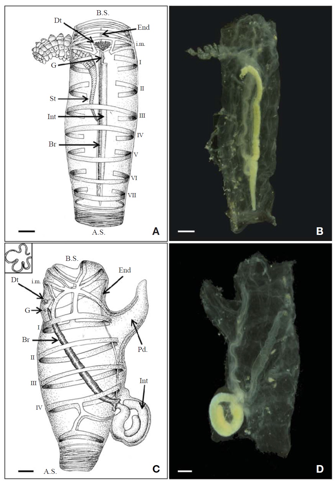
3*Cyclosalpa affinis (Chamisso, 1819) (Fig. 1A-D)
Material examined. 5 solitary zooids, 61 aggregate zooids, East Sea, 38° 29′N 131° 30′E, 15 Oct 2001; 2 aggregate zooids, East Sea, 38° 30′N 129° 49′E, 15 Oct 2001; 1 solitary zooid, 1 aggregate zooid, East Sea, 38° 30′N 130° 30′E, 15 Oct 2001; 2 solitary zooids, 1 aggregate zooid, East Sea, 36° 30′N 130° 29′E, 15 Oct 2001.
Description. Solitary zooid: 27-82 mm long, elongate, cylindrical body (Fig. 1A, B). Test thick and relatively stiff. Oral and atrial opening terminal. Dorsal tubercle strongly convoluted and totally ‘fan’ like shaped. Seven body muscles interrupted ventrally, MI and MII interrupted dorsally, MIII to MVII continuous dorsally. Light organs absent. Endostyle thin, extends from behind oral to MVI ventrally. Branchial septum extend from antero-dorsal surface to esophagus postero- ventrally. Developed stomach absent, straight intestine stretched along dorsal side of whole length of branchial septum. Anal opening just behind ganglion. Stolon arise posterior ventrally, stretch straight forward and break through test MI ventrally.
Aggregate zooid: 23-55 mm long, cylindrical (Fig. 1C, D). Test thick and soft. Peduncle anterior ventrally, not exceed 1/5 body length. Oral and atrial opening terminal. Light organs absent. Dorsal tubercle moderately convoluted. Four body muscles nearly parallel, interrupted ventrally, continuous dorsally. Endostyle thin, extends from behind oral to MIV posterior ventrally. Branchial septum extend from ganglion to gut. Gut wide looped, anal aperture near esophagus. Testis lies along intestine.
Distribution. Pacific Ocean, Atlantic Ocean, Indian Ocean.
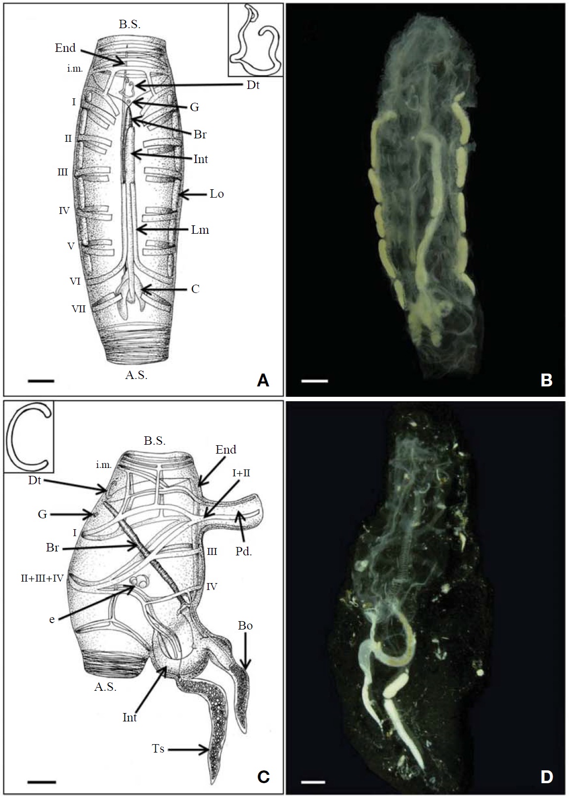
4*Cyclosalpa bakeri Ritter, 1905 (Fig. 2A-D)
Material examined. 51 aggregate zooids, East Sea, 37° 30′N 130°30°E, 18 Apr 2001; 11 solitary zooids, 387 aggregate zooids, East Sea, 38° 00′N 130° 30′E, 18 Apr 2001; 1 solitary zooid, Korea strait, 32° 29′N 127° 29′E, 24 Sep 2002; 1 solitary zooid, East Sea, 37° 32′N 130° 07′E, 30 Sep 2002; 8 aggregate zooids, Jeju, 33° 12′N 126° 36′E, 22 Jun 2009.
Description. Solitary zooid: 12-54 mm long, elongate, cylindrical body (Fig. 2A, B). Test flabby. Oral and atrial opening terminal. Dorsal tubercle ‘horseshoe’ like shaped. Seven body muscles interrupted ventrally and dorsally. MVI turns forward on each side, forming longitudinal muscle passes MV and MIV. Six pairs light organs well developed, five pairs between MI and MVI, one small pair before MI. Endostyle thin, extends from behind oral to front of gut. Branchial septum slender, extend from ganglion to MVI ven-
trally. Developed stomach absent, straight intestine along dorsal side of whole length of branchial septum. Two caecum presented posterior. Anal opening just behind MVI.
Aggregate zooid: 8-63 mm long, barrel shaped body (Fig. 2C, D). Test thin, flabby. Peduncle anterior ventrally, not exceed 1/3 body length. Oral and atrial opening terminal. Light organs absent. 4 body muscles continuous dorsally. Intermediate muscle and MI fuse dorsally. MII, MIII and MIV fuse dorsally. MI and MII fuse ventrally. MIV extend ventrally and fuse with MIII. Intermediate muscle, MI-MIV muscles continued into peduncle. Intestine muscle extends from MIV to side of alimentary canal. Dorsal tubercle Clike shaped. Endostyle thin, extends from behind oral to MIV posterior ventrally. Branchial septum slender, extends from ganglion to gut. Intestine u-like shaped, mostly lying outside of body, protuberated. One piece of intestine extended and end of this intestine, blood forming organ projected. Testis presented behind intestine. Embryo locates right side, between MIII and MIV.
Distribution. Pacific Ocean, Atlantic Ocean, Indian Ocean.
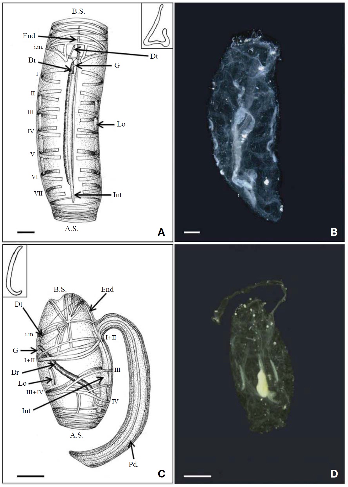
1*Cyclosalpa sewelli Metcalf, 1927 (Fig. 3A-D)
Material examined. 6 aggregate zooids, East Sea, 38° 00′N 130° 30′E, 18 Sep 2001; 1 aggregate zooid, South Sea, 35° 31′N 130° 22′E, 15 Oct 2001; 1 solitary zooid, 3 aggregate zooid, Korea strait, 32° 29′N 127° 29′E, 24 Sep 2002; 1 solitary zooid, Korea strait, 33° 59′N 128° 17′E, 24 Sep 2002; 4 aggregate zooids, South Sea, 35° 11′N 129° 53′E, 4 Dec 2002.
Description. Solitary zooid: 10-12 mm long, elongate, cylindrical body (Fig. 3A, B). Test thin, flabby (Fig. 3A, B). Oral and atrial opening terminal. Dorsal tubercle ‘triangle’ like shaped. Seven body muscles interrupted ventrally and dorsally. Longitudinal muscle absent. Four pairs light organs well developed, placed middle of either side between MII and MVI, spindle shaped. Endostyle thin, extends from behind oral to MVI ventrally. Branchial septum slender, extends from ganglion to MVI ventrally. Developed stomach absent, straight intestine stretched along dorsal side of whole length of branchial septum. Anal opening just behind neural ganglion.
Aggregate zooid: 7-15 mm long, cylindrical or barrel shape body (Fig. 3C, D). Test thin, flabby. Peduncle almost 1.5 times body length. Oral and atrial opening terminal. Elongate light organs on either side, between MII and MIII, almost 1/7-1/8 body length. 4 body muscles continuous dorsally. MI and MII fused dorsally and ventrally. MIII and MIV fused dorsally. Intermediate muscle, MI-MII muscles continued into peduncle. Dorsal tubercle bow-like shaped. Endostyle thin, extends from behind oral to MIV ventrally. Branchial septum slender, extends from ganglion to gut. Developed stomach absent, straight intestine stretched along the ventral side of whole length of endostyle. Anal opening near peduncle.
Distribution. Pacific Ocean, Atlantic Ocean, Indian Ocean.
2*Cyclosalpa polae Sigl, 1912 (Fig. 4A, B)
Material examined. 2 aggregate zooids, South Sea, 35° 11′N 129° 53′E, 4 Dec 2002.
Description. Aggregate zooid: 14-27 mm long, cylindrical or barrel shape body (Fig. 4A, B). Test thin, flabby. Peduncle almost body length. Oral and atrial opening terminal. Elongate light organs on either side, between MII and MIII, almost 1/4 body length. 4 body muscles continuous dorsally. MI, MII fused dorsally and ventrally. MIII, MIV fused dorsally. Intermediate muscle, MI-MII muscles continued into peduncle. Dorsal tubercle w-like shaped. Endostyle thin, extends from behind oral to MIV ventrally. Branchial septum slender, extends from ganglion to gut. Developed stomach absent, straight intestine stretched along ventral side of whole length of endostyle. Anal opening near peduncle.
Distribution. Pacific Ocean, Atlantic Ocean.
Remarks. Aggregate zooid of the species is the almost same as that of
Key to the life cycles of Salpida
1. Stolon present at ventral or posterior, egg and embryo absent; attachment organ absent ???????????????????solitary zooid
- Stolon absent, eggs or embryo present at right (if mirror image-sinistral individuals: left posterior); attachment organ (peduncle) present?????????????????????????????????aggregate zooid
Key to species of the genus Cyclosalpa from Korea
Solitary zooid
1. Longitudinal muscles present ?????????????
- Longitudinal muscle absent ????????????????????????????????????????2
2. Light organs present on either side ????
- Light organs absent ??????????????????????????
Aggregate zooid
1. Gut u-like shape ???????????????????????????????
- Gut wide looped or straight ????????????????????????????????????????2
2. Light organs absent ??????????????????????????
- Light organs present on either side ?????????????????????????????3
3. Dorsal tubercle w-like shaped ?????????????
- Dorsal tubercle bow-like shaped ???????
Korean name: 1*곧은장살파아과(신칭), 2*곧은장살파속(신칭), 3*무광곧은장살파(신칭), 4*꼬리곧은장살파(신칭)
Korean name: 1*스웰리곧은장살파(신칭), 2*북극곧은장살파(신칭)