검색 전체 메뉴
PDF
맨 위로
OA 학술지
First Record of Paranebalia longipes (Crustacea: Phyllocarida: Leptostraca) from South Korea
  • 비영리 CC BY-NC
  • 비영리 CC BY-NC
ABSTRACT
First Record of Paranebalia longipes (Crustacea: Phyllocarida: Leptostraca) from South Korea
KEYWORD
Paranebalia longipes , distribution , Geomundo Island , Munseom Island , Leptostraca , Korea
  • INTRODUCTION

    The genus Paranebalia Claus, 1880 (Crustacea: Phyllocarida: Leptostraca), is one among three genera belonging to the family Paranebaliidae Walker-Smith & Poore, 2001 that is currently composed of three species: P. longipes (Willemoes- Suhm, 1875), P. belizensis Modlin, 1991, and P. ayalai Escobar Briones & Alcocer Durand, 2003. Paranebalia speciesinhabit such as stones and dead corals on the low tide beaches in the lagoons (Willemoes-Suhm, 1875; Clark, 1932; Brattegard, 1970; Gamo and Takizawa, 1986), algal masses (Modlin, 1991; Roccatagliata et al., 2010), sponges and shell debris on the sandy mud substrates (Escobar Briones and Alcocer Durand, 2003; Roccatagliata et al., 2010).

    They are distinguished by the following characteristics: 1) eyes with denticles; 2) thoracopods stenopodous; 3) thoracopods 1-8 subequal in length and extending well beyond the ventral margin of carapace; 4) posterior margin of pleonite 5 smooth and that of pleonites 6-7 distinctly crenate; 5) protopods of pleopods 2-4 strongly serrulate along their posterior margins (Walker-Smith, 2000; Walker-Smith and Poore, 2001; Escobar Briones and Alcocer Durand, 2003; Haney and Martin, 2004; Roccatagliata et al., 2010).

    The genus Paranebalia is new to Korea. In this paper, we provide detail descriptions of the diagnostic characteristics and illustrations of the P. longipes. We also provided type locality and world distribution of the genus Paranebalia (see Discussion) (Fig. 1).

    MATERIALS AND METHODS

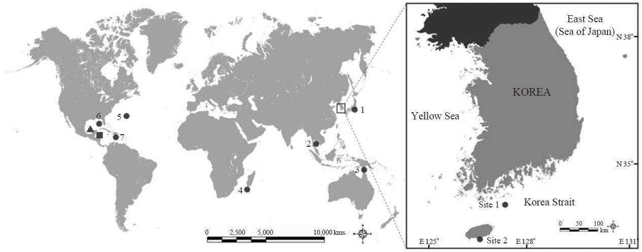
    Specimens were collected from the Geomundo Island and Munseom Island in South Korea. They were preserved directly in 95% ethyl alcohol after collection. All the examined specimens were deposited in the National Institute of Biological Resources (NIBR) and Inha University, South Korea.

    The specimens were examined and dissected under a stereomicroscope (Model SZX-7; Olympus, Tokyo, Japan). Illustrations of dissected appendages were made with a drawing tube connected to a light microscope (Model DM 2500, X50- 630; Leica, Wetzlar, Germany) and images were recorded with a microscope digital camera (Model Nikon D90; Nikon Corp., Tokyo, Japan). Drawings of the whole body was made using a drawing tube attached to a stereomicroscope (Olympus SZX-12). Images of whole body were photographed with a microscope digital camera (Model Moticam 2000; Motic Incorporation Ltd., Hong Kong, China), and developed with software (Model Helicon Focus; Helicon Soft Ltd., Kharkov, Ukraine). Measurements of appendages and whole body leng-

    th were done with a stage micrometer (Leica, Heidelberger, Germany) and an ocular micrometer.

    Total length (TL) was measured from the articulation between the rostrum and carapace to the posterior end of the uropods excluding setation. Dorsal carapace length (DCL) was considered the distance between the articulation of the rostrum and the margin of the posterodorsal cleft. Lateral carapace length (LCL) was considered the distance along the lateral surface between the anteriormost and posteriormost margin. Carapace height (CH) was measured between the dorsal and ventral margin. Rostrum length (RL) was measured along the midline.

    SYSTEMATIC ACCOUNTS

    Order Leptostraca Claus, 1880

    1*Family Paranebaliidae Walker-Smith & Poore, 2001

    2*Genus Paranebalia Claus, 1880

    3*Paranebalia longipes (Willemoes-Suhm, 1875) (Figs. 2-5)

    Nebalia longipes Willemoes-Suhm, 1875: 26, Pl. 5.

    Paranebalia longipes: Sars, 1887: 10, Pls. 1, 2; Claus, 1888: 124; Thiele, 1904: 14, Pls. 2, 4, figs. 25-36, 63, 64; Verrill, 1923: 209, Pl. 64, fig. 3; Clark, 1932: 231, Pls. 5, 6; Brattegard, 1970: 3, fig. 1; Wakabara, 1976: 300, table 1; Gamo and Takizawa, 1986: 31, figs. 2-4; Modlin, 1991: 603, table 1; Ledoyer, 1994: 21, fig. 1; Ledoyer, 2001: 17, fig. 2; Escobar Briones and Alcocer Durand, 2003: 347, table 1.

    Material examined. Korea: 3♂♂ and 5 ♀♀, Jeollanamdo, Yeosu-si, Isl. Geomundo, 34°02′N, 127°17′E, 17 Apr 2009, by SCUBA diving, depth 15-20 m, Hong SS; 1♀, Seowipo-si, Jeju-do, Isl. Munseom, 33°13′N, 126°32′E, 11 Feb 2012, collected from van Veen grab on bottom, depth 60 m, Kim MS.

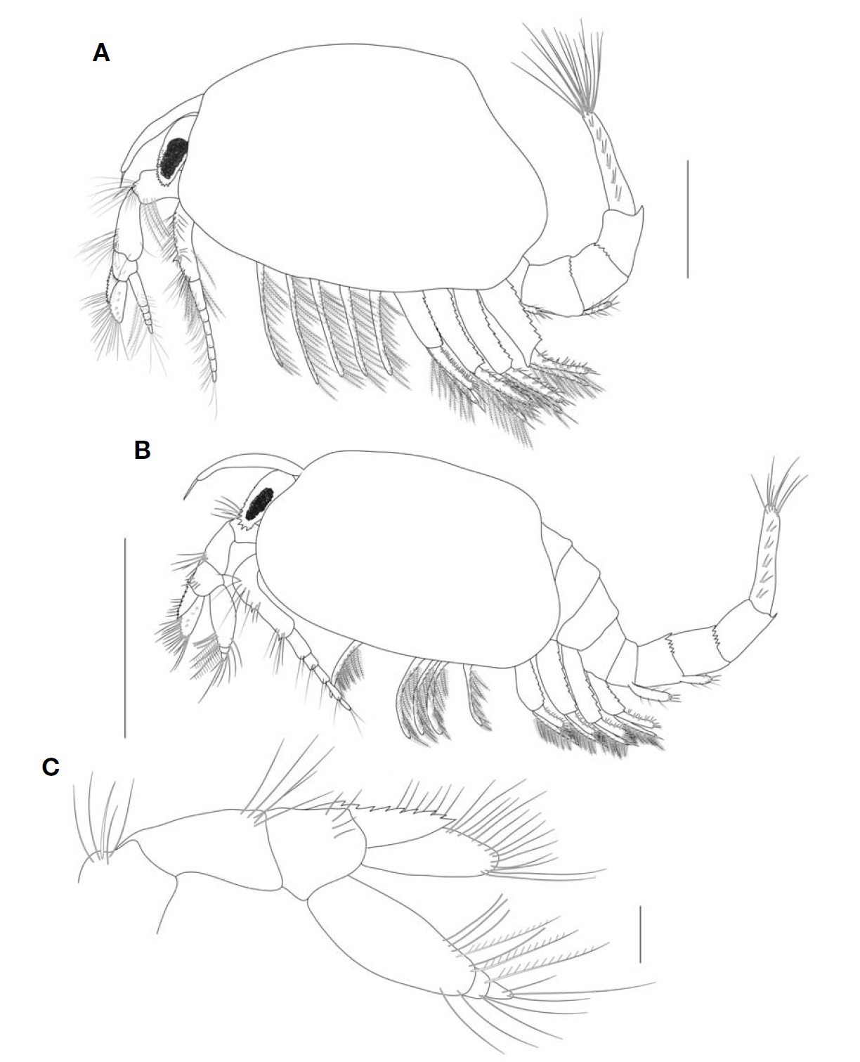
    Description. Female: Body (Fig. 5A) robust, transparent, light yellow colored in living or preserved specimens, TL 5.7 mm, DCL 2.3 mm, LCL 3.2 mm, CH 2.1 mm, RL 1.1 mm.

    Carapace (Fig. 5A) oval, extending back to middle of the pleonite 5 along sides; about 1.5 times longer than high.

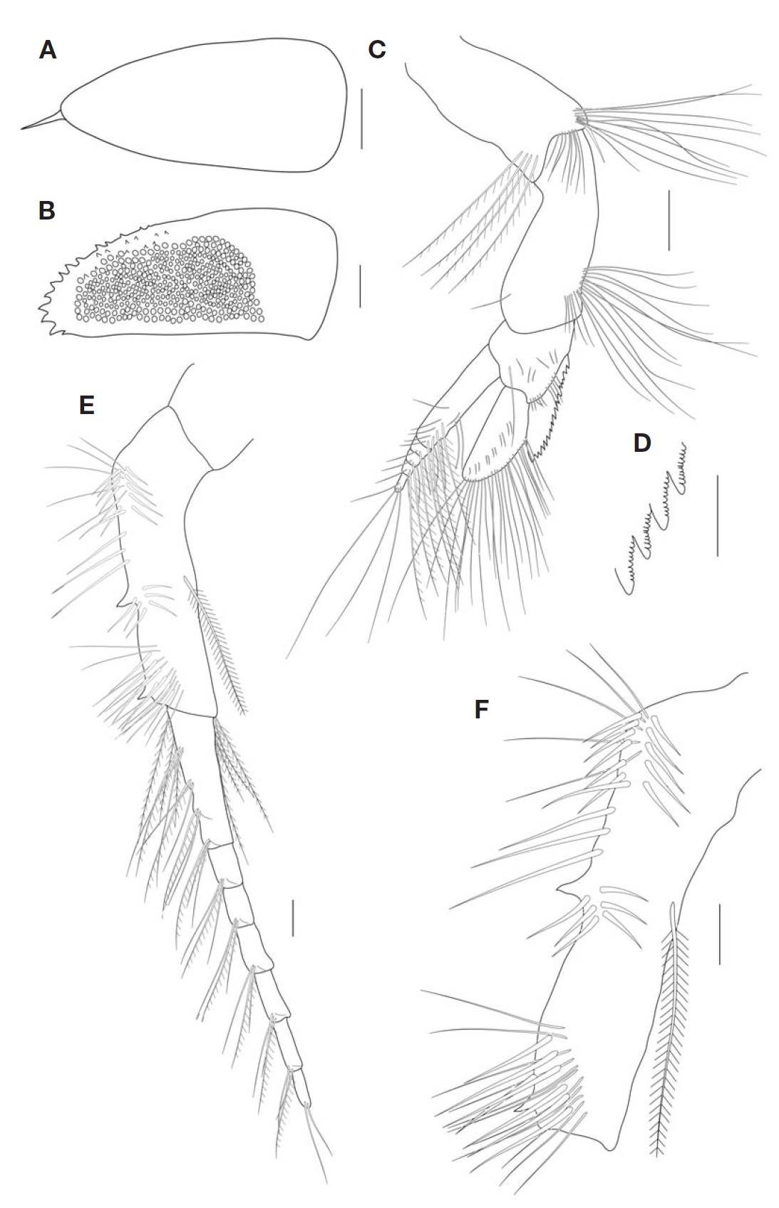
    Rostrum (Fig. 2A) relatively long, clearly past beyond distal margin of eyestalk; apex with 1 subterminal spine; about 2.5 times as long as wide; nearly triangular.

    Compound eye (Fig. 2B) more or less cylindrical; ommatidial part extending about 60% of TL of eyestalk; distal dorsal surface distinctly armed with many coarse teeth.

    Antennule (Fig. 2C) peduncle 4-articulated. First article (not illustrated) robust, shorter than eyestalk. Second article length/width ratio about 1.7, with 1) four plumose setae on lateral surface; 2) subterminal cluster of about 20 short and long simple setae, and one short spine-like seta. Third article as long as the second, widest distally, with cluster of about 17 simple setae on anterior distal margin, one short simple seta on posterior margin. Fourth article short with lateral

    flange having 19-22 roust teeth with crenate margins (Fig. 2D), with 10 simple setae on distal and lateral margins, one long seta and six shorter setae on medial margin. Antennular scale blade-like, 2.4 times as long as wide, with many short and long setae. Flagellum with five articles, distinctly shorter than peduncle; articles with 1) 6-7 aesthetascs; 2) 11 short simple setae and seven plumose setae, proximal article about 8.3 times longer than others.

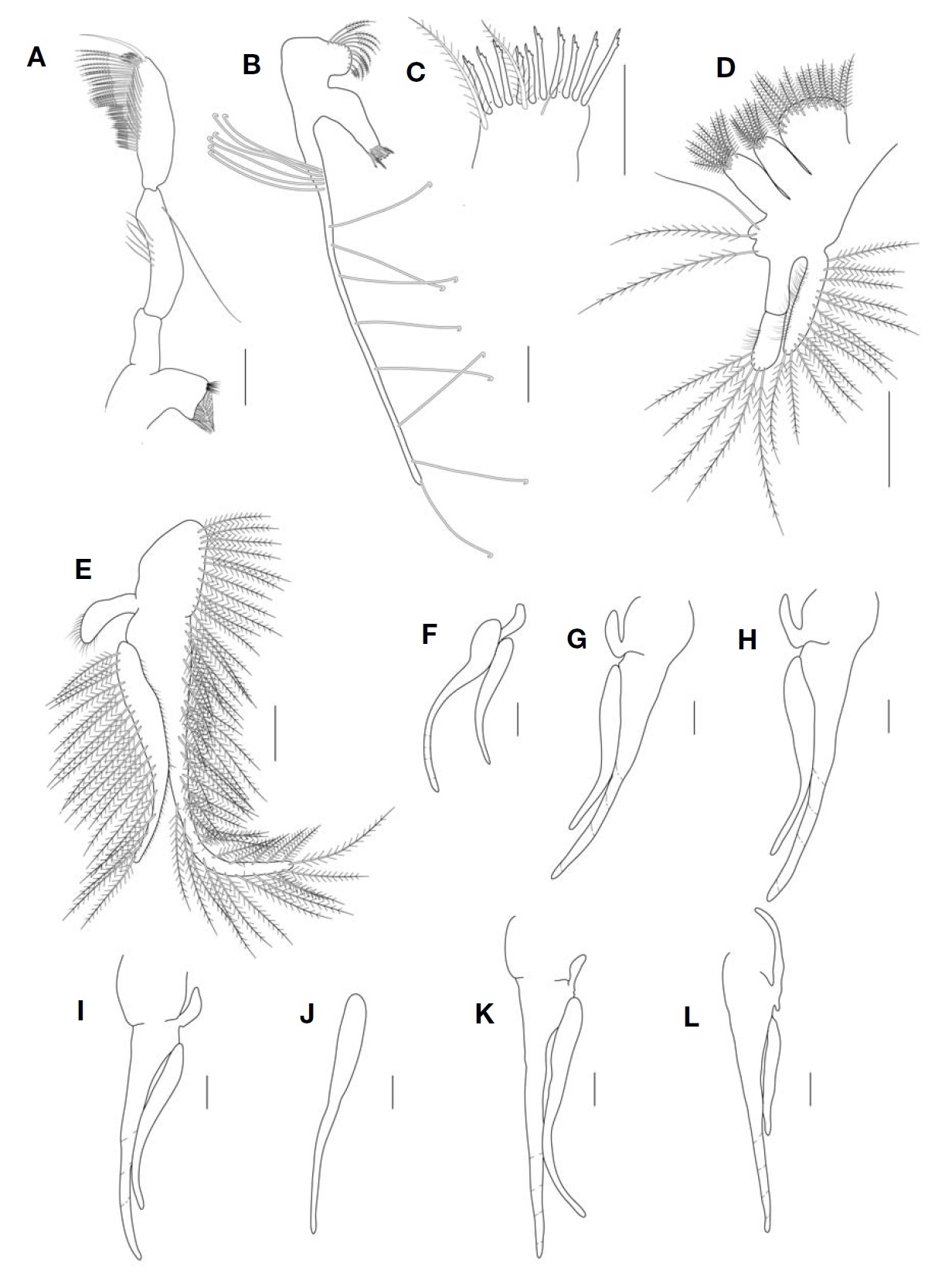
    Antenna (Fig. 2E) peduncle 3-articulated. First article short (not illustrated). Second article slightly rectangular. Third article longest, with different pattern of setae along medial anterior margin (Fig. 2F): 1) 18 spine-like setae and ten longer spine-like ones; 2) 16 long simple setae; two short acute process on lateral margins, one at about midlength, and second distally; one long plumose seta on posterior margin. Flagellum well-developed, composed of seven articles; article 1 with three plumose setae and ten simple setae, two distinctly shorter and thinner than the others, oriented posterior-

    ly; articles 2-6 with one plumose setae and three simple setae, one distinctly shorter and thinner than the others, respectively; article 7 with two simple setae distally.

    Mandible (Fig. 3A) palp 3-segmented; second article slightly curved, with one long subterminal seta and five simple setae on medial margin. Third article as long as second, pos-

    [Table 1.] Morphological comparison between Korean Paranebalia longipes and other regions

    label

    Morphological comparison between Korean Paranebalia longipes and other regions

    terior margin with three types of setae: 1) row of distally plumose setae arising from ending of proximal quarter extending to proximal half; 2) row of longer setae extending along distal half, distally plumose; 3) about 3-4 curved, dentate setae on distal margin, and single terminal long spinelike seta.

    First maxilla (Fig. 3B) proximal endite with rounded medial margin bearing distally plumose setae. Distal endite (Fig. 3C) subrectangular, distal margin with two long plumose setae, one short simple seta, and 10-11 robust spines with three teeth along distal posterior margin also provided with some smaller teeth. Palp with proximal cluster of 5-6 setae and 8-12 widely spaced setae along its whole length.

    Second maxilla (Fig. 3D) protopod with four endites with plumose setae, endite 1 the largest; endites 2 and 3 rectangular, nearly subequal in size; endite 4 the smallest. Endopod 2-segmented, longer than exopod, first article slightly longer than second; lateral and medial margins with numerous plumose setae and short simple setae. Exopod clearly longer than the first article of endopod; lateral and medial margin with 13 plumose setae and numerous short simple setae.

    Thoracopods (Fig. 3E-L) of female all similar in shape and changing in size. Marginal setation is the same on all thoracopods. Endopod clearly longer than exopod; with numerous plumose setae along anterior and medial margins. Exopod with plumose setae along posterior margin. Epipod small, narrow, with short setae, but thoracopod 8 epipod distinctly longer than those of thoracopods 1-7 (Thoracopod 6 endopod and epipod broken) (Fig. 3J).

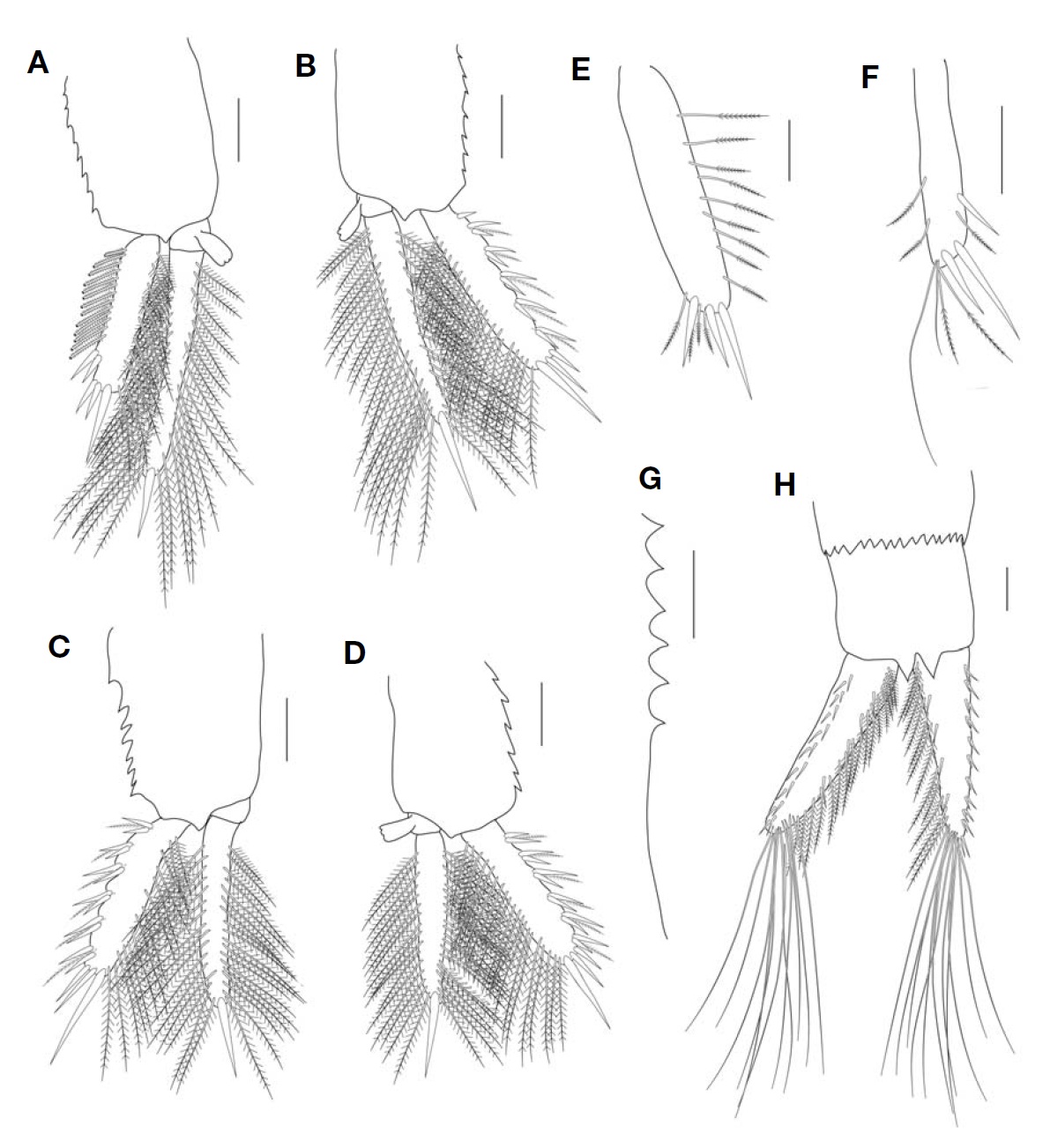
    Pleopod 1 (Fig. 4A) composed of protopod, endopod and exopod. Protopod with 9-11 serrations at posterior margin. Endopod 2-segmented, longer than exopod; first article much shorter, bearing appendix interna; second article with 28-32 plumose setae along both medial and lateral margins. Exopod about 0.6 times length of endopod; with a row of about 12- 14 serrate spines along lateral margin (‘spine-row’), each spine with small tridentate tip, central tooth bifid; five robust spines on distolateral margin; with numerous plumose setae along medial margin.

    Pleopods 2-4 similar (Fig. 4B-D), protopods with 7-9 serrations at posterior margin. Endopod slightly longer than exopod, 2-segmented; proximal article short, provided with appendix interna; lateral and medial margins of distal article each with about 35-40 plumose setae, ending in one robust spine. Exopod with a row of 5-6 robust spine pairs, each pair composed of one long and shorter spine, with one plumose seta between spine pairs; medial border with plumose setae, three spines distally, increasing distally in length.

    Pleopods 5-6 (Fig. 4E, F). Pleopod 5 uniramous, with three robust spines along distolateral and terminal margins; 13-15 distally plumose setae along medial and distal margins, setae appearing jointed at midlength. Pleopod 6 uniramous, with three robust spines along distolateral and terminal margins. Medial and terminal margins with plumose setae distally, same as those of pleopod 5.

    Pleonites (Fig. 4G) 6 and 7, with acute denticles distally along only posterior dorsal margin.

    Uropods (Figs. 4H, 5A), 1.4 times length of pleonite 8, with 18-20 robust setae along lateral margin; about 7-10 simple setae and 11-16 plumose setae on lateral inner margin; 11 long simple setae on distal margin.

    Description (male). Body (Fig. 5B) smaller than female, TL 3.7 mm, DCL 1.2 mm, LCL 1.6 mm, CH 1.0 mm, RL 0.7mm, but morphologically similar to female except for antennular flagellum. Antennular flagellum (Fig. 5C) distinctly inflated, articulations between each segments visible only terminally; flange on fourth article with fewer teeth.

    Habitat. At the type locality, this species is collected from stones and dead corals on the beach at low tide in a lagoon communicating with open sea through a narrow passage, and depth was 5-9 m (Willemoes-Suhm, 1875; Clark, 1932; Brattegard, 1970; Gamo and Takizawa, 1986). This species collected from the South Korea on algal and sponge debris at 15-20 m deep.

    Remarks. The Nebalia longipes was firstly described by Willemoes-Suhm (1875) based on the specimens collected from the Bermuda Islands in Atlantic Ocean. Later, Claus (1880) transferred this species to the genus Paranebalia because of their unique characteristics (e. g. denticulate eyes). Paranebalia longipes is mainly distributed worldwide and has been reported from Gulf of Mexico, Caribbean Sea (Willemoes-Suhm, 1875; Sars, 1887; Thiele, 1904; Verrill, 1923; Clark, 1932; Brattegard, 1970; Haney and Martin, 2004; Martin and Haney, 2009; Roccatagliata et al., 2010), Japan, Gulf of Siam, Torres Strait, Walters Shoal, Madagascar Mauritius Islands (Thiele, 1904; Gamo and Takizawa, 1986; Ledoyer, 1994, 2001). In contrast, P. belizensis and P. ayalai are endemic species, which reported only from their type localities: the former species was reported from Twin Cays, Belize; the latter was reported from Campeche Bay, Gulf of Mexico (Modlin, 1991; Escobar Briones and Alcocer Durand, 2003; Roccatagliata et al., 2010) (Fig. 1).

    P. longipes is distinguished from other species by several characteristics. P. belizensis is very similar to P. longipes, but this species have different number of antennal flagellum, antennular flagellum, and denticles on flange of article 4 at antennule. Additionally, P. ayalai is distinctly distinguished from P. longpies and P. belizensis, by one characteristic that is difference in margin of protopod of pleopods. P. ayalai have smooth margin on protopod of pleopods, whereas other species have serrated margin (Modlin, 1991; Escobar Briones and Alcocer Durand, 2003).

    In general, P. longipes from the South Korea is well congruent with the original descriptions and illustrations of Willemoes- Suhm (1875). However, Korean specimens somewhat differ from those of other localities in the following characteristics: 1) the antennule fourth article with lateral flange having 19-22 robust teeth; 2) the antennular flagellum with five articles and the antennal flagellum composed of seven articles; and 3) proximal/distal length of the endopod is nearly 0.6 on second maxilla. A comparison between Korean specimens and other regions is present in Table 1.

참고문헌
  • 1. Brattegard T 1970 Marine biological investigations in the Bahamas. 13. Leptostraca from shallow water in the Bahamas and Southern Florida [Sarsia] Vol.44 P.1 google
  • 2. Clark AE 1932 Nebaliella caboti n. sp., with observations on other Nebaliacea. [Transactions of the Royal Society of Canada] Vol.26 P.217-235 google
  • 3. Claus C 1880 Grindzuge der Zoologie Vol. 1. P.1-822 google
  • 4. Claus C 1888 Uber den Organismus der Nebaliden und die systematische Stellung der Leptostraken. [Arbeiten aus dem Zoologischen der Universitat Wien und der ZoologischenStation in Triest] Vol.8 P.1-148 google
  • 5. Claus C 1888 Uber den Organismus der Nebaliden und die systematische Stellung der Leptostraken. [Arbeiten aus demZoologischen der Universitat Wien und der ZoologischenStation in Triest] Vol.8 P.1-148 google
  • 6. Escobar Briones E, Alcocer Durand J 2003 Paranebalia ayalai, a new leptostracan species (Crustacea: Phyllocarida: Nebaliidae) from the southwestern Gulf of Mexico. In: Agustin Ayala-Casta~nares: universitario, impulsor de la investigacion cientifica (Ed., Soto LA). P.347-352 google
  • 7. Gamo S, Takizawa Y 1986 On Paranebalia longipes (Willemoes- Suhm, 1875) (Crustacea, Leptostraca, Nebaliacea) from Sagami Bay. [Bulletin of the Biogeographical Societyof Japan] Vol.41 P.31-36 google
  • 8. Hale HM 1929 The Crustaceans of South Australia. P.202-380 google
  • 9. Haney TA, Martin JW 2004 A new genus and species of leptostracan (Crustacea: Malacostraca: Phyllocarida) from Guana Island, British Virgin Islands, and review of leptostracan genera. [Journal of Natural History] Vol.38 P.447-469 google
  • 10. Ledoyer M 1994 Leptostraces (Crustacea) du banc Walters (Ocean Indien: 32°S-44°SE) recoltes au cours de la campagne MD 08/Benthos du MS 《Marion Dufresne》, en 1976. [Marine Life] Vol.4 P.21-24 google
  • 11. Ledoyer M 2001 Leptostraces (Crustacea) du Sud-Ouest de l’ocean Indien tropical (Madagascar, ile Maurice). [MarineLife] Vol.11 P.17-20 google
  • 12. Martin JW, Haney TA 2009 Leptostraca (Crustacea) of the Gulf of Mexico. In: Gulf of Mexico: origin, waters and biota. Vol. 1 (Eds., Felder DL, Camp DK). P.895-899 google
  • 13. Modlin RF 1991 Paraneballa belizensis, a new species from shallow waters off Belize, central America (Crustacea: Malacostraca: Leptostraca). [Proceeding of the BiologicalSociety of Washington] Vol.104 P.603-612 google
  • 14. Roccatagliata D, Chiesa IL, Raffo MP, Simes EG 2010 On the occurrence of the genus Paranebalia (Crustacea: Phyllocarida: Leptostraca) in northern Patagonia [Zootaxa] Vol.2349 P.65-68 google
  • 15. Sars GO 1887 Report on the Phyllocarida collected by H.M.S. Challenger during the years 1873-76. Reports on the Scientific Results of the Voyage of H.M.S. Challenger during the years 1873-76 [Zoology] Vol.19 P.1-38 google
  • 16. Thiele J 1904 Die Leptostraken. Wissenschaftliche Ergebnisse der Deutschen Tiefsee-Expedition auf dem Dampfer “Valdivia” 1898-1899 Vol.8 P.1-26 google
  • 17. Verrill AE 1923 Crustacea of Bermuda: Schizopoda, Cumacea, Stomatopoda and Phyllocarida. [Transactions of the ConnecticutAcademy of Arts and Sciences] Vol.26 P.181-211 google
  • 18. Wakabara Y 1976 Paranebalia fortunata n. sp. from New Zealand (Crustacea, Leptostraca, Nebaliacea). [Journal ofthe Royal Society of New Zealand] Vol.6 P.297-300 google
  • 19. Walker-Smith GK 2000 Levinebalia maria, a new genus and new species of Leptostraca (Crustacea) from Australia. [Memoirs of Museum Victoria] Vol.58 P.137-148 google
  • 20. Walker-Smith GK, Poore GCB 2001 A phylogeny of the Leptostraca (Crustacea) with keys to families and genera. [Memoirsof Museum Victoria] Vol.58 P.383-410 google
  • 21. Willemoes-Suhm RV 1875 II. On some Atlantic Crustacea from the ‘Challenger’ Expedition. [Transactions of the LinneanSociety of London] Vol.1 P.23-59 google
OAK XML 통계
이미지 / 테이블
  • [ Fig. 1. ]  Distribution of genus Paranebalia based on previous records and on the present study. P. longipes (●): 1, Japan; 2, Gulf of Siam; 3, Torres strait; 4, Madagascar; 5, Bermuda islands; 6, Gulf of Mexico; 7, Caribbean Sea (Willemoes-Suhm, 1875; Sars, 1887; Thiele, 1904; Verrill, 1923; Hale, 1929; Clark, 1932; Brattegard, 1970; Gamo and Takizawa, 1986; Ledoyer, 1994, 2001; Haney and Martin, 2004; Martin and Haney, 2009; Roccatagliata et al., 2010). P. ayalai (▲): Campeche Bay, Gulf of Mexico (Escobar Briones and Alcocer Durand, 2003). P. belizensis (■): Twin cays, Belize (Modlin, 1991).
    Distribution of genus Paranebalia based on previous records and on the present study. P. longipes (●): 1, Japan; 2, Gulf of Siam; 3, Torres strait; 4, Madagascar; 5, Bermuda islands; 6, Gulf of Mexico; 7, Caribbean Sea (Willemoes-Suhm, 1875; Sars, 1887; Thiele, 1904; Verrill, 1923; Hale, 1929; Clark, 1932; Brattegard, 1970; Gamo and Takizawa, 1986; Ledoyer, 1994, 2001; Haney and Martin, 2004; Martin and Haney, 2009; Roccatagliata et al., 2010). P. ayalai (▲): Campeche Bay, Gulf of Mexico (Escobar Briones and Alcocer Durand, 2003). P. belizensis (■): Twin cays, Belize (Modlin, 1991).
  • [ Fig. 2. ]  Paranebalia longipes, female. A, Rostrum, dorsal view; B, Eye, lateral view; C, Antennule, lateral view; D, Antennule fourth article, serration on denticles of flange; E, Antenna, lateral view; F, Antenna, third article, external side, lateral view. Scale bars: A, C, E=0.2 mm, B, D, F=0.1 mm.
    Paranebalia longipes, female. A, Rostrum, dorsal view; B, Eye, lateral view; C, Antennule, lateral view; D, Antennule fourth article, serration on denticles of flange; E, Antenna, lateral view; F, Antenna, third article, external side, lateral view. Scale bars: A, C, E=0.2 mm, B, D, F=0.1 mm.
  • [ Fig. 3. ]  Paranebalia longipes, female. A, Mandibular palp; B, First maxilla; C, First maxilla, distal endite; D, Second maxilla; E, Thoracopod 1, showing setation; F, Thoracopod 2; G, Thoracopod 3; H, Thoracopod 4; I, Thoracopod 5; J, Thoracopod 6; K, Thoracopod 7; L, Thoracopod 8. Scale bars: A, B, D-L=0.2 mm, C=0.1 mm.
    Paranebalia longipes, female. A, Mandibular palp; B, First maxilla; C, First maxilla, distal endite; D, Second maxilla; E, Thoracopod 1, showing setation; F, Thoracopod 2; G, Thoracopod 3; H, Thoracopod 4; I, Thoracopod 5; J, Thoracopod 6; K, Thoracopod 7; L, Thoracopod 8. Scale bars: A, B, D-L=0.2 mm, C=0.1 mm.
  • [ Fig. 4. ]  Paranebalia longipes, female. A, Pleopod 1, lateral view (only half of setae of spine row illustrated); B, Pleopod 2, lateral view; C, Pleopod 3, lateral view; D, Pleopod 4, lateral view; E, Pleopod 5; F, Pleopod 6; G, Pleonite 7, posterior lateral border, denticles; H, Uropod, ventral view. Scale bars: A-D, H=0.2 mm, E-G=0.1 mm.
    Paranebalia longipes, female. A, Pleopod 1, lateral view (only half of setae of spine row illustrated); B, Pleopod 2, lateral view; C, Pleopod 3, lateral view; D, Pleopod 4, lateral view; E, Pleopod 5; F, Pleopod 6; G, Pleonite 7, posterior lateral border, denticles; H, Uropod, ventral view. Scale bars: A-D, H=0.2 mm, E-G=0.1 mm.
  • [ Fig. 5. ]  Paranebalia longipes. A, Female, lateral view; B, Male, lateral view; C, Male, antennule, lateral view. Scale bars: A, B=1 mm, C=0.1 mm.
    Paranebalia longipes. A, Female, lateral view; B, Male, lateral view; C, Male, antennule, lateral view. Scale bars: A, B=1 mm, C=0.1 mm.
  • [ Table 1. ]  Morphological comparison between Korean Paranebalia longipes and other regions
    Morphological comparison between Korean Paranebalia longipes and other regions
(우)06579 서울시 서초구 반포대로 201(반포동)
Tel. 02-537-6389 | Fax. 02-590-0571 | 문의 : oak2014@korea.kr
Copyright(c) National Library of Korea. All rights reserved.