검색 전체 메뉴
PDF
맨 위로
OA 학술지
Ethanolic Extract of Chondria crassicaulis Inhibits the Expression of Inducible Nitric Oxide Synthase and Cyclooxygenase-2 in LPS-Stimulated RAW 264.7 Macrophages
  • 비영리 CC BY-NC
  • 비영리 CC BY-NC
ABSTRACT
Ethanolic Extract of Chondria crassicaulis Inhibits the Expression of Inducible Nitric Oxide Synthase and Cyclooxygenase-2 in LPS-Stimulated RAW 264.7 Macrophages
KEYWORD
Chondria crassicaulis , anti-inflammation , iNOS , COX-2 , TNF-α , IL-1β , IL-6
  • Introduction

    Macrophages are major inflammatory cells and immune effector cells. Activation of macrophages occurs in inflamed tissues and is induced by exposure to interferon-γ, tumor ne-crosis factor (TNF)-α, interleukin (IL)-1β, IL-6, and micro-bial lipopolysaccharide (LPS) (Xie et al., 1993). Activated macrophages produce excessive amounts of inflammatory mediators such as nitric oxide (NO) and prostaglandins (PGs), in addition to various pro-inflammatory cytokines (Nathan, 1992; Zhang and Ghosh, 2000). Excessive produc-tion of inflammatory mediators and cytokines contributes to the pathogenesis of chronic diseases such as atherosclerosis, inflammatory arthritis, and cancer (Libby, 2006; Packard and Libby, 2008; Solinas et al., 2010). Substances that inhibit the production of these molecules are considered to be potential anti-inflammatory agents.

    The expression of inflammatory proteins such as cyclo-oxyganase-2 (COX-2) and inducible nitric oxide synthase (iNOS) and pro-inflammatory cytokines is primarily con-trolled at the transcription level (D’Acquisto et al., 1997; Kim and Moudgil, 2008). Transcriptional induction of COX-2 and iNOS is largely dependent on the cooperative activity of multiple transcription factors, including nuclear factor-κB (NF-κB) and activator protein 1 (AP-1), which act on cog-nate cis-acting elements in the COX-2 and iNOS promoters (D’Acquisto et al., 1997; Marks-Konczalik et al., 1998). NF-κB plays pivotal roles in the immediate early stages of im-mune, acute-phase, and inflammatory responses, as well as in cell survival (Makarov, 2001). Under unstimulated condi-tions, NF-κB is located in the cytoplasm as an inactive com-plex with inhibitor of κB (IκB)-α. LPS treatment activates the IκB-α kinase complex, resulting in the phosphorylation, ubiquitination, and degradation of IκB-α and the subsequent translocation of NF-κB to the nucleus, where it promotes the transcription of target genes.

    Marine algae have been identified as rich sources of structurally diverse bioactive compounds with great phar-maceutical potential (Abad et al., 2008; Blunt et al., 2010). A variety of biological compounds, including phlorotannins and fucoxanthin, were isolated from marine algae and char-acterized in terms of their biological activities (Kim et al., 2005, 2009, 2011; Woo et al., 2009). In Korea, Chondria crassicaulis is one of the most commonly consumed red sea-weeds. As part of our ongoing investigation into the isolation of lipophilic compounds with anti-inflammatory activities from marine algae, we detected an anti-inflammatory activ-ity in the ethanolic extract of C. crassicaulis. To the best of our knowledge, no previous study has reported on the anti-inflammatory activity, or other biological activities, of C. crassicaulis. This led us to investigate the anti-inflammatory action of C. crassicaulis ethanol extract (CCE) in cultured RAW 264.7 cells. The findings demonstrate that CCE may serve as a source of nutraceuticals for preventing or treating inflammation-related diseases.

    Materials and Methods

      >  Plant material and reagents

    C. crassicaulis was collected along the coast of Busan, Korea, in January 2009, with voucher specimens being de-posited in the laboratory of H.R. Kim. The samples were rinsed with tap water to remove salt and were dried in a cold-air drier (Shilla Ref. Co., Busan, Korea) at 60℃ for 40 h. Dried samples were ground with a hammer mill, and the resulting powder was stored at -20℃ until use. Antibodies against iNOS, COX-2, NF-κB, lamin, and actin were pur-chased from Santa Cruz Biotechnology (Santa Cruz, CA, USA). IκB-α and p-IκB-α were obtained from Cell Signaling Technology (Danvers, MA, USA). Horseradish peroxidase-conjugated anti-rabbit and anti-mouse secondary antibodies were purchased from Santa Cruz Biotechnology.

      >  Preparation of CCE

    Dried powder from C. crassicaulis (100 g) was extracted three times through incubation with 96% (v/v) ethanol for 3 h at 70℃. The extracts were combined, concentrated in a rotary vacuum evaporator (Eyela, Tokyo, Japan) at 40℃, and lyophilized. Dried CCE (9.5 g) was dissolved in 100% dimethyl sulfoxide (DMSO) and further diluted with culture medium before treatment. The final concentration of DMSO in the cell culture medium was less than 0.1% (v/v).

      >  Cell culture and viability assay

    RAW 264.7 murine macrophages (ATCC, Rockville, MD, USA) were cultured at 37℃ in Dulbecco’s modified Eagle’s medium (Gibco, Grand Island, NY, USA) supple-mented with 10% fetal bovine serum, penicillin (100 U/mL), and streptomycin sulfate (100 μg/mL) in a humidified atmo-sphere containing 5% CO2. Cell viability was determined by a 3-(4,5-dimethyl-2-yl)-5-(3-carboxymethoxyphenyl)-2-(4-sulfophenyl)-2H-tetrazolium (MTS) assay using a CellTiter 96 AQueous One Solution Cell Proliferation Assay Kit (Pro-mega, Madison, WI, USA) according to the manufacturer’s manual. Briefly, cells were inoculated at a density of 1 × 10⁵ cells/well into 96-well plates and cultured at 37℃ for 24 h. The culture medium was replaced with 200 μL of serial dilu-tions of CCE (0-200 μg/mL), and the cells were incubated for 24 h. The culture medium was removed and replaced by 95 μL of fresh culture medium plus 5 μL of MTS solution. After 1 h, the absorbance at 490 nm was measured using an Ultraspec 2100 Pro microplate reader (GE Healthcare Bio-Sciences Co., Piscataway, NJ, USA).

      >  Measurement of NO

    RAW 264.7 cells (1 × 10⁶ cells/mL) were transferred to 12-well plates, incubated with 0-200 μg/mL CCE for 1 h, and stimulated with 1 μg/mL LPS for 24 h. Cell culture medium was collected after centrifugation at 2,000 g for 10 min. The nitrite concentration in the culture medium was measured as an indicator of NO production. Then, 100 μL of the culture supernatant was mixed with the same volume of Griess re-agent (0.1% naphtylethylene diamine dihydrochloride and 1% sulfanilamide in 5% H3PO4). The absorbance of the resulting mixture at 540 nm was measured with an Ultraspec 2100 Pro microplate reader. The concentration of nitrite was calculated using sodium nitrite as a standard.

      >  Enzyme-linked immunosorbent assay (ELISA)

    ELISA was used to assess the inhibitory effect of CCE on the production of PGE2 and the cytokines TNF-α, IL-1β, and IL-6 in LPS-stimulated RAW 264.7 cells. RAW 264.7 cells (1 × 10⁶ cells/mL) were transferred to 12-well plates, treated with 0-200 μg/mL CCE for 1 h, and stimulated with LPS (1 μg/mL) for 24 h. After incubation, the supernatants were collected and assayed with the appropriate ELISA kit (R&D Systems, Min-neapolis, MN, USA) according to the manufacturer’s protocol.

      >  Western blot analysis

    RAW 264.7 cells were washed twice with ice-cold phos-phate-buffered saline (PBS), lysed through incubation in buffer (50 mM Tris-HCl [pH 7.5], 150 mM NaCl, 1% Non-idet-40, 1% Tween 20, 0.1% sodium dodecyl sulfate, 10 μg/mL leupeptin, 50 mM NaF, 1 mM PMSF) on ice for 30 min. After centrifugation at 18,000 g for 10 min, the protein con-tent of the supernatant was measured. Protein aliquots (40 μg) were separated by sodium dodecyl sulfate polyacrylamide gel electrophoresis (10%) and transferred to nitrocellulose membranes (Millipore, Bedford, MA, USA). The membranes were washed with Tris-buffered saline (10 mM Tris, 150 mM NaCl) supplemented with 0.05% (v/v) Tween 20 (TBST) and then blocked with TBST containing 5% nonfat dried milk (w/v). The membranes were incubated at 4℃ with antibod-ies specific for COX-2 (dilution 1:2,000; incubation period 10 h), iNOS (1:2,000; 10 h), p-IκB-α (1:2,000; 15 h), IκB-α (1:2,000; 15 h), NF-κB (1:2,000; 15 h), and actin (1:5,000; 10 h). The membranes were then incubated with horseradish peroxidase-conjugated secondary antibodies for 2 h at room temperature. The membranes were washed three times with TBST at room temperature. Immunoreactivity was detected using ECL reagent (GE Healthcare Bio-Sciences Co.). Equal protein loading was assessed by measuring the actin protein levels.

      >  Transient transfection and luciferase assay

    One microgram of murine NF-κB promoter/luciferase DNA (Stratagene, Santa Clara, CA, USA) was transiently transfect-ed with 200 ng of control pRL-TK DNA (Promega) into RAW 264.7 cells (2 × 10⁶ cells/well) in a 24-well plate using Lipo-fectamine/Plus reagent (Invitrogen, Carlsbad, CA, USA) for 24 h. Cells pretreated with 0-200 μg/mL CCE were stimulated with LPS (μg/mL) for 6 h. Each well was then washed twice with cold PBS and harvested in 100 μL of lysis buffer (0.5 mM HEPES [pH 7.8], 1% Triton N-101, 1 mM CaCl2 1 mM MgCl2) and analyzed for luciferase activity using a lucifer-ase assay kit. Luminescence was measured using a TopCount microplate scintillation and luminescence counter in single-photon counting mode (0.1 min/well) following adaptation for 5 min in the dark. Luciferase activity was normalized to the expression of pRL-TK.

      >  Preparation of cytosolic and nuclear extracts

    RAW 264.7 cells were treated with various concentrations of CCE for 1 h and stimulated with LPS (1 μg/mL) for 30 min. Cells were washed twice with cold PBS, scraped into 200 μL of cold PBS, and pelleted through centrifugation of 300 g for 5 min. Cell pellets were resuspended in hypotonic buffer

    (10 mM HEPES/KOH, 10 mM KCl, 2 mM MgCl2, 0.1 mM EDTA, 1 mM DTT, 0.5 mM PMSF, pH 7.9) and incubated on ice for 15 min. After vortexing for 10 s, homogenates were centrifuged at 13,000 g for 10 min. The resulting supernatant (cytosolic extract) was collected. The pellet was gently resus-pended in 20 μL of complete lysis buffer (50 mM HEPES/KOH, 50 mM KCl, 1 mM DTT, 300 mM NaCl, 0.1 mM EDTA, 10% glycerol, 0.5 mM PMSF, pH 7.9) and centrifuged at 13,000 g for 20 min at 4℃. The resulting supernatant was used as a nuclear extract.

      >  Statistical analysis

    Data were expressed as the mean ± SD. Data were ana-lyzed by one-way analysis of variance (ANOVA), and pairs of groups were then analyzed by Student’s t-test. Differences were considered significant at P < 0.05. All analyses were per-formed using SPSS for Windows version 10.07 (SPSS Inc., Chicago, IL, USA).

    Results and Discussion

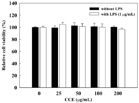
      >  Effect of CCE on RAW 264.7 cell viability

    To determine whether CCE causes cytotoxicity in RAW 264.7 cells, we assessed the effects of various concentrations of CCE on RAW 264.7 cell viability by MTS assay (Fig. 1). We found that CCE, alone or combined with LPS (1 μg/mL), did not affect the viability of RAW 264.7 cells at concentra-tions of up to 200 μg/mL. At a concentration of 400 μg/mL, CCE reduced the viability of RAW 264.7 cells to 78.8 ± 4.8% when applied alone and to 82.5 ± 5.1% when applied with LPS. Thus, we used CCE at concentrations of ≤200 μg/mL, which did not affect cell viability, in further studies of anti-inflammatory activity and possible mechanisms of action in RAW 264.7 cells.

    The results of several studies strongly suggest that phenolic compounds derived from botanical sources are able to sup-press inflammation via removal of reactive oxygen species by antioxidants (Rahman et al., 2006; Kim and Kim, 2010). We previously described the anti-oxidative and anti-inflammatory activities of phloroglucinol derivatives of Ecklonia stolonifera (Kim et al., 2009; Kim et al., 2011). The effects of different solvents used to extract polyphenols from plant material have been tested (Pinelo et al., 2004). Although the extraction yield is dependent on the solvent used and the method of extraction, extracts from various red seaweeds show a relationship be-tween antioxidant activity and polyphenolic content (Ganesan et al., 2008). Ethanol extraction gave a relatively high radi-cal scavenging activity and a high yield of polyphenol com-pounds (Lopez et al., 2011). Thus, we used ethanol to extract anti-inflammatory compounds from C. crassicaulis. We sub-sequently assessed the anti-inflammatory activity of the result-ing ethanolic extract in LPS-stimulated RAW 264.7 cells.

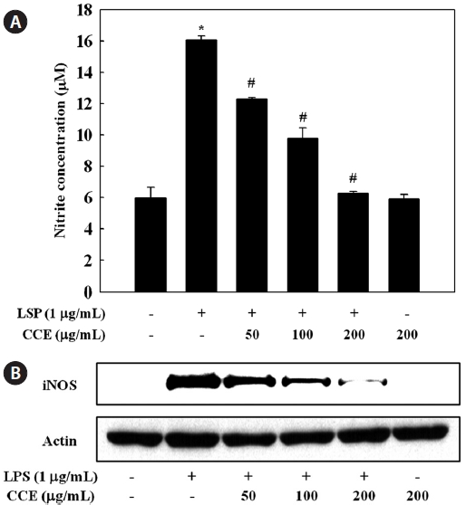
      >  Effect of CCE on the production of NO in LPS-stimulated cells

    To determine the effect of CCE on NO production in LPS-stimulated RAW 264.7 cells, we measured culture medium ni-trite concentrations using the Griess reagent. RAW 264.7 cells were pretreated with various concentration of CCE (50-200 μg/mL) for 1 h and stimulated with LPS for 24 h. Production of NO, measured as nitrite, was increased by LPS. CCE sig-nificantly and dose-dependently suppressed the production of NO by LPS-stimulated cells (P < 0.05) (Fig. 2A). Since iNOS is the enzyme responsible for the production of NO, we ana-lyzed the effect of CCE on iNOS protein expression in LPS-stimulated RAW 264.7 cells by Western blotting. As shown in Fig. 2B, CCE strongly and dose-dependently suppressed the expression of iNOS protein. These results indicate that CCE-mediated inhibition of NO production in LPS-stimulated macrophages was associated with downregulation of iNOS. The MTS assay showed that the CCE caused no cytotoxicity at concentrations up to 200 μg/mL CCE in RAW 264.7 cells (Fig. 1). Thus, the inhibitory effect of CCE on NO production was not attributable to cytotoxic effects.

    NO is synthesized from ?-arginine and molecular oxygen in a reaction catalyzed by NOS. Under pathological conditions, a significant increase in iNOS-derived NO contributes to the induction of inflammation, acting synergistically with other inflammatory mediators (Nathan, 1992). iNOS is strongly induced following exposure to bacterial endotoxin and pro-inflammatory cytokines (Guha and Mackman, 2001). Com-pounds able to reduce NO production or iNOS activity may be attractive anti-inflammatory agents, and for this reason, the suppressive effects of natural marine compounds on NO pro-duction have been intensively studied with the aim of devel-oping anti-inflammatory drugs (Abad et al., 2008; Jung et al., 2009; Heo et al., 2010; Jin et al., 2010; Kim and Kim, 2010). The present results suggest that CCE-mediated inhibition of NO production in LPS-stimulated macrophages was associ-ated with downregulation of the iNOS protein.

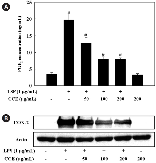
      >  Effect of CCE on PGE2 production in LPS-stimulated cells

    To evaluate the effect of CCE on PGE2 production in LPS-stimulated RAW 264.7 cells, culture medium PGE2 concentra-tions were determined by ELISA. The stimulation of PGE2 production in RAW 264.7 cells by LPS was suppressed by CCE treatment (P < 0.05) (Fig. 3A). Since COX-2 is the en-zyme responsible for the production of PGE2, we analyzed the effect of CCE on COX-2 protein expression in LPS-stimulated RAW 264.7 cells by Western blotting. As shown in Fig. 3B, CCE strongly and dose-dependently suppressed the expres-sion of COX-2 protein.

    COXs regulate the conversion of arachidonic acid to PGE2 and are rate-limiting enzymes in the biosynthesis of PGs (Du-

    bois et al., 1998). COX-1 is constitutively expressed in many tissues, while COX-2 is known as an inducible enzyme that, in most cases, generates large amounts of PGs. COX-2 is highly expressed in inflammation-related cell types including macro-phages and mast cells stimulated with pro-inflammatory cy-tokines and/or LPS (Nathan, 1992; Vane et al., 1994). Recent studies have shown that in vivo and in vitro treatments with natural compounds reduce inflammation by suppressing iNOS and COX-2 (Shin et al., 2010; Chung et al., 2011; Pan et al., 2011). The present results suggest that CCE-mediated inhi-bition of PGE2 production in LPS-stimulated macrophages was associated with downregulation of COX-2. This is, to our knowledge, the first study to address the inhibition of iNOS and COX-2 expression by CCE.

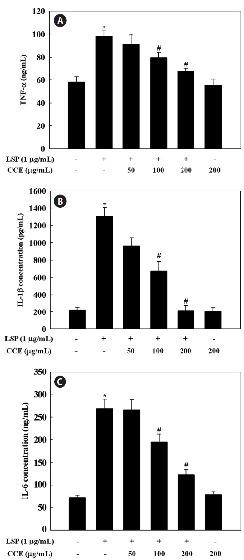
      >  Effect of CCE on the production of TNF-α, IL-1β, and IL-6 in LPS-stimulated cells

    Since CCE was found to inhibit the inflammatory mediators NO and PGE2 in a dose-dependent manner, we investigated the inhibitory effect of CCE on LPS-induced TNF-α, IL-1β,

    and IL-6 release by ELISA. Stimulation of RAW 264.7 cells with LPS significantly increased the levels of TNF-α, IL-1β, and IL-6. However, CCE, at concentrations ranging from 50 to 200 μg/mL, dose-dependently inhibited cytokine production in LPS-stimulated RAW 264.7 cells (P < 0.05) (Fig. 4). The

    inhibinhibitory effect of CCE on TNF-α, IL-1β, and IL-6 produc-tion was not attributable to cytotoxic effects, since cell viabil-ity was not altered by CCE at the concentrations used in this study (Fig. 1).

    Pro-inflammatory cytokines such as TNF-α, IL-1β, and IL-6 are small secreted proteins that regulate immunity and inflammation. Bacterial LPS stimulates macrophages to re-lease TNF-α. Secreted TNF-α or LPS then induces cells to release IL-1β and IL-6 (Beutler and Cerami, 1989). TNF-α induces several physiological processes, including septic shock, inflammation, and cytotoxicity (Dinarello, 1999). IL-1β is a major pro-inflammatory cytokine, which is mainly re-leased by macrophages and is believed to play a key role in the pathophysiology of endometriosis (Lebovic et al., 2000). IL-1β is also important in the initiation and enhancement of inflammatory responses to microbial infection (Kim and Moudgil, 2008). IL-6 is a pivotal pro-inflammatory cytokine that is mainly synthesized by macrophages. It plays a role in the acute-phase immune response (Yoshimura, 2006) and is regarded as an endogenous mediator of LPS-induced fever. The results of the present study suggest that CCE significantly suppressed LPS-induced TNF-α, IL-1β, and IL-6 secretion, which supports the idea that CCE inhibits the initial phase of a LPS-stimulated inflammatory response.

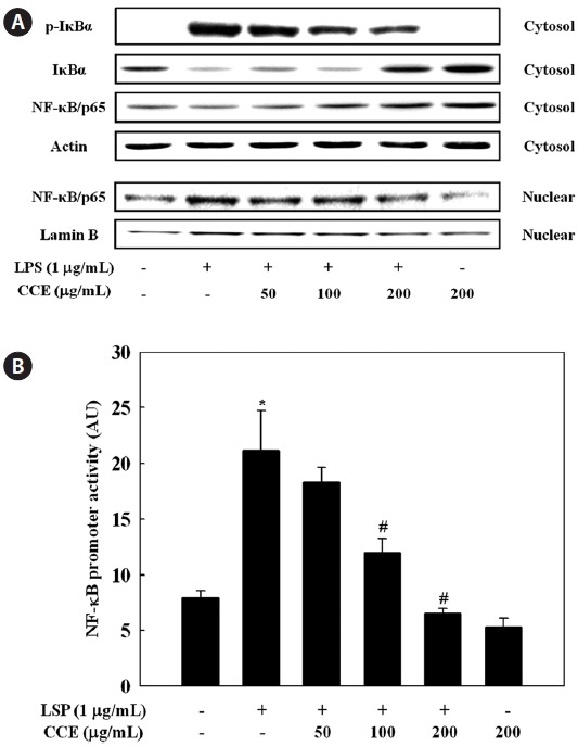
      >  Effect of CCE on NF-κB activation in LPS-stimulat-ed cells

    We further investigated whether CCE could inhibit the LPS-stimulated degradation of IκB-α in RAW 264.7 cells by Western blotting. LPS treatment resulted in degradation of IκB-α, a response that was dose-dependently reversed by CCE pretreatment (Fig. 5A). To investigate translocation of the p65 subunit of NF-κB from the cytosol to the nucleus after its release from IκBs, we examined NF-κB levels in cytosolic and nuclear extracts. CCE pretreatment dose-dependently increased cytosolic NF-κB levels (Fig. 5A). CCE dose-de-pendently reduced the nuclear p65 level, and in parallel in-creased the cytosolic p65 level. Considering the inhibitory effects of CCE on LPS-induced NF-κB activation, we next determined the effect of CCE on NF-κB promoter activity in LPS-stimulated macrophage cells. To achieve this, cells were transiently transfected with a luciferase construct containing the murine NF-κB promoter, pretreated for 2 h with various concentrations of CCE, and treated with LPS (1 μg/mL) for 6 h. Luciferase assay data show that CCE treatment significantly suppressed LPS-induced NF-κB promoter-driven luciferase expression in macrophages (P < 0.05) (Fig. 5B). These re-sults suggest that CCE-mediated inhibition of iNOS, COX-2, and pro-inflammatory cytokine expression in LPS-stimulated macrophages was regulated by an NF-κB pathway.

    The induction of inflammatory mediators such as NO and PGE2, and of pro-inflammatory cytokines such as TNF-α, IL-1β, and IL-6, is dependent on NF-κB activation (Li and Ver-

    ma, 2002). NF-κB is known to play a pivotal role in the regu-lation of cell survival genes and to coordinate the expression of pro-inflammatory enzymes and cytokines such as iNOS, COX-2, TNF-α, IL-1β, and IL-6 (Xie et al., 1993; D’Acquisto et al., 1997; Marks-Konczalik et al., 1998; Makarov, 2001). NF-κB is associated with and tightly controlled by an inhibi-tory subunit, IκB, which retains NF-κB in the cytoplasm in an inactive form. Activation of NF-κB by LPS involves the phosphorylation of IκB-α kinase, which phosphorylates IκB-α at Ser32 and Ser36, leading to the subsequent degradation of IκB-α and inducing translocation of NF-κB into the nucleus (Chen et al., 1995). In the present study, we observed that deg-radation of IκB-α in response to LPS treatment was reversed by CCE treatment, suggesting that CCE protected IκB-α from proteolytic degradation. Moreover, we found that transloca-tion of NF-κB into the nucleus was dose-dependently inhib-ited by CCE. From these data, the CCE-mediated downregula-tion of LPS-induced iNOS, COX-2, TNF-α, IL-1β, and IL-6 expression in RAW 264.7 cells is likely largely dependent on the ability of CCE to inhibit NF-κB signaling. This is, to the best of knowledge, the first report addressing CCE’s negative regulation of LPS-activated NF-κB signaling.

    In conclusion, we demonstrated that CCE inhibited pro-duction of the inflammatory mediators NO and PGE2 and the pro-inflammatory cytokines TNF-α, IL-1β, and IL-6 in LPS-stimulated RAW 264.7 macrophages. Moreover, the inhibitory effects of CCE were found to be associated with the inactivation of NF-κB signaling through inhibition of IκB degradation. Verification of CCE’s anti-inflammatory activity and mechanisms of action in cells will be beneficial to the fur-ther application of CCE in functional foods in the treatment of inflammatory diseases. These results suggest the need to perform additional studies to identify the compounds in CCE that contribute to its anti-inflammatory activity.

참고문헌
  • 1. Abad MJ, Bedoya LM, Bermejo P 2008 Natural marine anti-inflam-matory products. [Mini Rev Med Chem] Vol.8 P.740-754 google
  • 2. Beutler B, Cerami A 1989 The biology of cachectin/TNF--a prima-ry mediator of the host response. [Annu Rev Immunol] Vol.7 P.625-655 google cross ref
  • 3. Blunt JW, Copp BR, Munro MH, Northcote PT, Prinsep MR 2010 Marine natural products. [Nat Prod Rep] Vol.27 P.165-237 google cross ref
  • 4. Chen Z, Hagler J, Palombella VJ, Melandri F, Scherer D, Ballard D, Maniatis T 1995 Signal-induced site-specific phosphorylation targets IκBα to the ubiquitin-proteasome pathway. [Genes Dev] Vol.9 P.1586-1597 google cross ref
  • 5. Chung EY, Kim BH, Hong JT, Lee CK, Ahn B, Nam SY, Han SB, Kim Y 2011 Resveratrol down-regulates interferon-γ-inducible inflammatory genes in macrophages: molecular mechanism via de-creased STAT-1 activation. [J Nutr Biochem] Vol.22 P.902-909 google cross ref
  • 6. D’Acquisto F, Iuvone T, Rombola L, Sautebin L, Di Rosa M, Car-nuccio R 1997 Involvement of NF-κB in the regulation of cy-clooxygenase-2 protein expression in LPS-stimulated J774 macro-phages. [FEBS Lett] Vol.418 P.175-178 google cross ref
  • 7. Dinarello CA 1999 Cytokines as endogenous pyrogens. [J Infect Dis] Vol.179 P.S294-S304. google cross ref
  • 8. Dubois RN, Abramson SB, Crofford L, Gupta RA, Simon LS, Van De Putte LB, Lipsky PE 1998 Cyclooxygenase in biology and disease. [FASEB J] Vol.12 P.1063-1073 google cross ref
  • 9. Ganesan P, Kumar CS, Bhaskar N 2008 Antioxidant properties of methanol extract and its solvent fractions obtained from selected Indian red seaweeds. [Bioresour Technol] Vol.99 P.2717-2723 google
  • 10. Guha M, Mackman N 2001 LPS induction of gene expression in human monocytes. [Cell Signal] Vol.13 P.85-94 google cross ref
  • 11. Heo SJ, Yoon WJ, Kim KN, Ahn GN, Kang SM, Kang DH, Affan A, Oh C, Jung WK, Jeon YJ 2010 Evaluation of anti-inflammatory effect of fucoxanthin isolated from brown algae in lipopolysaccha-ride-stimulated RAW 264.7 macrophages. [Food Chem Toxicol] Vol.48 P.2045-2051 google cross ref
  • 12. Jin M, Suh SJ, Yang JH, Lu Y, Kim SJ, Kwon S, Jo TH, Kim JW, Park YI, Ahn GW, Lee CK, Kim CH, Son JK, Son KH, Chang HW 2010 Anti-inflammatory activity of bark of Dioscorea batatas DECNE through the inhibition of iNOS and COX-2 expressions in RAW 264.7 cells via NF-κB and ERK1/2 inactivation. [Food Chem Toxicol] Vol.48 P.3073-3079 google cross ref
  • 13. Jung WK, Ahn YW, Lee SH, Choi YH, Kim SK, Yea SS, Choi I, Park SG, Seo SK, Lee SW, Choi IW 2009 Ecklonia cava ethanolic extracts inhibit lipopolysaccharide-induced cyclooxygenase-2 and inducible nitric oxide synthase expression in BV2 microglia via the MAP kinase and NF-κB pathways. [Food Chem Toxicol] Vol.47 P.410-417 google cross ref
  • 14. Kim AR, Shin TS, Lee MS, Park JY, Park KE, Yoon NY, Kim JS, Choi JS, Jang BC, Byun DS, Park NK, Kim HR 2009 Isolation and identification of phlorotannins from Ecklonia stolonifera with antioxidant and anti-inflammatory properties. [J Agric Food Chem] Vol.57 P.3483-3489 google cross ref
  • 15. Kim AR, Lee MS, Shin TS, Hua H, Jang BC, Choi JS, Byun DS, Utsuki T, Ingram D, Kim HR 2011 Phlorofucofuroeckol A inhibits the LPS-stimulated iNOS and COX-2 expressions in macrophages via inhibition of NF-κB Akt and p38 MAPK. [Toxicol In Vitro] Vol.25 P.1789-1795 google cross ref
  • 16. Kim EY, Moudgil KD 2008 Regulation of autoimmune inflamma-tion by pro-inflammatory cytokines. [Immunol Lett] Vol.120 P.1-5 google cross ref
  • 17. Kim MM, Kim SK 2010 Effect of phloroglucinol on oxidative stress and inflammation. [Food Chem Toxicol] Vol.48 P.2925-2933 google cross ref
  • 18. Kim YC, An RB, Yoon NY, Nam TJ, Choi JS 2005 Hepatoprotec-tive constituents of the edible brown alga Ecklonia stolonifera on tacrine-induced cytotoxicity in Hep G2 cells. [Arch Pharm Res] Vol.28 P.1376-1380 google cross ref
  • 19. Lebovic DI, Bentzien F, Chao VA, Garrett EN, Meng YG, Taylor RN 2000 Induction of an angiogenic phenotype in endometriotic stromal cell cultures by interleukin-1β. [Mol Hum Reprod] Vol.6 P.269-275 google cross ref
  • 20. Li Q, Verma IM 2002 NF-κB regulation in the immune system. [Nat Rev Immunol] Vol.2 P.725-734 google cross ref
  • 21. Libby P 2006 Inflammation and cardiovascular disease mechanisms. [Am J Clin Nutr] Vol.83 P.456S-460S google cross ref
  • 22. Lopez A, Rico M, Rivero A, de Tangil MS 2011 The effects of solvents on the phenolic contents and antioxidant activity of Sty-pocaulon scoparium algae extracts. [Food Chem] Vol.125 P.1104-1109 google
  • 23. Makarov SS 2001 NF-κB in rheumatoid arthritis: a pivotal regulator of inflammation hyperplasia and tissue destruction. [Arthritis Res] Vol.3 P.200-206 google cross ref
  • 24. Marks-Konczalik J, Chu SC, Moss J 1998 Cytokine-mediated transcriptional induction of the human inducible nitric oxide syn-thase gene requires both activator protein 1 and nuclear factor κB-binding sites. [J Biol Chem] Vol.273 P.22201-22208 google cross ref
  • 25. Nathan C 1992 Nitric oxide as a secretory product of mammalian cells. [FASEB J] Vol.6 P.3051-3064 google cross ref
  • 26. Packard RR, Libby P 2008 Inflammation in atherosclerosis: from vascular biology to biomarker discovery and risk prediction. [Clin Chem] Vol.54 P.24-38 google
  • 27. Pan MH, Hong HM, Lin CL, Jhang AZ, Tsai JH, Badmaev V, Nagab-hushanam K, Ho CT, Chen WJ 2011 Se-methylselenocysteine inhibits lipopolysaccharide-induced NF-κB activation and iNOS induction in RAW 264.7 murine macrophages. [Mol Nutr Food Res] Vol.55 P.723-732 google
  • 28. Pinelo M, Rubilar M, Sineiro J, Nunez MJ 2004 Extraction of anti-oxidant phenolics from almond hulls (Prunus amygdalus) and pine sawdust (Pinus pinaster). [Food Chem] Vol.85 P.267-273 google cross ref
  • 29. Rahman I, Biswas SK, Kirkham PA 2006 Regulation of inflamma-tion and redox signaling by dietary polyphenols. [Biochem Pharma-col] Vol.72 P.1439-1452 google cross ref
  • 30. Shin JS, Park YM, Choi JH, Park HJ, Shin MC, Lee YS, Lee KT 2010 Sulfuretin isolated from heartwood of Rhus verniciflua in-hibits LPS-induced inducible nitric oxide synthase cyclooxygen-ase-2 and pro-inflammatory cytokines expression via the down-regulation of NF-κB in RAW 264.7 murine macrophage cells. [Int Immunopharmacol] Vol.10 P.943-950 google cross ref
  • 31. Solinas G, Marchesi F, Garlanda C, Mantovani A, Allavena P 2010 Inflammation-mediated promotion of invasion and metastasis. [Cancer Metastasis Rev] Vol.29 P.243-248 google cross ref
  • 32. Vane JR, Mitchell JA, Appleton I, Tomlinson A, Bishop-Bailey D, Croxtall J, Willoughby DA 1994 Inducible isoforms of cy-clooxygenase and nitric-oxide synthase in inflammation. [Proc Natl Acad Sci U S A] Vol.91 P.2046-2050 google cross ref
  • 33. Woo MN, Jeon SM, Shin YC, Lee MK, Kang MA, Choi MS 2009 Anti-obese property of fucoxanthin is partly mediated by altering lipid-regulating enzymes and uncoupling proteins of visceral adi-pose tissue in mice. [Mol Nutr Food Res] Vol.53 P.1603-1611 google cross ref
  • 34. Xie QW, Whisnant R, Nathan C 1993 Promoter of the mouse gene encoding calcium-independent nitric oxide synthase confers induc-ibility by interferon γ and bacterial lipopolysaccharide. [J Exp Med] Vol.177 P.1779-1784 google cross ref
  • 35. Yoshimura A 2006 Signal transduction of inflammatory cytokines and tumor development. [Cancer Sci] Vol.97 P.439-447 google cross ref
  • 36. Zhang G, Ghosh S 2000 Molecular mechanisms of NF-κB acti-vation induced by bacterial lipopolysaccharide through Toll-like receptors. [J Endotoxin Res] Vol.6 P.453-457 google cross ref
OAK XML 통계
이미지 / 테이블
  • [ Fig. 1. ]  Effect of Chondria crassicaulis ethanolic extract (CCE) on the viability of RAW 264.7 cells. Cells were treated with indicated concentration of CCE alone or CCE with lipopolysaccharide (LPS) (1 μg/mL) for 24 h. The cell viability was determined by MTS assay. Each column represents the means ± SDs from three independent experiments.
    Effect of Chondria crassicaulis ethanolic extract (CCE) on the viability of RAW 264.7 cells. Cells were treated with indicated concentration of CCE alone or CCE with lipopolysaccharide (LPS) (1 μg/mL) for 24 h. The cell viability was determined by MTS assay. Each column represents the means ± SDs from three independent experiments.
  • [ Fig. 2. ]  Effect of Chondria crassicaulis ethanolic extract (CCE) on lipopolysaccharide (LPS)-induced nitrite production and inducible nitric oxide synthase (iNOS) expression in RAW 264.7 cells. (A) Cells pretreated with different concentrations (50 100 200 μg/mL) of CCE for 1 h were stimulated with LPS (1 μg/mL) for 24 h. The culture medium was used to measure the amount of nitrite to evaluate NO production by Griess reagents. (B) Cells pretreated with indicated concentration of CCE for 1 h were stimulated with LPS (1 μg/mL) for 16 h. Equal amounts of total proteins were subjected to 10% sodium dodecyl sulfate polyacrylamide gel electrophoresis. The expression of iNOS and actin protein was detected by Western blotting using responding antibodies. *P < 0.05 indicates significant differences from the control group. #P < 0.05 indicates significant differences from the LPS-treated group.
    Effect of Chondria crassicaulis ethanolic extract (CCE) on lipopolysaccharide (LPS)-induced nitrite production and inducible nitric oxide synthase (iNOS) expression in RAW 264.7 cells. (A) Cells pretreated with different concentrations (50 100 200 μg/mL) of CCE for 1 h were stimulated with LPS (1 μg/mL) for 24 h. The culture medium was used to measure the amount of nitrite to evaluate NO production by Griess reagents. (B) Cells pretreated with indicated concentration of CCE for 1 h were stimulated with LPS (1 μg/mL) for 16 h. Equal amounts of total proteins were subjected to 10% sodium dodecyl sulfate polyacrylamide gel electrophoresis. The expression of iNOS and actin protein was detected by Western blotting using responding antibodies. *P < 0.05 indicates significant differences from the control group. #P < 0.05 indicates significant differences from the LPS-treated group.
  • [ Fig. 3. ]  Effect of Chondria crassicaulis ethanolic extract (CCE) on lipopolysaccharide (LPS)-stimulated prostaglandin E2 (PGE2) production and cyclooxygenase-2 (COX-2) expression in RAW 264.7 cells. (A) Cells pretreated with different concentrations (50 100 200 μg/mL) of CCE for 1 h were stimulated with LPS (1 μg/mL) for 24 h. The culture medium was used to measure the amount of PGE2 production by enzyme-linked immunosorbent assay. (B) Cells pretreated with indicated concentration of CCE for 1 h were stimulated with LPS (1 μg/mL) for 16 h. Equal amounts of total proteins of harvest cells were subjected to 10% sodium dodecyl sulfate polyacrylamide gel electrophoresis. The expression of iNOS and actin protein was detected by Western blotting using responding antibodies. *P < 0.05 indicates significant differences from the control group. #P < 0.05 indicates significant differences from the LPS-treated group.
    Effect of Chondria crassicaulis ethanolic extract (CCE) on lipopolysaccharide (LPS)-stimulated prostaglandin E2 (PGE2) production and cyclooxygenase-2 (COX-2) expression in RAW 264.7 cells. (A) Cells pretreated with different concentrations (50 100 200 μg/mL) of CCE for 1 h were stimulated with LPS (1 μg/mL) for 24 h. The culture medium was used to measure the amount of PGE2 production by enzyme-linked immunosorbent assay. (B) Cells pretreated with indicated concentration of CCE for 1 h were stimulated with LPS (1 μg/mL) for 16 h. Equal amounts of total proteins of harvest cells were subjected to 10% sodium dodecyl sulfate polyacrylamide gel electrophoresis. The expression of iNOS and actin protein was detected by Western blotting using responding antibodies. *P < 0.05 indicates significant differences from the control group. #P < 0.05 indicates significant differences from the LPS-treated group.
  • [ Fig. 4. ]  Effects of Chondria crassicaulis ethanolic extract (CCE) on pro-inflammatory cytokine productions in lipopolysaccharide (LPS)-stimulated RAW 264.7 cells. RAW 264.7 cells pretreated with indicated concentration of CCE for 1 h were stimulated with LPS (1 μg/mL) for 24 h. Tumor necrosis factor (TNF)-α (A) interleukin (IL)-1β (B) and IL-6 (C) in the cultured supernatant were measured by enzyme-linked immunosorbent assay. Data are mean ± SDs. of three independent experiments. *P < 0.05 indicates significant differences from the control group. #P < 0.05 indicates significant differences from the LPS-treated group.
    Effects of Chondria crassicaulis ethanolic extract (CCE) on pro-inflammatory cytokine productions in lipopolysaccharide (LPS)-stimulated RAW 264.7 cells. RAW 264.7 cells pretreated with indicated concentration of CCE for 1 h were stimulated with LPS (1 μg/mL) for 24 h. Tumor necrosis factor (TNF)-α (A) interleukin (IL)-1β (B) and IL-6 (C) in the cultured supernatant were measured by enzyme-linked immunosorbent assay. Data are mean ± SDs. of three independent experiments. *P < 0.05 indicates significant differences from the control group. #P < 0.05 indicates significant differences from the LPS-treated group.
  • [ Fig. 5. ]  Effect of Chondria crassicaulis ethanolic extract (CCE) on activation of nuclear factor-κB (NF-κB) in lipopolysaccharide (LPS)-stimulated RAW 264.7 cells. RAW 264.7 cells pretreated with indicated concentration of CCE for 1 h were stimulated with LPS (1 μg/mL) for 30 min. The expression of inhibitor of κB-α and NF-κB (A) were determined by a Western blot analysis. (B) RAW 264.7 cells were co-transfected with 1 μg of NF-κB promoter-containing luciferase DNA along with 20 ng of control pRL-TK DNA for 24 h. Transfected cells pretreated with indicated concentration of CCE for 1 h were stimulated with LPS (1 μg/mL) for 6 h. Data are mean ± SDs of three independent experiments. *P < 0.05 indicates significant differences from the control group. #P < 0.05 indicates significant differences from the LPS-treated group.
    Effect of Chondria crassicaulis ethanolic extract (CCE) on activation of nuclear factor-κB (NF-κB) in lipopolysaccharide (LPS)-stimulated RAW 264.7 cells. RAW 264.7 cells pretreated with indicated concentration of CCE for 1 h were stimulated with LPS (1 μg/mL) for 30 min. The expression of inhibitor of κB-α and NF-κB (A) were determined by a Western blot analysis. (B) RAW 264.7 cells were co-transfected with 1 μg of NF-κB promoter-containing luciferase DNA along with 20 ng of control pRL-TK DNA for 24 h. Transfected cells pretreated with indicated concentration of CCE for 1 h were stimulated with LPS (1 μg/mL) for 6 h. Data are mean ± SDs of three independent experiments. *P < 0.05 indicates significant differences from the control group. #P < 0.05 indicates significant differences from the LPS-treated group.
(우)06579 서울시 서초구 반포대로 201(반포동)
Tel. 02-537-6389 | Fax. 02-590-0571 | 문의 : oak2014@korea.kr
Copyright(c) National Library of Korea. All rights reserved.