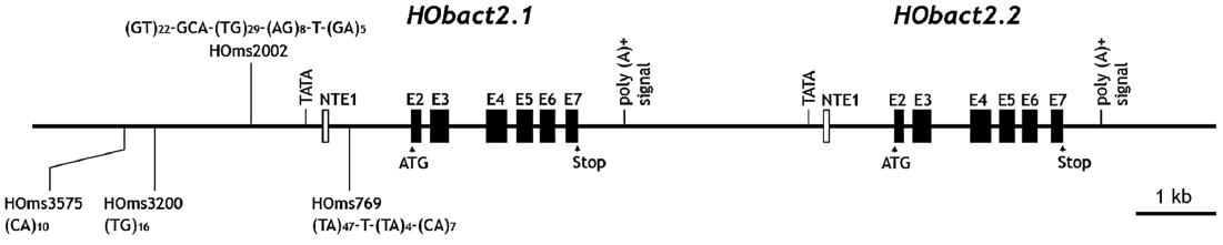
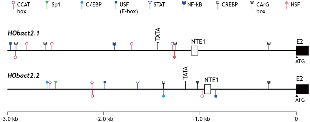
검색 전체 메뉴
PDF
맨 위로
OA 학술지
Tail-to-Head Tandem Duplication and Simple Repetitive Sequences of the Cytoplasmic Actin Genes in Greenling Hexagrammos otakii (Teleostei; Scorpaeniformes)
  • 비영리 CC BY-NC
  • 비영리 CC BY-NC
ABSTRACT
Tail-to-Head Tandem Duplication and Simple Repetitive Sequences of the Cytoplasmic Actin Genes in Greenling Hexagrammos otakii (Teleostei; Scorpaeniformes)
KEYWORD
Cytoplasmic actin , Gene duplication , SRS , Tissue distribution , Hexagrammos otakii
  • Introduction

    Cytoskeletal actin provides a mechanical support for the cell and mediates cell motility and organelle movement, which are associated with several dynamic cellular activities such as morphological and developmental changes, macromolecule transport, and chemotaxis (Reisler and Egelman, 2007). Fur-thermore, actin is involved in signal transduction (Percipalle and Visa, 2006), RNA localization (Kustermans et al., 2008), apoptosis (Franklin-Tong and Gourlay, 2008), and nitric oxide (NO) regulation (Ji et al., 2007). In addition, the 5′-flanking region of cytoskeletal actin genes is capable of driving the ex-pression of downstream structural genes when it is included in a chimeric transgene construct as a regulatory element (Nam et al., 2008).

    In teleosts, genetic determinants of cytoskeletal actin have been isolated from various species belonging to a wide array of taxonomic positions. Although the coding regions of cyto-skeletal actin are well characterized, their genomic organiza-tion and upstream regulatory regions require further investi-gation (see Venkatesh et al., 1996; Cho et al., 2011). Teleosts experienced whole-genome duplication in their evolutionary history and many teleost species may exhibit additional, lo-cus-specific gene duplications in their genomes (Jaillon et al., 2004). Hence, the teleost group likely encodes diverse paralog isoforms in their genomes, and some of these paralogs may undergo subfunctionalization or neofunctionalization. In ver-tebrates, the actin multigene family evolved into six or more isoforms from a single ancestral form (Miwa et al., 1991; Venkatesh et al., 1996), and previous studies have suggest-ed that a greater spectrum of actin isoforms may exist in the fish genome compared to representative mammalian species (Venkatesh et al., 1996). Duplicated (diverged) actin genes are known to exist in the teleost genomes, but their organization has not been extensively studied.

    Greenling Hexagrammos otakii (Teleostei; Scorpaeni-formes) is a commercially important marine food fish in Korea and China, and its market demand has been gradually increas-ing. This species is also a candidate for a marine launching program in Korea because of its nonmigratory and gathering behavior around artificial reefs. Although the culturing tech-niques and artificial manipulation required throughout its life cycle have not been established, various studies have been performed to better understand their physiology, with a focus on reproduction, growth, metabolism, and stress (Nam and Kim, 2002; Kang et al., 2004; Kim et al., 2009). Discovery and characterization of useful genetic markers and gene se-quences are essential to gain a deeper insight into the physiol-ogy of a species. As a part of our long-term research to col-lect genomic and gene information from this species to aid its genetic breeding program, the objective of this study was to characterize the genomic organization and gene structure of the duplicated cytoplasmic actin isoforms.

    Materials and Methods

      >  Fish specimens and nucleic acid libraries

    Greenling specimens were purchased from a local fish farm and transferred to a laboratory culture station. Genomic DNA samples were obtained from a fin clip or whole blood using the conventional SDS/proteinase K method followed by etha-nol precipitation. To construct the greenling cDNA library, total RNA was extracted from liver and brain tissues using the RNeasy Midi Kit (Qiagen, Hilden, Germany). After puri-fication of the poly(A)+ RNA fraction using the mRNA Isola-tion Kit (Promega, Madison, WI, USA), an aliquot (5 μg) of poly(A)+ RNA was used as the template for cDNA synthesis. Construction of the cDNA libraries was performed using the Lambda Zap cDNA Synthesis Kit (Stratagene, La Jolla, CA, USA) according to the manufacturer’s instruction. The ge-nomic DNA library was constructed using whole blood DNA purified from a male adult using the Lambda Gem 11 Cloning System (Promega) according to manufacturers’ protocol.

      >  Isolation of genomic genes using DNA library screening

    Based on our preliminary expressed sequence tag (EST) analysis of greenling brain and liver cDNA libraries (unpub-lished data), an EST clone with significant homology to ver-tebrate cytoplasmic actin was identified and used as a probe to perform a series of filter screenings of the greenling ge-nomic DNA library. The EST clone (0.6 kb) was PCR-labeled with digoxygenin-11-dUTP (Roche Applied Science, Man-heim, Germany). Approximately 5 × 10⁵ phage plaques were screened by plaque hybridization using the DIG DNA Label-ing and Detection Kit (Roche Applied Science). The selected phages were amplified using KW251Escherichia coli strain and the phage DNA was purified using the Wizard Lambda Phage Prep Kit (Promega). Purified phage DNA was digested with SacI and subcloned into a pBluescript KS II phagemid vector (Stratagene). The sequence of the DNA inserts in each subclone was determined using the transposon-mediated shotgun sequencing method (EZ-Tn5 KAN-2 Kit; Epicentre, Madison, WI, USA) according to the manufacturer’s instruc-tions. Based on the contig assembly using Sequencher (Gene Codes, Ann Arbor, MI, USA), overlapping genomic clones were PCR-isolated and sequenced to validate the representa-tive sequence of the cytoplasmic actin gene.

      >  Isolation of cDNA sequences

    Based on genomic sequence analysis, type-specific reverse transcription (RT)-PCR primer pairs (HOact2.1c-1F/1R and HOact2.2c-1F/1R) were designed to amplify the complete open reading frame (ORF) of each isoform. Total RNA (500 ng) from the brain or gill was used as a template for RT-PCR isolation. Oligonucleotide primers and thermal cycling condi-tions used in this study are summarized to Table 1. The am-plification product was sequenced after TA cloning into the pGEM-T Easy Vector (Promega). Based on the ORF-contain-ing sequences, the 5′- and 3′-end untranslated region (UTR) sequences of each isoform were obtained by vectorette PCR, which was performed using template DNA prepared from greenling brain or liver cDNA libraries (excised stocks) and two vector primers (SK and T7). Binding sites are located at either end of the multiple cloning sites in the phagemid vec-tor (pBluescript SK; Stratagene) used for construction of the cDNA libraries. Isoform-specific primers included in the vec-torette PCR as either forward or reverse primers paired with SK or T7 vector primers were HOact2.1-vec1 and HOact2.2-vec1, respectively. Based on the contig assembly of ORF and ends sequences, we determined the full-length cDNA se-quence of each isoform.

      >  Bioinformatic sequence analysis

    Using NCBI GenBank BLAST, we searched for orthologs of the full-length cDNA sequences. Putative ORFs of each isoform were identified using ORF Finder ( http://www.ncbi.nlm.nih.gov/gorf/gorf.html ). Predicted molecular mass and theoretical pI values were calculated using the ProtParam tool ( http://www.expasy.org/tools/protparam.html ). Multiple sequence alignments were performed using a ClustalW al-gorithm ( http://www.genome.jp/tools/clustalw/ ). Genomic structure and organization were compared using representa-tive orthologs and/or paralogs obtained from the public data-bases, including GenBank and Ensembl ( http://www.ensembl.org/ ). Putative transcription factor (TF)-binding motifs in the 5′-flanking upstream region of the actin isoforms were iden-tified using TFSEARCH ( http://www.cbrc.jp/research/db/TFSEARCH.html ) and Transcription Element Search System (TESS; http://www.cbil.upenn.edu/cgi-bin/tess/tess ). Identifi-cation of repetitive sequences was performed using the Unipro UGENE ( http://ugene.unipro.ru/ ).

      >  Typing of simple sequence repeats (SSRs)

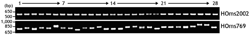
    We identified microsatellite-like, SSRs in the noncoding re-gion of one actin isoform. To identify polymorphism among greenling individuals, two PCR primer pairs (HOms769 1F/1R and HOms2002 1F/1R) were designed to amplify each of the two microsatellite regions. In total, 48 individuals ran-domly obtained from three different domestic markets were subjected to microsatellite typing. Genomic DNA was extract-ed from a caudal fin clip (as described above) and 200 ng of purified DNA was used as a template for PCR amplification. The PCR product (5 μL) was separated on 1.5% agarose gel and visualized by ethidium bromide staining to examine any

    [Table 1.] Oligonucleotide primers used in this study

    label

    Oligonucleotide primers used in this study

    polymorphism in PCR product length among individuals.

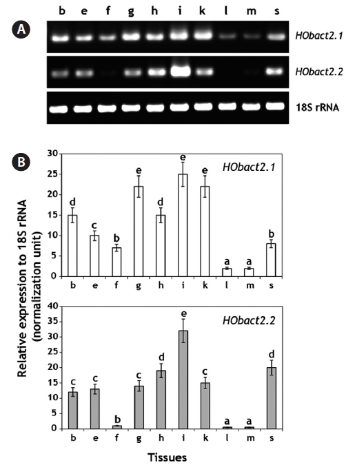
      >  Tissue distribution and basal expression of actin transcripts using a RT-PCR assay

    We performed real-time RT-PCR to examine the tissue dis-tribution and basal expression of each actin isoform transcript. Total RNA was extracted from ten somatic tissues including the brain, eye, fin, gill, heart, intestine, kidney, liver, muscle, and spleen, which were obtained from 12 juvenile individu-als (average body weight, 456 ± 52.0 g), as described above. Total RNA (4 μg) was reverse-transcribed (37℃ for 60 min) into cDNA in a reaction volume of 40 μL using the Omniscript Reverse Transcription Kit (Qiagen) according to the manufac-turer’s instructions. A conserved reverse primer for teleost 18S rRNA (Fi18SrRNA-1R primer) was also included in the RT reaction at a 0.1 μM final concentration to prepare the nor-malization standard across tissues. The RT product (cDNA) was diluted fourfold (for actins) or 40-fold (for 18S rRNA) with sterile distilled water, and 2 μL of the diluted cDNA was used as a template for PCR amplification. Based on the pre-liminary optimization of thermal cycling conditions and PCR efficiency (E = 10-1/slop - 1 in the standard curve for each gene), each actin isoform and 18S rRNA control were amplified us-ing the primers qHOact2.1-1F/1R (242 bp), qHOact2.2-1F/1R (218 bp), and q18S-1F/1R (261 bp), respectively. The reaction was performed on the iCycler iQ Real-Time Detection System

    (Bio-Rad, Hercules, CA, USA) using the 2× iQ SYBR Green Supermix (Bio-Rad) with a reaction volume of 25 μL. Based on the amplification assays (performed in triplicate), the rela-tive transcript levels of each actin isoform across tissues were normalized against the levels of the 18S rRNA control using the following formula: relative expression = [(1 + E18S rRNA)Ct18S rRNA]-1/[(1 + EACTIN)CtACTIN]-1, in which E is the PCR effi-ciency and Ct is the threshold cycle number (Kubista et al., 2006; Schmittgen and Livak, 2008). Differences in the rela-tive expression levels of each actin transcript among tissues were assessed by analysis of variance (ANOVA) followed by Duncan’s multiple range test at P = 0.05. The “endpoint” semiquantitative RT-PCR was conducted with the same prim-er pairs, and the amplification products were visualized on a 1.5% agarose gel using ethidium bromide staining.

    Results and Discussion

      >  Gene structure, genomic organization, and cDNA sequences of greenling cytoplasmic actins

    Based on the sequence analysis of genomic DNA library clones followed by PCR isolation, the 14,813-bp genomic re-gion containing two cytoplasmic actin genes was read at least twice in both directions. In the genomic region, two copies of the cytoplasmic actin gene existed tandemly in a tail-to-head organization. These two actin copies were named HOactb2.1 and HOactb2.2 (Fig. 1). Based on sequence comparison with its corresponding cDNA version (see below), HOactb2.1 has six translated exons (exons 2-7; 126, 240, 247, 192, 182, 144 bp including the stop codon) and one non-translated exon 1 (NET1; 69 bp). These seven exons are interrupted by six in-trons (1026, 104, 507, 109, 95, 181 bp, respectively, for in-trons 1-6). HOactb2.2 is also composed of seven exons (six translated exons and one NTE1; 66 bp) with a similar exon/intron organization. The length of each translated HOactb2.2 exon is identical to that from HOactb2.1. Furthermore, the in-tron lengths of the second isoform (797, 102, 509, 109, 95, and 180, respectively, for introns 1-6) are also similar with those from HOactb2.1, except intron 1 following NTE1. The puta-tive polyadenylation signal (AATAAA) is predicted at 503 and 477 bp after the stop codon, respectively, in HOactb2.1 and HOactb2.2. The distance between the two actin gene copies (estimated based on the distance from the HOactb2.1 poly-adenylation signal to the HOactb2.2TATA box) is only 2.3 kb. The full-length cDNA sequence of HOactb2.1 obtained by RT-PCR isolation and vectorette consists of 72 bp of 5′-UTR, 1,125 bp of a single ORF encoding a polypeptide of 375 amino acids (aa), and 552 bp of a 3′-UTR including the stop codon and 21 bp poly(A)+ tail. HOactb2.2 also encodes a 375-aa polypeptide with a homogenous cDNA structure com-posed of a 69-bp 5′-UTR, 1,125-bp ORF, and 525-bp 3′-UTR. Putative polyadenylation signals (AATAAA; also detected in the genomic sequence) are present 26 bp before the poly(A)+ tail in both isoforms. At the nucleotide level, the two isoforms share 99.3% sequence identity in the coding region with only 8 nucleotide substitutions. The noncoding regions (5′- and 3′-UTRs) have more nucleotide differences. When comparing the 69-bp 5′-UTR in HOactb2.1 and HOactb2.2, nucleotide substitutions occur at 10 positions (85.5% sequence identity), while the 3′-UTR has nucleotide differences at 46 positions (19 substitutions and 27 deletions) in the 531-bp aligned region (91.3% identity). At the amino acid level, the two isoforms exhibit only one amino acid substitution (Thr323 ↔ Ser323) in the 375-aa polypeptide (99.7% identity). Consequently, they have similar protein characteristics including molecular weight (41.8 kDa) and theoretical pI value (5.3). The amino acid sequences of both isoforms share considerable homology to previously known vertebrate orthologs (data not shown).

    All actin isoforms have remarkably similar amino acid sequences and polypeptide lengths across diverse metazoan organisms, demonstrating their high evolutionary constraints (Reece et al., 1992). However, compared to mammals, the te-leostean group possesses a wider spectrum of actin isoforms, in which several isoforms show no clear orthology to known mammalian isoforms (Venkatesh et al., 1996; Kim et al., 2008). For example, of the various cytoplasmic actin isoforms identified in teleosts, the major β-cytoplasmic-1 genes are clearly homologous to the mammalian isoforms (actb1 and/or

    actg1). Other isoforms are distinct from the mammalian coun-terparts, suggesting that the teleost group actin multigenes experienced different evolutionarily pathway(s) from that of mammals (Kim et al., 2008). In this study, the two greenling actin genes belong to the teleost minor cytoplasmic actin group (actb2) based on multiple sequence alignments and molecular phylogeny studies (data not shown). The number and position of the HOactb2.1 and HOactb2.2 introns exactly correspond to Takifugu rubripes β-cytoplasmic actin-2, characterized by Venkatesh et al. (1996). Furthermore, the similarity in coding nucleotide sequences, intron lengths, and head-to-tail arrange-ment between HOactb2.1 and HOactb2.2 unambiguously demonstrates that the two isoforms evolved by a recent gene duplication event. Our bioinformatic analyses also indicated that similar locus-specific duplication of actin gene copies occurred in the stickleback (Gasterosteus aculeatus; Gaster-osteiformes) Ensembl scaffold (ENSGACG00000001330 and ENSGACG00000001357 in scaffold_225) and in ze-brafish (Danio rerio; Cypriniformes) chromosome 25 (ENS-DARG00000070646 and ENSDARG00000070647).

      >  Characteristics of the 5’-upstream region

    The 5′-flanking upstream regions (3 kb upstream from the translation start codon ATG) of HOactb2.1 and HOactb2.2 were analyzed to identify potential TF-binding motifs (Fig. 2). Each isoform contained binding motifs for various TFs in both the 5′-flanking region and intron 1 following the non-translated exon. In the proximal promoter region, HOactb2.1 and HOactb2.2 possess a conserved TATA box at -1,348 and -1,065 bp from the ATG site (i.e., -250 and -199 bp from the transcription start site; TSS), respectively. In both isoforms, one CAAT (CCAAT) and CArG [CC(A/T)6GG] box are pre-dicted between the TSS and TATA box, although the relative order and distance between these boxes are not identical be-tween HOactb2.1 and HOactb2.2. These essential boxes are considered key elements to allow for constitutive and ubiq-uitous expression of the cytoplasmic actin genes in vertebrate tissues (Reece et al., 1992; Kim et al., 2008). The two isoforms exhibit four (HOactb2.1) and three (HOactb2.2) additional copies of CAAT boxes in their upstream regions. Fairly con-sistent with other beta-cytoskeletal teleost actin genes, HO-actb2.1 and HOactb2.2 have one CArG box (a trans-acting factor-binding site) at a conserved position in intron 1, which is thought to function as an enhancer in the regulation of the cytoplasmic actin (Liu et al., 1991; Noh et al., 2003). Addi-tionally, HOactb2.1 (but not HOactb2.2) has two more CArG boxes in its distal promoter region. Besides these known TF binding sites, both HOactb2.1 and HOactb2.2 likely pos-sess motifs associated with stress and immune responses. They include binding sites for upstream stimulatory factor (USF; CANNTG), CAAT enhancer-binding protein (C/EBP; TTDNGNAA), stimulating protein (Sp1; GGCRGGG), heat shock factor (HSF; RGAANRTTC), nuclear factor kappa B (NF-κB; GGGRNNYYCC), signal transducers and activator of transcription (STAT; TTCNNNGAA), and cAMP-response element-binding protein (CREBP; TGACGY). Although these TFs are known to regulate immune- and stress-responsive genes (Anderson, 2000; Truksa et al., 2007; Cho et al., 2009), whether they modulate HOactb2.1 and HOactb2.2 regulation is not clear. Thus, examination of their gene expression un-der various stimulatory challenges is required to evaluate the involvement of these bioinformatically identified TFs in the transcription of H. otakii actin genes. Although the regulatory machinery (including these responsive TFs) has not been em-pirically characterized in fish cytoplasmic actins, our findings are similar to previous bioinformatic observations of other fish cytoskeletal actin genes that predicted different stress-respon-

    sive TF-binding motifs in their regulatory regions (Lee et al., 2009; Cho et al., 2011). In addition, our data are partially sup-ported by the fact that mRNA expression of cytoskeletal actin genes is modulated depending on the developmental stage, stress state, or pathological condition of the mammal. Thus, caution is required when using actin gene expression as an in-variant, internal control for gene expression assays (Chapon-nier and Gabbiani, 2004; Filby and Tyler, 2007; Small et al., 2008).

      >  Simple sequence repeats

    Of the two H. otakii actin isoforms, HOactb2.1 contained SSRs in the 5′-flanking region and intron 1. The HOactb2.1 upstream region contained four microsatellite loci (three in the promoter region and one in intron 1). All SSRs found in HOactb2.1 were dinucleotide repeats: [(TA)47-T-(TA)4-(CA)7 at position -769 to -653 bp from the ATG start codon], [(GT)22-GCA-(TG)29-(AG)8-T-(GA)5 at -2,002 to -1,871 bp ], [(TG)16 at -3,200 to -3,169 bp] and [(CA)10 at -3,575 to -3,556 bp] (see also Fig. 1). As a preliminary experiment, we selected two longer SSR regions located at positions -769 (HOms769) and -2,002 (HOms2002), and evaluated whether they were polymorphic among individuals. Using the primer pairs HOms769 1F/1R (for HOms769 locus) and HOms2002 1F/1R (for HOms2002 locus), the two microsatellite loci were inde-pendently amplified using PCR. As shown in Fig. 3, the SSR in intron 1 (HOms769) was highly polymorphic, even based on the conventional electrophoretic separation of the ampli-fication products, whereas the SSR in the promoter region (HOms2002) was much less variable among individuals. Mi-crosatellite SSR markers are useful to assess genetic diversity and develop molecular breeding techniques in fish due to their high level of polymorphism and codominant inheritance (Liu and Cordes, 2004). Microsatellite loci have also been isolated from a dinucleotide-enriched greenling genomic library (Chen et al., 2009). The HOms769 locus identified in this study could be added to a polymorphic microsatellite marker set to address the genetic diversity of the species. Evaluating its potential use for cross-species amplification in the closely related Hexa-grammos species would also be valuable. However, the pos-sible number of alleles for this locus in different H. otakii pop-ulations should be characterized using genotyping analyses.

      >  Tissue distribution and basal expression levels

    Based on the real-time RT-PCR assay, each greenling actin isoform was detected ubiquitously in all the tissues examined, although the basal expression levels varied among tissues (Fig. 4). HOactb2.1 transcripts were actively expressed in the intes-tine, kidney, and gills, while its expression level was lowest in the liver and skeletal muscle (P < 0.05). Other tissues, includ-

    ing the brain, eye, fin, heart, and spleen, showed a moderate level of HOactb2.1 mRNA expression. HOactb2.2 transcripts showed a more intestine-enriched distribution (P < 0.05) than HOactb2.1. Overall, the two isoforms had a similar, but not identical, pattern of tissue expression with high levels in the intestine and low levels in the liver and skeletal muscle. The ubiquitous distribution pattern of H. otakii actin transcripts is not unexpected considering its essential housekeeping roles in most cell types. Furthermore, their overall pattern (high expression in the intestine and kidney and low expression in the skeletal muscle and liver) agrees with the expression of cytoplasmic actin genes from other teleost species (Kim et al., 2008; Lee et al., 2009). Meanwhile, Venkatesh et al. (1996) suggested that actin gene copies that experienced significant subfunctionalization after gene duplication would have dif-ferent tissue distribution patterns, as exemplified by diverse actin paralogs in the pufferfish genome. However, based on the similar tissue expression patterns of HOactb2.1 and HO-actb2.2, the two greenling actin copies (which recently duplicated) have not significantly differentiated in their basic func-tions. Nevertheless, further study to evaluate any differential expression in response to various experimental challenges are required since the two H. otakii actin isoforms have different TF-binding motifs in their regulatory regions.

    In summary, two tandemly existing cytoplasmic actin genes were isolated and characterized from a greenling (H. otakii). They have a similar genomic organization and gene structure with a high degree of sequence identity at both the nucleo-tide and amino acid levels. However, in spite of their similar gene structures, the distribution pattern of TF-binding motifs in the regulatory region was different between the two isoform copies. Only one isoform (HObact2.1) had multiple SSRs in either the 5′-flanking region or intron 1. One of these SSRs is highly polymorphic among individuals. The mRNA tissue distribution pattern of the two actin isoforms was very simi-lar, which agrees with vertebrate cytoskeletal and non-muscle actins. Collectively, the two actin isoforms in the H. otakii ge-nome evolved from a recent gene duplication event and have not yet experienced significant subfunctionalization in their essential housekeeping roles.

참고문헌
  • 1. Anderson KV 2000 Toll signaling pathways in the innate immune re-sponse. [Curr Opin Immunol] Vol.12 P.13-19 google cross ref
  • 2. Chaponnier C, Gabbiani G 2004 Pathological situations character-ized by altered actin isoform expression. [J Pathol] Vol.204 P.386-395 google cross ref
  • 3. Chen SL, Miao GD, Shao CW, Tian YS, Liao X 2009 Isolation and characterization of polymorphic microsatellite loci from fat green-ling (Hexagrammos otakii). [Conserv Genet] Vol.10 P.1429-1431 google cross ref
  • 4. Cho YS, Lee SY, Kim KH, Kim SK, Kim DS, Nam YK 2009 Gene structure and differential modulation of multiple rockbream (Ople-gnathus fasciatus) hepcidin isoforms resulting from different bio-logical stimulations. [Dev Comp Immunol] Vol.33 P.46-58 google cross ref
  • 5. Cho YS, Lee SY, Kim YK, Kim DS, Nam YK 2011 Functional ability of cytoskeletal β-actin regulator to drive constitutive and ubiquitous expression of a fluorescent reporter throughout the life cycle of transgenic marine medaka Oryzias dancena. [Transgenic Res] Vol.20 P.1333-1355 google cross ref
  • 6. Filby AL, Tyler CR 2007 Appropriate ‘housekeeping’ genes for use in expression profiling the effects of environmental estrogens in fish. [BMC Mol Biol] Vol.8 P.10 google cross ref
  • 7. Franklin-Tong VE, Gourlay CW 2008 A role for actin in regulating apoptosis/programmed cell death: evidence spanning yeast plants and animals. [Biochem J] Vol.413 P.389-404 google cross ref
  • 8. Jaillon O, Aury JM, Brunet F, Petit JL, Stange-Thomann N 2004 Genome duplication in the te-leost fish Tetraodon nigroviridis reveals the early vertebrate proto-karyotype. [Nature] Vol.431 P.946-957 google cross ref
  • 9. Ji Y, Ferracci G, Warley A, Ward M, Leung KY, Samsuddin S, Leveque C, Queen L, Reebye V, Pal P, Gkaliagkousi E, Seager M, Ferro A 2007 β-Actin regulates platelet nitric oxide synthase 3 activity through interaction with heat shock protein 90. [Proc Natl Acad Sci U S A] Vol.104 P.8839-8844 google cross ref
  • 10. Kang HW, Chung EY, Kim JH 2004 Sexual maturation and spawn-ing characteristics in greenling Hexagrammos otakii of the west coast in Korea. [J Aquac] Vol.17 P.30-38 google
  • 11. Kim KY, Lee SY, Cho YS, Bang IC, Kim DS, Nam YK 2008 Characterization and phylogeny of two β-cytoskeletal actins from Hemibarbus mylodon (Cyprinidae Cypriniformes) a threatened fish species in Korea. [DNA Seq] Vol.19 P.87-97 google
  • 12. Kim Y, Kim PK, Kim HJ, Jo JY, Han W, Park J 2009 Effects of water temperature and ambient ammonia concentration on oxygen consumption and ammonia excretion of greenling Hexagrammos otakii Jordan et Starks. [Korean J Fish Aquat Sci] Vol.42 P.373-379 google cross ref
  • 13. Kubista M, Andrade JM, Bengtsson M, Forootan A, Jonak J, Lind K, Sindelka R, Sjoback R, Sjogreen B, Strombom L, Stahlberg A, Zoric N 2006 The real-time polymerase chain reaction. [Mol As-pects Med] Vol.27 P.95-125 google cross ref
  • 14. Kustermans G, Piette J, Legrand-Poels S 2008 Actin-targeting nat-ural compounds as tools to study the role of actin cytoskeleton in signal transduction. [Biochem Pharmacol] Vol.76 P.1310-1322 google cross ref
  • 15. Lee SY, Kim KH, Nam YK 2009 Molecular characterization of rockbream (Oplegnathus fasciatus) cytoskeletal β-actin gene and its 5’-upstream regulatory region. [Fish Aquat Sci] Vol.12 P.90-97 google cross ref
  • 16. Liu ZJ, Cordes JF 2004 DNA marker technologies and their appli-cations in aquaculture genetics. [Aquaculture] Vol.238 P.1-37 google cross ref
  • 17. Liu ZJ, Moav B, Faras AJ, Guise KS, Kapuscinski AR, Hackett P 1991 Importance of the CArG box in regulation of β-actin-encoding genes. [Gene] Vol.108 P.211-217 google cross ref
  • 18. Miwa T, Manabe Y, Kurokawa K, Kamada S, Kanda N, Bruns G, Ueyama H, Kakunaga T 1991 Structure chromosome loca-tion and expression of the human smooth muscle (enteric type) γ-actin gene: evolution of six human actin genes. [Mol Cell Biol] Vol.11 P.3296-3306 google
  • 19. Nam YK, Kim DS 2002 Molecular cloning and alternative splicing of growth hormone transcripts in greenling Hexagrammos otakii. [J Korean Fish Soc] Vol.35 P.676-681 google
  • 20. Nam YK, Maclean N, Hwang G, Kim DS 2008 Autotransgenic and allotransgenic manipulation of growth traits in fish for aquaculture: a review. [J Fish Biol] Vol.72 P.1-26 google cross ref
  • 21. Noh JK, Cho KN, Han EH, Kim A, Lee JS, Kim DS, Kim CG 2003 Genomic cloning of mud loach Misgurnus mizolepis (Cyprini-formes Cobitidae) β-actin gene and usefulness of its promoter re-gion for fish transgenesis. [Mar Biotechnol] Vol.5 P.244-252 google cross ref
  • 22. Percipalle P, Visa N 2006 Molecular functions of nuclear actin in transcription. [J Cell Biol] Vol.172 P.967-971 google cross ref
  • 23. Reece KS, McElroy D, Wu R 1992 Function and evolution of ac-tins. [Evol Biol] Vol.26 P.1-34 google
  • 24. Reisler E, Egelman EH 2007 Actin structure and function: what we still do not understand. [J Biol Chem] Vol.282 P.36133-36137 google cross ref
  • 25. Schmittgen TD, Livak KJ 2008 Analyzing real-time PCR data by the comparative C(T) method. [Nat Protoc] Vol.3 P.1101-1108 google cross ref
  • 26. Small BC, Murdock CA, Bilodeau-Bourgeois AL, Peterson BC, Waldbieser GC 2008 Stability of reference genes for real-time PCR analyses in channel catfish (Ictalurus punctatus) tissues under varying physiological conditions. [Comp Biochem Physiol B Biochem Mol Biol] Vol.151 P.296-304 google cross ref
  • 27. Truksa J, Lee P, Beutler E 2007 The role of STAT AP-1 E-box and TIEG motifs in the regulation of hepcidin by IL-6 and BMP-9: lessons from human HAMP and murine Hamp1 and Hamp2 gene promoters. [Blood Cells Mol Dis] Vol.39 P.255-262 google cross ref
  • 28. Venkatesh B, Tay BH, Elgar G, Brenner S 1996 Isolation charac-terization and evolution of nine pufferfish (Fugu rubripes) actin genes. [J Mol Biol] Vol.259 P.655-665 google cross ref
OAK XML 통계
이미지 / 테이블
  • [ Table 1. ]  Oligonucleotide primers used in this study
    Oligonucleotide primers used in this study
  • [ Fig. 1. ]  Gene structure and genomic organization of two tandemly duplicated cytoplasmic actin genes (HObact2.1 and HObact2.2) in the Hexagrammos otakii genome. Coding exons (E2-E7) are indicated by closed vertical boxes while a non-translated exon 1 (NTE1) by open vertical boxes. Translation start site (ATG codon) and stop codon of each actin isoform are noted by arrow heads. Putative TATA box and polyadenylation signal are also shown. The di-nucleotide microsatellite-like sequences found in HObact2.1 promoter and intron 1 are indicated along with their core sequences.
    Gene structure and genomic organization of two tandemly duplicated cytoplasmic actin genes (HObact2.1 and HObact2.2) in the Hexagrammos otakii genome. Coding exons (E2-E7) are indicated by closed vertical boxes while a non-translated exon 1 (NTE1) by open vertical boxes. Translation start site (ATG codon) and stop codon of each actin isoform are noted by arrow heads. Putative TATA box and polyadenylation signal are also shown. The di-nucleotide microsatellite-like sequences found in HObact2.1 promoter and intron 1 are indicated along with their core sequences.
  • [ Fig. 2. ]  Distribution patterns of potential transcription factor (TF) binding motifs in upstream regions of Hexagrammos otakii actin genes (HObact2.1 and HObact2.2). For each isoform copy -3.0 kb region from the ATG codon in the exon 2 (E2) was bioinformatically analyzed to identify putative TF binding motifs. Non-translated exon 1 (NTE1) indicates the non-translated exon 1; Sp1 stimulating protein; C/EBP CAAT enhancer-binding protein; USF upstream stimulatory factor; STAT signal transducers and activator of transcription; NF-κB nuclear factor kappa B; CREBP cAMP-response element-binding protein; HSF heat shock factor.
    Distribution patterns of potential transcription factor (TF) binding motifs in upstream regions of Hexagrammos otakii actin genes (HObact2.1 and HObact2.2). For each isoform copy -3.0 kb region from the ATG codon in the exon 2 (E2) was bioinformatically analyzed to identify putative TF binding motifs. Non-translated exon 1 (NTE1) indicates the non-translated exon 1; Sp1 stimulating protein; C/EBP CAAT enhancer-binding protein; USF upstream stimulatory factor; STAT signal transducers and activator of transcription; NF-κB nuclear factor kappa B; CREBP cAMP-response element-binding protein; HSF heat shock factor.
  • [ Fig. 3. ]  Representative ethidium bromide-stained agarose gel showing the PCR products amplified from either two selected microsatellite loci (HOms769 in intron 1 or HOms2002 in the promoter region) found in the HObact2.1 isoform. Only the results from 28 individuals are shown out of 48 individuals were assessed. The numbers on the top indicate the fish identification number.
    Representative ethidium bromide-stained agarose gel showing the PCR products amplified from either two selected microsatellite loci (HOms769 in intron 1 or HOms2002 in the promoter region) found in the HObact2.1 isoform. Only the results from 28 individuals are shown out of 48 individuals were assessed. The numbers on the top indicate the fish identification number.
  • [ Fig. 4. ]  Tissue distributions and basal expression levels of the two Hexagrammos otakii actin gene (HObact2.1 and HObact2.2) transcripts as assessed by semi-quantitative reverse transcription (RT)-PCR (A) and real-time RT-PCR (B) based on the normalization against 18S rRNA control. Tissue abbreviations are brain (b) eye (e) fin (f) gill (g) heart (h) intestine (i) kidney (k) liver (l) skeletal muscle (m) and spleen (s). In real-time RT-PCR analysis histograms with the same letters are not significantly different (P < 0.05) based on the ANOVA followed by Duncan’s multiple range tests.
    Tissue distributions and basal expression levels of the two Hexagrammos otakii actin gene (HObact2.1 and HObact2.2) transcripts as assessed by semi-quantitative reverse transcription (RT)-PCR (A) and real-time RT-PCR (B) based on the normalization against 18S rRNA control. Tissue abbreviations are brain (b) eye (e) fin (f) gill (g) heart (h) intestine (i) kidney (k) liver (l) skeletal muscle (m) and spleen (s). In real-time RT-PCR analysis histograms with the same letters are not significantly different (P < 0.05) based on the ANOVA followed by Duncan’s multiple range tests.
(우)06579 서울시 서초구 반포대로 201(반포동)
Tel. 02-537-6389 | Fax. 02-590-0571 | 문의 : oak2014@korea.kr
Copyright(c) National Library of Korea. All rights reserved.