검색 전체 메뉴
PDF
맨 위로
OA 학술지
Development of a Denaturing High-Performance Liquid Chromatography (DHPLC) Assay to Detect Parasite Infection in Grass Shrimp Palaemonetes pugio
  • 비영리 CC BY-NC
  • 비영리 CC BY-NC
ABSTRACT
Development of a Denaturing High-Performance Liquid Chromatography (DHPLC) Assay to Detect Parasite Infection in Grass Shrimp Palaemonetes pugio
KEYWORD
Liquid chromatography assay , Grass shrimp Palaemonetes pugio , Trematode Microphallus turgidus , Bopyrid isopod Probopyrus pandalicola
  • Introduction

    Tremendous endeavors have been carried out to monitor coastal ecosystem pollution. Because the impact on humans and ecosystems is ambiguous, biomonitoring such as the ‘Mussel Watch’ Program (Kim et al., 2008), is a powerful method to measure the dynamics of lethal chemicals in the environment. Thus far, to assess the pollution level, many species have been subjected to biomonitoring programs, including micro/macrophytes (Ali et al., 1999; Burridge and Bidwell, 2002), zooplankton (Ritterhoff and Zauke, 1997; Kahle and Zauke, 2003), bivalve mollusks (Boening, 1999; O'Connor, 2002; Cho 2006), fish (Cho et al., 2003; Van der Oost et al., 2003 ). Grass shrimp Palaemonetes pugio, a common inhabitant of East and Gulf coast estuaries in the United States, has been used as one of several promising species for environmental monitoring (Leight et al., 2005). Grass shrimp is an important ecosystem species, as it plays an important role in accelerating the breakdown of detritus, and also transferring energy from producer to the top levels of the estuarine food chain (Anderson, 1985). It also serves as a detritus decomposer, primary and secondary consumer, as well as crucial dietary component for carnivore fish, birds, mammals, and larger crustaceans (Heard, 1982; Anderson, 1985; Key et al., 2006).

    The grass shrimp is also well known as an intermediate host for parasites such as, the trematode Microphallus turgidus (Pung et al., 2002) and bopyrid isopod Probopyrus pandalicola (Chaplin-Ebanks and Curran, 2007). Human infections of flukes are most common in Asia, Africa, South America, and the Middle East. Flukes can be classified into two groups, on the basis of the system which they infect in the vertebrate host. Thus far, isopods are not harmful to humans. Infection of these parasites results in detrimental effects to metabolic and reproductive activities of the host animal in aquatic ecosys-tems (Kahn et al., 2003; Chaplin-Ebranks and Curran, 2005).

    Recently, denaturing high-performance liquid chromatograph (DHPLC) has been introduced in marine biology as a versatile and competent tool for the identification of planktonic bivalve larvae (Wang et al., 2006) and bacterial composition in environmental samples (Barlaan et al., 2005). The DHPLC technique has also been also successfully used to detect parasites in blue crabs (Troedsson et al., 2008a, 2008b). The 18S ribosomal subunit has been used as an attractive target for DHPLC assay because it contains highly conserved regions throughout eukaryotic organisms but also has many high variation regions (Troedsson et al., 2008a). The DHPLC technique can be a prominent tool for diagnosing disease or parasitic infections, especially for unidentified parasitic species. In this study, we developed a DHPLC assay by 18S universal primers to detect parasitic infection of the grass shrimp, which is an indicator species of estuarine ecosystem health.

    Materials and Methods

      >  Experimental animal

    Using isolated 18S ribosomal DNA from the grass shrimp Palaemonete pugio and its parasites (trematode Microphallus turgidus and bopyrid isopod Probopyrus pandalicola), we developed a DHPLC assay for separating and detecting parasitic infections in grass shrimp. For grass shrimp, uninfected animals were collected from the Skidaway River in February 2008. Each parasite was dissected from infected grass shrimp sampled in May 2008.

      >  Small subunit ribosomal DNA (SSU rDNA) and sequencing

    For DNA extraction, fresh muscle of grass shrimp and its parasites were carefully dissected under a stereomicroscope and then digested with proteinase-K using the DNeasy Blood & Tissue Kit (Qiagen, Hilden, Germany) following manufacture’s instruction. Partial 18S rDNA sequences were amplified by PCR-amplification using a universal primer set: UnivF- 15 (5′-CTGCCAGTAGTCATATGC-3′) and UnivR-1765 (5′-ACCTTGTTACGACTTTAC-3′). PCR amplification was performed using a Gene 9700 Thermo cycler (Applied Biosystems, Foster City, CA, USA) with the following parameters: 95℃ for 3 min, followed by 25 cycles at 94℃ for 30 s, 45℃ for 30 s, and 72℃ for 60 s, with an additional extension at 72℃ for 7 min.

    After gel electrophoresis using a 1% agarose gel, the 18S rDNA fragments were purified using Quantum Prep Freeze ‘N Squeeze DNA Gel Extraction Spin Columns (Bio-Rad, Hercules, CA, USA) and then cloned using the TOPO TA Cloning Kit (Invitrogen, Carlsbad, CA, USA). Plasmid DNA was extracted and purified using a QIAprep Spin Miniprep Kit (Qiagen). Sequence analysis was performed using an automated capillary sequencer (CEQ-8000; Beckman Coulter, Fullerton, CA, USA) with a CEQ DTCS Quick-Start Sequencing Kit (Beckman Coulter) and M13F and M13R sequencing primers. Sequences were assembled using ContigExpress of Vector NTI Advance 10.3.0 (Invitrogen). Phylogenic analysis was also carried out using Basic Local Alignment Search Tool (BLAST; Zhang et al., 2000), and a phylogenic tree was constructed using the pairwise alignment neighbor-joining (NJ) method.

      >  PCR amplicons for the DHPLC assay

    To develop the DHPLC assay, 18S rDNAs of each species were amplified using a set of universal primers: UnivF-1131 (5′-AAACTYAAAGRAATTGACGG-3′) and UnivR-1629R (5′-GACGGGCGGTGTGTRC-3′). The PCR reaction mixture contained 0.5 μM of each primer, 200 μM of dNTP (Promega, Madison, WI, USA), 0.3 unit of Discoverase dHPLC DNA polymerase (Invitrogen), 2 mM of MgSO4, and 1× Discoverase PCR buffer (Invitrogen) in a final volume of 20 μL with 2 μL of 25 ng/μL template. PCR amplification was performed using the following steps: 94℃ for 30 s, followed by 25 cycles at 94℃ for 30 s, 54℃ for 30 s, and 68℃ for 30 s, with an additional extension at 68℃ for 5 min. Following electrophoresis using a 1% agarose gel, the PCR amplicons were used as a template for the DHPLC assay.

      >  Factors influencing chromatographic characteristics

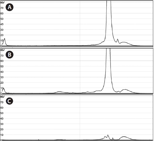
    To investigate chromatographic dynamics, the oven temperature- dependent thermodynamic characteristics of each double-stranded PCR product were tested in varying temperatures up to 75% of the helical fraction predicted by Navigator software (Transgenomic, Omaha, NE, USA).

    PCR amplicons were equally mixed together and then applied to DHPLC separation by varying the buffer concentration and oven temperature. Then 5 μL of template (16.5 ng of each PCR product) was injected into a DNASep HT Cartridge (Transgenomic). A systematic 11 × 9 matrix factorial analysis was conducted using 11 steps of oven temperature (55-59℃) and 9 steps of buffer B concentration (56-64%) with 68% of the fixed ending concentration. The total elution time was set as 10 min. Separation buffers were purchased from Transgenomic: WAVE Optimized Buffer A (0.1 M triethylammonimum acetate [TEAA]) and WAVE Optimized Buffer B (0.1 M TEAA in 25% (v/v) acetonitrile). The flow rate was also tested from 0.35 to 0.9 mL/min to achieve the highest resolution (R) of the chromatograph. Chromatographic detection was performed using fluorescence detection (Ex = 495 nm and Em = 537 nm, HSX-3500) after staining the DNA fragments with SYBR Gold (Molecular Probes, Sunnyvale, CA, USA). To optimize the efficiency of separation, a chromatographic resolution (R) was calculated by multiplying the retention time (RT), distance of each peak (Δ1 and Δ2), and peak area (a1, a3, and a3) as shown below:

    image

      >  Development of the blocking probe

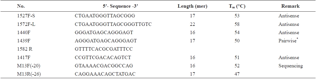
    To facilitate detection of low copies of parasitic DNA sequences, species-specific blocking probes targeting 18S rDNA fragments for the DHPLC assay were designed using Primer Premier 5.0 (Biosoft International, Palo Alto, CA, USA). The blocking probe was synthesized from peptide nucleic acid (PNA), which was designed to clamp PCR primers and thus prevent the activity of polymerases. After confirmation of the sequence using C3 spacer, PNA were synthesized for the blocking probe (Panagene, Daejeon, Korea).

      >  Verification of the DHPLC assay

    To validate the DHPLC assay using wild samples, genomic DNA was extracted from whole bodies using liquid nitrogen grinding. Then 2 ng of extracted genomic DNA was applied to PNA-PCR in 20 μL of reaction mix containing 0.5 μM of each primer, 200 μM of dNTP (Promega), 0.3 unit of Discoverase dHPLC DNA polymerase (Invitrogen), 1× Discoverase PCR buffer (Invitrogen) containing 2 mM of MgSO4, 1 μM of the PNA probe, and 2 ng of template. PNA-PCR amplicons were separated using a DNASep HT Cartridge (Transgenomic). To identify the composition of species, the peaks eluted from DHPLC separation were collected by fraction collector (FCW- 200; Transgenomic) and applied to sequencing following the method described above. Phylogenetic analysis of each peak was performed using the BLASTN search and NJ method in GenBank (Zhang et al., 2000).

      >  Statistical analysis

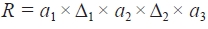
    Linear regression was carried out for thermodynamic variation of chromatographs in response to oven temperatures and amounts of amplicons relative to the concentration of the blocking probe. For determining the peak of variables, a nonlinear regression for R was carried out for buffer B and oven temperature. The statistical analyses were performed using SigmaPlot (Systat Software, Inc., Chicago, IL, USA). To optimize the conditions for DHPLC separation, variables from an 11 × 9 matrix factorial experiment were analyzed against the contour of R using Surfer 8.05 (Golden Software, Inc., Golden, CO, USA).

    Results

      >  SSU rDNA gene and chromatographic dynamics of single amplicons

    Nearly full-length 18S rDNA sequences of grass shrimp and its parasites were PCR-amplified (1,857 bp for Palaemonetes pugio, 1,924 bp for Microphallus turgidus, and 2,286 bp for Probopyrus pandalicola) using universal primer pairs (UnivF-15 and UnivR-1765). According to a phylogenetic analysis using a Blastn search, the rDNA sequences from the parasites M. turgidus and P. pandalicola were 95% and 97% similar to Microphallus primas and Probopyrus pacificiensis, respectively. Palaemonetes pugio, however, showed a higher similarity (99%) to its congener, Palaemonetes vulgaris. The genes were submitted to GenBank (EU848423 for P. pugio, EU848422 for P. pandalicola, and EU825773 for M. turgidus). For the DHPLC assay, 527 to 659-bp amplicons were achieved using the universal primer set of Univ-1131F and Univ-1629R (527 bp for P. pugio, 582 bp for M. turgidus, and 659 bp for P. pandalicola).

    To investigate the thermodynamic characteristics of the rDNA sequences, 5 μL of the 527-bp 18S rDNA amplicon of P. pugio was used for the DHPLC assay with a fixed linear gradient (60-70%) and a varying range of oven temperature (57-62℃). No significant changes were observed in the RT and peak sharpness below 60℃. However, significant changes were observed in the RT and peak sharpness above 61℃ (Fig. 1). Also, the RT of the 659-bp 18S rDNA amplicon of M. turgidus decreased with increasing oven temperature above 58.5℃ (Fig. 2), but it rarely changed below 58.5℃. For P. pugio and P. pandalicola, the RT of the 18S rDNA amplicons rarely changed upon varying the oven temperature (data not shown).

      >  Optimization of DHPLC conditions for separating the 18S rDNA amplicons

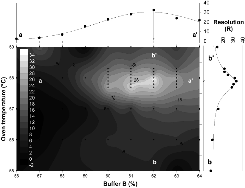
    To optimize DHPLC separation, 11×9 matrix factorial analyses were carried out using two variables: oven temperature (56-59℃) and percent buffer B (56-64%). The contour analysis showed that the highest R value was achieved at about 57.9℃ and 62% of buffer B. The distribution of R was further analyzed by regression analysis to confirm the peak of R. At 57.9℃, R was nonlinearly regressed against buffer B with the highest peak at 62% of buffer (line a-a′; r2 = 0.9683, F = 82.3495, and P = 0.0001). R was also nonlinearly regressed against oven temperature with the highest peak at 57.9℃ (line b-b′; r2 = 0.9501, F = 48.5924, and P = 0.0001) (Fig. 3). Among the investigated variables, oven temperature appeared to be the more important factor for separation, which showed a steeper peak around the optimal range of the variables. I also investigated the influence of flow rate against R using six different flow rates (0.35-0.85 mL/min at 1 mL/min intervals). While no significant change occurred in R, the RT decreased with increasing flow rates (data not shown). Based on these findings, the RT of the separation was set to 0.45 mL/min, and the chromatographs were positioned in the middle of the elution time.

      >  Development of the blocking probe

    To develop the blocking probe sequence, we tested several species-specific sequences of 18S rDNA of P. pugio (Table 1). The amplification of sequences with very low copy numbers was highly affected by sequence composition but not by melting temperature (Tm). Among the single probes, PP- 1417F showed the highest peak of parasitic genes (Fig. 4). The efficiency of parasitic gene amplification was not positively correlated with Tm, but it seemed to be closely related to the binding position. The efficiency of parasitic gene amplification was arranged in order of PP-1417F > PP-1439F = PP-1440F > PP-1527F-S >> PP-1527F-L. While a Tm difference (1.6℃) was observed, PP-1439F and PP-1440F showed no difference in the efficiency of amplifying parasitic genes. However, binding of PP-1527F-S and PP-1527F-L to the same position resulted in differences in parasitic gene amplification, which may result in false priming within the DHPLC amplicons of the parasitic sequences. Alternatively, compared to single blocking probe, pairwise binding probes were more efficient in PCR amplification of parasitic genes. Although PP-1439F/1528R pairwise blocking probes showed the best amplification due to cost efficiency, we selected PP-1417F for PNA synthesis with a modification of the lysine residue at the N-terminus to increase its solubility (Lys-CCG TTC GAC ACA GTC T-CONH2).

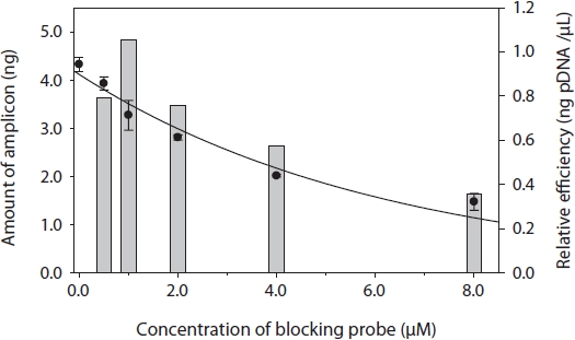
    To determine the optimal concentration of the blocking probe, the blocking efficiency of PP-1417F was investigated using 0-8 μM of blocking probe and cloned plasmid DNA of P. pugio. The amount of targeted 18S rDNA PCR amplicons decreased exponentially upon increasing concentrations of the blocking probe. To investigate the cost-efficient concentration, the relative blocking efficiency was calculated by dividing the suppressed amount of amplicons by the primer concentration (bar chart in Fig. 5). The most efficient concentration of the blocking probe was 1 μM, which resulted in approximately 25% suppression relative to 0 μM. The relative blocking efficiency continuously decreased with increasing concentrations of the blocking probe.

    [Table 1.] Information of blocking probe candidates for PNA development

    label

    Information of blocking probe candidates for PNA development

      >  DHPLC separation for the wild-type sample

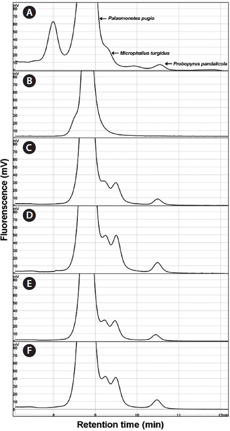
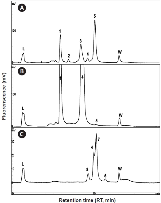
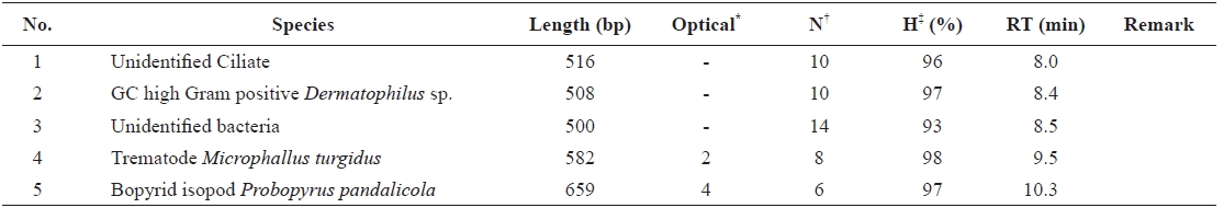
    Using the optimized DHPLC assay, genomic DNA from the wild sample was applied to DHPLC separation after PNA-PCR with 1 μM of the PNA blocking probe. The PNAPCR- DHPLC assay showed enhanced PCR amplification of sequences with very low copy numbers. To evaluate our optimized DHPLC assay, each gene fragment was collected and sequenced for the species identified. PP08-116, which was infected by the bopyrid isopod P. pandalicola had seven detectable peaks, of which four fragments were identified as having similar sequences to a 367-bp 18S rDNA fragment from P. pugio, a 508-bp sequence of a high GC Gram-positive bacteria 16S rDNA fragment, a 527-bp 18S rDNA fragment of P. pugio, and a 659-bp 18S rDNA fragment of P. pandalicola (Fig. 6A). However, only the 527-bp fragment of P. pugio and a 659-bp fragment of P. pandalicola were observed in the non-blocking PCR-DHPLC (Fig. 6B). A shift in the RT was observed in the wild-type sample elution relative to the optimized assay using cloned plasmid DNA (Fig. 6C). Although a slight RT shift occurred in comparison to the optimized assay using cloned plasmid DNA, all peaks were highly reproducible.

    Discussion

    Detection of symbionts and/or parasites is crucial for understanding ecosystem homeostasis in coastal ecosystems. Recently, PCR methods have provided useful tools to address these matters and expand its application to various diseases and parasitism (Lightner and Redman, 1998). PCR diagnosis has mostly been used to evaluate the presence of DNA from

    certain species with high sensitivity. To overcome these drawbacks, universal primers have been developed to target SSU rDNA of diverse phylum (Bower et al., 2004; Blankenship and Yayanos, 2005). When coupled with universal primers, DHPLC can be a competent method due to its high sensitivity for separation, which can detect as small as a 2-bp difference in sequence composition (Troedsson et al., 2008b).

    Due to the ubiquitous distribution and its role in estuarine ecosystems, the grass shrimp has drawn much interest for scientists in its use as a bioindicator (Key et al., 2006). Grass shrimp is also known to serve as an intermediate host for some parasites. Therefore, the grass shrimp is a useful indicator species for monitoring parasitism in estuarine ecosystems. In this study, we developed a faster, cheaper, and simpler surveillance method compared with gel-based separation technologies used to clone plasmid DNA from model hosts and parasites.

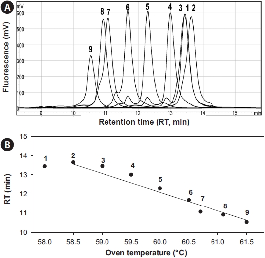
    The optimal separation conditions were determined to be an oven temperature of 57.9℃ and 62-68% of buffer B. The most important factor was the oven temperature, which skewed the regression curve more than the buffer concentration (Fig. 3), which is inconsistent with Troedsson et al. (2008a). Chromatographic behavior of each fragment was highly related with the helicity of the fragments, which suggested that <95% helicity will generate multiple peaks from a single fragment. Use of the temperature 57.9℃ accounted for 96% for P. pugio, 97% for P. pandalicola, and 97% for M. turgidus in the helical fraction of each fragment. In addition, the flow rate is considered another important variable for optimizing our new method (Troedsson et al., 2008a). However, in this study, the flow rate did not affect the R value. Therefore, to maximize the separation capacity around the peak, we adjusted the flow rate to position the chromatograph in the middle of the chromatographic window.

    I used a PNA blocking probe to suppress PCR amplification of host genes and enhance amplification. The sequence of PNA was developed using a C3 spacer oligonucleotide because C3 is inexpensive and can be quickly synthesized. In another study, PNA showed two to three orders of magnitude greater efficiency compared with C3 in detecting sequences with very low copy numbers (Cho et al., unpublished). When developing the PNA sequence, many variables had to be considered, such as the Tm, concentration, and kinetics (Ørum, 2000). In addition, our study showed that the position of hybridization was also important to obtain a high blocking efficiency: the closer the probe was bound to the PCR primer, the greater the resulting blocking efficiency and enhanced gene amplification of very low copy number DNA fragments. Thirteen- to 18- mers of PNAs are commonly used for PNA hybridization. The concentration of PNA was tested by cloning plasmid DNA of the targeted host gene, which showed approximately 25% suppression compared to non-blocking PCR. The plasmid concentration for this experiment was approximately 106 copies, which is reasonable compared to PNA-PCR for genomic DNA. In PNA-PCR, we used 2 ng of genomic DNA as a template. In the chromatograph of PP08-116, the peak area of targeted gene fragments was much smaller than the peaks of other parasitic genes, regardless of cycle numbers, which were suppressed by 90% compared to the peak of P. pandalicola.

    [Table 2.] Detection of symbionts using the developed DHPLC assay

    label

    Detection of symbionts using the developed DHPLC assay

    I used a universal primer set targeting highly conserved regions of 18S rDNA for PCR amplification. This universal primer set has been utilized as reliable primers from several studies (Gruebl et al., 2002; Troedsson et al., 2008a, 2008b). The detection limit of PNA-PCR in this study was relatively lower than those of species-specific PCR (103-102 copies) (data not shown), which might be acceptable for a surveillance tool of parasitism because a species-specific PCR assay does not verify the existence of various parasitism infections (Burreson, 2008). Although blocking probes can suppress the amplification of host gene fragments, this does not mean that the predominant host gene is not amplified. To overcome this problem, selective targeting of universal primers or nonphylum universal primers (Bower et al., 2004), which amplify only parasites or weakly including target species, can be considered. Several phylum-specific universal primers have been developed and extensively used not only for DHPLC assays but also for several gel electrophoresis-related methods (Iwen et al., 2002; Baker et al., 2003; Blankenship and Yayanos, 2005; Sherwood and Presting, 2007). Development of phylum- specific universal primers requires massive information on related species sequences and data processing. A combination of universal primers and blocking probe can therefore provide extensive applications of DHPLC for surveillance of parasitism.

    The optimized DHPLC assay showed a well-established chromatograph of isopod-infected grass shrimp with a high peak of the parasite. The peak of the host gene was suppressed over 90% more than other parasite genes. Five peaks in total were observed from PNA-PCR from genomic DNA of wildtype samples infected by isopods, including a 508-bp 16S rDNA of GC Gram-positive bacteria at 8.407 min and a 659- bp 18S rDNA of P. pandalicola at 10.127 min (Table 2).

    Estuarine systems are ecologically important environments due to their species diversity and high productivity. However, the dynamics or energetics of an ecosystem have mainly focused on macrobiota (i.e., production and/or standing stock biomass) and infectious agents, which are perceived to contribute negligible biomass to ecosystems (Poulin, 1999). Recently, however, the biomass of free-living and parasitic spe-cies was estimated to exceed that of the top predators in an estuarine ecosystem (Kuris et al., 2008). This implies a high possibility of infection of estuarine living animals from diverse parasites. Therefore, the monitoring of parasitism dynamics in estuarine/coastal ecosystems can be a crucial to maintain the homeostasis of estuarine/coastal ecosystems. From this perspective, the DHPLC assay developed in this study for grass shrimp provides useful information about parasitic dynamics in estuarine ecosystems.

참고문헌
  • 1. Ali MB, Tripathi RD, Rai UN, Pal A, Singh SP. 1999 Physicochemical characteristics and pollution level of Lake Nainital (U.P., India): role of macrophytes and phytoplankton in biomonitoring and phytoremediation of toxic metal ions. [Chemosphere] Vol.39 P.2171-2182 google
  • 2. Anderson GA. 1985 Species Profiles: Life Histories and Environmental Requirements of Coastal Fishes and Invertebrates (Gulf of Mexico): Grass Shrimp. US Fish and Wildlife Service, Biological Report 82 (11.35). P.13 google
  • 3. Baker GC, Smith JJ, Cowan DA. 2003 Review and re-analysis of domain-specific 16S primers. [J Microbiol Methods] Vol.55 P.541-555 google
  • 4. Barlaan EA, Sugimori M, Furukawa S, Takeuchi K. 2005 Profiling and monitoring of microbial populations by denaturing high-performance liquid chromatography. [J Microbiol Meth] Vol.61 P.399-412 google
  • 5. Blankenship LE, Yayanos AA. 2005 Universal primers and PCR of gut contents to study marine invertebrate diets. [Mol Ecol] Vol.14 P.891-899 google
  • 6. Boening DW. 1999 An evaluation of bivalves as biomonitors of heavy metals pollution in marine waters. [Environ Monit Assess] Vol.55 P.459-470 google
  • 7. Bower SM, Carnegie RB, Goh B, Jones SR, Lowe GJ, Mak MW. 2004 Preferential PCR amplification of parasitic protistan small subunit rDNA from metazoan tissues. [J Eukaryot Microbiol] Vol.51 P.325-332 google
  • 8. Burreson EM. 2008 Misuse of PCR assay for diagnosis of mollusc protistan infections. [Dis Aquat Organ] Vol.80 P.81-83 google
  • 9. Burridge TR, Bidwell J. 2002 Review of the potential use of brown algal ecotoxicological assays in monitoring effluent discharge and pollution in southern Australia. [Mar Pollut Bull] Vol.45 P.140-147 google
  • 10. Chaplin-Ebanks SA, Curran MC. 2005 The effect of the parasitic isopod, Probopyrus pandalicola (Packard, 1879), on tidal activity patters of the grass shrimp, Palaemonetes pugio Holthuis, 1949. [Crustaceana] Vol.78 P.1053-1061 google
  • 11. Cho SM. 2006 Effect of polycyclic aromatic hydrocarbons (PAHs) on the Pacific oyster, Crassostrea gias. google
  • 12. Cho SM, Kurihara R, Strussmann CA, Uozumi M, Yamakawa H, Yamasaki T, Soyano K, Shiraishi H, Morita M, Hashimoto S. 2003 Histological abnormalities in the gonads of konoshiro gizzard shad (Konosirus punctatus) from coastal area of Japan. [Environ Sci] Vol.10 P.25-36 google
  • 13. Gruebl T, Frischer ME, Sheppard M, Neumann M, Maurer AN, Lee RF. 2002 Development of an 18S rRNA gene-targeted PCR-based diagnostic for the blue crab parasite Hematodinium sp. [Dis Aquat Org] Vol.49 P.61-70 google
  • 14. Heard RW. 1982 Guide to Common Tidal Marsh Invertebrates of the Northeastern Gulf of Mexico. google
  • 15. Iwen PC, Hinrichs SH, Rupp ME. 2002 Utilization of the internal transcribed spacer regions as molecular targets to detect and identify human fungal pathogens. [Med Mycol] Vol.40 P.87-109 google
  • 16. Kahle J, Zauke GP. 2003 Trace metals in Antarctic copepods from the Weddell Sea (Antarctica). [Chemosphere] Vol.51 P.409-417 google
  • 17. Kahn RN, Spiers JA, Pung OJ. 2003 Effects of the trematode Microphallus turgidus on locomotion and prey capture in the grass shrimp Palaemonetes pugio. [J Helminthol] Vol.77 P.327-330 google
  • 18. Key PB, Wirth EF, Fulton MH. 2006 A review of grass shrimp, Palaemonetes spp., as a bioindicator of anthropogenic impacts. [Environ Bioindic] Vol.1 P.115-128 google
  • 19. Kim Y, Powell EN, Wade TL, Presley BJ. 2008 Relationship of parasites and pathologies to contaminant body burden in sentinel bivalves: NOAA Status and Trends 'Mussel Watch' Program. [Mar Environ Res] Vol.65 P.101-127 google
  • 20. Kuris AM, Hechinger RF, Shaw JC, Whitney KL, Aguirre-Macedo L, Boch CA, Dobson AP, Dunham EJ, Fredensborg BL, Huspeni TC, Lorda J, Mababa L, Mancini FT, Mora AB, Pickering M, Talhouk NL, Torchin ME, Lafferty KD. 2008 Ecosystem energetic implications of parasite and free-living biomass in three estuaries. [Nature] Vol.454 P.515-518 google
  • 21. Leight AK, Scott GI, Fulton MH, Daugomah JW. 2005 Long term monitoring of grass shrimp Palaemonetes spp. Population metrics at sites with agricultural runoff influences. [Integr Comp Biol] Vol.45 P.143-150 google
  • 22. Lightner DV, Redman RM. 1998 Shrimp diseases and current diagnostic methods. [Aquaculture] Vol.164 P.201-220 google
  • 23. O'Connor TP. 2002 National distribution of chemical concentrations in mussels and oysters in the USA. [Mar Environ Res] Vol.53 P.117-143 google
  • 24. Ørum H. 2000 PCR clamping. [Curr Issues Mol Biol] Vol.2 P.27-30 google
  • 25. Poulin R. 1999 The functional importance of parasites in animal communities: many roles at many levels? [Int J Parasitol] Vol.29 P.903-914 google
  • 26. Pung OJ, Khan RN, Vives SP, Walker CB. 2002 Prevalence, geographic distribution, and fitness effects of Microphallus turgidus (Trematoda: Microphallidae) in grass shrimp (Palaemonetes spp.) from coastal Georgia. [J Parasitol] Vol.88 P.89-92 google
  • 27. Ritterhoff J, Zauke GP. 1997 Trace metals in field samples of zooplankton from the Fram Strait and the Greenland Sea. [Sci Total Environ] Vol.199 P.255-270 google
  • 28. Sherwood AR, Presting GG. 2007 Universal primers amplify a 23S rDNA plastid marker in eukaryotic algae and cyanobacteria. [J Phycol] Vol.43 P.605-608 google
  • 29. Troedsson C, Lee RF, Stokes V, Walters TL, Simonelli P, Frischer ME. 2008a Development of a denaturing high-performance liquid chromatography method for detection of protist parasites of metazoans. [Appl Environ Microbiol] Vol.74 P.4336-4345 google
  • 30. Troedsson C, Lee RF, Walters T, Stokes V, Brinkley K, Naegele V, Frischer ME. 2008b Detection and discovery of crustacean parasites in blue crabs (Callinectes sapidus) by using 18S rRNA gene-targeted denaturing high-performance liquid chromatography. [Appl Environ Microbiol] Vol.74 P.4346-4353 google
  • 31. Van der Oost R, Beyer J, Vermeulen NPE. 2003 Fish bioaccumulation and biomarkers in environmental risk assessment: a review. [Environ Toxicol Pharmcol] Vol.13 P.57-149 google
  • 32. Wang S, Bao Z, Zhang L, Li N, Zhan A, Guo W, Wang X, Hu J. 2006 A new strategy for species identification of planktonic larvae: PCR-RFLP analysis of the internal transcribed spacer region of ribosomal DNA detected by agarose gel electrophoresis or DHPLC. [J Plankton Res] Vol.28 P.375-384 google
  • 33. Zhang Z, Schwartz S, Wagner L, Miller W. 2000 A greedy algorithm for aligning DNA sequences. [J Comput Biol] Vol.7 P.203-214 google
OAK XML 통계
이미지 / 테이블
  • [ Fig. 1. ]  Influence of temperature on chromatographic characteristics of 527 bp Palaemonete pugio 18S rDNA amplicon. The amplicons were eluted in denaturing high-performance liquid chromatography under 60-70% of buffer B and 0.35 mL/min flow rate at given oven temperature: (A) 57℃, (B) 60℃, and (C) 61℃, respectively. No significant changes were observed up to 61℃, while a significant change observed at 61℃ in retention time and peak shape.
    Influence of temperature on chromatographic characteristics of 527 bp Palaemonete pugio 18S rDNA amplicon. The amplicons were eluted in denaturing high-performance liquid chromatography under 60-70% of buffer B and 0.35 mL/min flow rate at given oven temperature: (A) 57℃, (B) 60℃, and (C) 61℃, respectively. No significant changes were observed up to 61℃, while a significant change observed at 61℃ in retention time and peak shape.
  • [ Fig. 2. ]  Changes of chromatograph (A) and retention time (B) of Microphallus trugidus. 582 bp 18S rDNA amplicon in response to oven temperature. Numbers (1-9) at (A) are corresponded to each data at (B). Above 58.5℃, significant decrease was observed at retention time (RT) (r2 = 0.9146, F = 86.6999 and P < 0.0001)
    Changes of chromatograph (A) and retention time (B) of Microphallus trugidus. 582 bp 18S rDNA amplicon in response to oven temperature. Numbers (1-9) at (A) are corresponded to each data at (B). Above 58.5℃, significant decrease was observed at retention time (RT) (r2 = 0.9146, F = 86.6999 and P < 0.0001)
  • [ Fig. 3. ]  Optimization of denaturing high-performance liquid chromatography separation for mixture of 18S rDNA amplicons with 5 by 9 factorial matrix study of buffer B (%) and oven temperature. Closed circles (●) indicate the case of experimental cases. Non-linear regression analysis were carried out between R and buffer B at 57.9℃ (Line a-a′) and R and oven temperature at 62% of buffer B (Line b-b′). R was highly correlated with buffer B (r2 = 0.9683, F = 82.3495 and P = 0.0001) and oven temperature (r2 = 0.9501, F = 48.5924 and P = 0.0001), which was maximized at 62% (buffer B) and 57.9℃ (oven temperature). The optimal condition for the separation of mixed 18S rDNA amplicons was 62-68% of buffer B% and 57.9℃ of oven temperature.
    Optimization of denaturing high-performance liquid chromatography separation for mixture of 18S rDNA amplicons with 5 by 9 factorial matrix study of buffer B (%) and oven temperature. Closed circles (●) indicate the case of experimental cases. Non-linear regression analysis were carried out between R and buffer B at 57.9℃ (Line a-a′) and R and oven temperature at 62% of buffer B (Line b-b′). R was highly correlated with buffer B (r2 = 0.9683, F = 82.3495 and P = 0.0001) and oven temperature (r2 = 0.9501, F = 48.5924 and P = 0.0001), which was maximized at 62% (buffer B) and 57.9℃ (oven temperature). The optimal condition for the separation of mixed 18S rDNA amplicons was 62-68% of buffer B% and 57.9℃ of oven temperature.
  • [ Table 1. ]  Information of blocking probe candidates for PNA development
    Information of blocking probe candidates for PNA development
  • [ Fig. 4. ]  Efficiency of blocking probes. To develop the sequence of peptide nucleic acid blocking probe, 6 candidates were tested using C3 spacer oligonucleotide: (A) PP-1527F-S, (B) PP-1527F-L, (C) PP-1440F, (D) PP-1439F/1582R, (E) PP-1439F, and (F) PP1417F. Information of each probe refers to Table 1. Among the single blocking probe, PP1417F showed the height peak of parasitic genes although pairwise blocker showed slightly better efficiency. P. pugio, Palaemonetes pugio; M. turgidus, Microphallus turgidus; P. pandalicola, Probopyrus pandalicola.
    Efficiency of blocking probes. To develop the sequence of peptide nucleic acid blocking probe, 6 candidates were tested using C3 spacer oligonucleotide: (A) PP-1527F-S, (B) PP-1527F-L, (C) PP-1440F, (D) PP-1439F/1582R, (E) PP-1439F, and (F) PP1417F. Information of each probe refers to Table 1. Among the single blocking probe, PP1417F showed the height peak of parasitic genes although pairwise blocker showed slightly better efficiency. P. pugio, Palaemonetes pugio; M. turgidus, Microphallus turgidus; P. pandalicola, Probopyrus pandalicola.
  • [ Fig. 5. ]  Influence of blocking probe concentration on amount of PCR amplification for target 18S rDNA fragment (●) and relative blocking efficiency against unit amount of blocking probe (bar). Using cloned 18S rDNA pladmid DNA of Palaemonetes pugio, PCR amplification were carried out with varying concentration of blocking probe (PP-1417F-C3). The amplicons were eluted and quantified by denaturing high-performance liquid chromatography. Amount of PCR amplicon was exponentially decreased with increase of blocking probe (r2 = 0.9414, F = 81.3607 and P = 0.0008). In relative efficiency, the highest efficiency was observed at 1 μM of blocking probe with 1.05 ng/μL.
    Influence of blocking probe concentration on amount of PCR amplification for target 18S rDNA fragment (●) and relative blocking efficiency against unit amount of blocking probe (bar). Using cloned 18S rDNA pladmid DNA of Palaemonetes pugio, PCR amplification were carried out with varying concentration of blocking probe (PP-1417F-C3). The amplicons were eluted and quantified by denaturing high-performance liquid chromatography. Amount of PCR amplicon was exponentially decreased with increase of blocking probe (r2 = 0.9414, F = 81.3607 and P = 0.0008). In relative efficiency, the highest efficiency was observed at 1 μM of blocking probe with 1.05 ng/μL.
  • [ Fig. 6. ]  Chromatograph of denaturing high-performance liquid chromatography assay with PNA blocking probe (A) and without blocking probe (B) for wild genomic DNA of Palaemonetes pugio and optimized separation with cloned plasmid DNAs (C). L, injection peak; W, washing peak; 1, unidentified 268 bp amplicon; 2, 367 bp 18S rDNA fragment of P. pugio; 3, 508 bp of high GC Gram positive bacteria 16S rDNA fragment; 4, 527 bp 18S rDNA fragment of P. pugio; 5, 659 bp 18S rDNA fragment of Probopyrus pandalicola; 7, 582 bp 18S rDNA fragment of Microphallus turgidus; 8, unidentified peak. A significant suppression of PCR amplification was observed at 527 bp P. pugio fragment but significantly enhanced amplification of other parasites in PNA-PCR chromatograph (A).
    Chromatograph of denaturing high-performance liquid chromatography assay with PNA blocking probe (A) and without blocking probe (B) for wild genomic DNA of Palaemonetes pugio and optimized separation with cloned plasmid DNAs (C). L, injection peak; W, washing peak; 1, unidentified 268 bp amplicon; 2, 367 bp 18S rDNA fragment of P. pugio; 3, 508 bp of high GC Gram positive bacteria 16S rDNA fragment; 4, 527 bp 18S rDNA fragment of P. pugio; 5, 659 bp 18S rDNA fragment of Probopyrus pandalicola; 7, 582 bp 18S rDNA fragment of Microphallus turgidus; 8, unidentified peak. A significant suppression of PCR amplification was observed at 527 bp P. pugio fragment but significantly enhanced amplification of other parasites in PNA-PCR chromatograph (A).
  • [ Table 2. ]  Detection of symbionts using the developed DHPLC assay
    Detection of symbionts using the developed DHPLC assay
(우)06579 서울시 서초구 반포대로 201(반포동)
Tel. 02-537-6389 | Fax. 02-590-0571 | 문의 : oak2014@korea.kr
Copyright(c) National Library of Korea. All rights reserved.