We report observation of laser desorption/ionization (LDI) of peptides from flat surfaces of tungsten silicide (WSi2). In contrast to MALDI (matrix-assisted laser desorption/ionization) and SALDI (surface-assisted laser desorption/ionization) mass spectrometry, this study did not utilize any matrices and surface nanostructures. In this work, LDI on WSi2 surfaces is demonstrated to cover a mass range up to 1,600 Da (somatostatin; monoisotopic mass = 1637.9 Da). In addition, it exhibited a high sensitivity, which could detect peptides, which could detect peptides of low femtomole levels (20 fmol for angiotensin II). The observed LDI process was discussed to be largely thermal, more specifically, due to laser-induced surface heating that is most likely promoted by the low thermal diffusivity (κ) of WSi2 substrate.
Matrix-assisted laser desorption/ionization (MALDI) is a powerful means of soft ionization for mass spectrometry of thermally labile biomolecules. However, the use of organic crystals as matrix creates certain limitations on applications. It includes interference of matrix peaks in the small mass region (< 500 Da) and sweet spots. Careful matrix selection for given analytes is also important for successful mass analysis. To address such issues, extensive efforts were made to develop matrix-free laser desorption/ionization (LDI) in recent years, which can be an alternative method of soft ionization to the MALDI method.1
DIOS (desorption/ionization on porous silicon) mass spectrometry was the first demonstration of LDI, which utilized specially prepared Si surfaces.2 In DIOS mass spectrometry, LDI of small molecules was found to occur on the nanoporous surfaces of crystalline Si (c-Si) formed by photo-electrochemical etching. However, without such surface nanostructures of a sub-micrometer dimension, the DIOS process was hardly observed to take place. Since the advent of DIOS mass spectrometry, surfaces possessing various nanostructures have been examined for their LDI capabilities. In the researches, Si-based nanostructures were most popularly investigated, the examples of which include porous Si surfaces fabricated by etching,3 clathrate-structured Si surfaces,4 Si nanowires,5 Si microcolumn and nanoposts arrays.6,7 In addition to Si, nanostructured surfaces made of other materials, such as nanostructured Au thin film,8 selfassembled germanium nanodots,9 and zinc oxide nanowires,10 also exhibited a promising level of sensitivity and mass range for mass spectrometry. Likewise, utilization of surface nanostructures has been a key component in the previous developments of high-efficiency LDI surfaces.
On the other hand, few cases of LDI occurring on flat surfaces are known,
>
Preparation of WSi2 surfaces
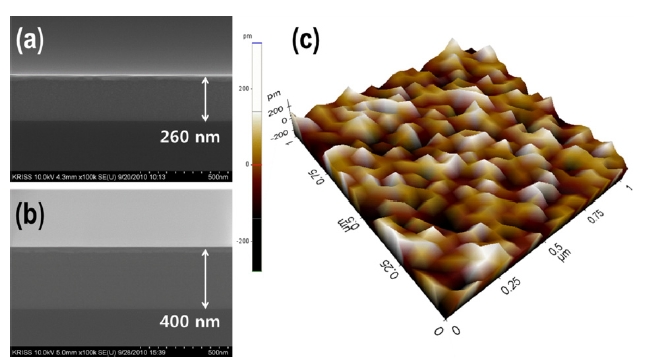
WSi2 on Si(001) substrates were prepared by the Ar+ magnetron sputtering method. During the sputtering process, the flow of Ar gas was maintained at 50 mTorr and the working pressure was 1.7 mTorr. Various thicknesses of WSi2 were examined, including 65 nm, 260 nm, and 400 nm. For additional heat treatment, the samples were heated at 873 K in vacuum (2 × 10?6 Torr) for 1 hr. The prepared WSi2 surfaces were then characterized using a fieldemission scanning electron microscope (FESEM, S-4800, Hitachi), an X-ray diffractometer (θ-2θ, Cu-Kα, Bruker D8), and an atomic force microscopy (AFM, XE-150, PSIA). The AFM (
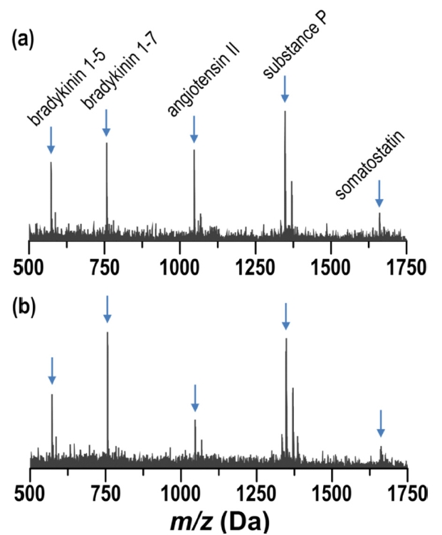
LDI mass spectrometry was performed using a MALDI-TOF mass spectrometer (Autoflex III; Bruker-Daltonics, Leipzig, Germany) in reflectron mode. Before experiments, WSi2 chips were sonicated in isopropyl alcohol for 30s, washed in deionized water, and then dried under a gentle flow of nitrogen. A peptide solution was prepared in 10% methyl alcohol to a concentration of 10 pmol/μL. It consisted of bradykinin 1-5 (monoisotopic mass: 572.7 Da), bradykinin 1-7 (756.9 Da), angiotensin II (1046.2 Da), substance P (1347.6 Da), and somatostatin (1637.9 Da). The chemicals were commercially obtained from Sigma-Aldrich and used without purification. For LDI experiments, a 0.2 μl drop of the peptide solution (2 pmol) was taken and pipetted onto WSi2 surfaces, and then dried in ambient conditions. The WSi2 surfaces were moderately hydrophilic, so the sample drop somewhat spread on the surfaces. Thus, the actual sample consumption used to obtain a LDI mass spectrum was much lower than the applied quantity of 2 pmol.
Although efficient LDI from various nanostructured
surfaces is known, understanding of the LDI mechanism has been very limited. However, some of previous investigations recognized a possible contribution of laser-induced surface heating to the LDI process.3,10 It was viewed that the physical existence of surface nanostructures might provide heat conduction barriers for absorbed laser energy; thus, the absorbed energy is momentarily trapped in surface nanostructures, which gives rise to a rapid surface heating. Such a rapid increase in surface temperature is known to promote thermal desorption of thermally labile molecules, which thus can contribute to LDI of biomolecules.13
A recent work on a-Si demonstrated that a rapid surface heating for LDI could be achieved even without employing surface nanostructures by selecting appropriate surface materials.11 Because of the low thermal conductivity (K = 5.5W m?1 K?1), the thermal diffusivity (κ = K/Cp) of a-Si is very small (0.03 cm2 s?1; heat capacity (Cp) for a-Si = 1.65 J cm?3 K?1). During the period of laser irradiation (t~7 ns), the absorbed energy can diffuse only for about 90 nm (D) into the bulk substrate (depth of conductive heating: (D) = (κt/π)1/2). As the heat energy is localized near the surface due to the low thermal diffusivity, the surface temperature on a-Si can drastically increase to higher than 1000 K within 10 nsec. On the other hand, because of the higher thermal conductivity (150 W m?1 K?1) of c-Si, the transient increase in surface temperature is predicted to be only about 300 K,
much lower than on a-Si, which may explain that LDI of biomolecules on flat c-Si surfaces has not been successful before.
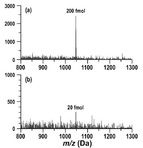
In this study, we examined WSi2 surfaces for flat LDI substrates. WSi2 possesses a moderate thermal conductivity of 18.3Wm-1 K?1. However, as typical in metal silicides, it has a very large heat capacity (309.3 J cm?3 K?1), which gives a much low thermal diffusivity of 0.0006 cm2 s?1 that predicts a conduction depth (D) of 11 nm. Thereby, it can be suggested that the heat capacity of WSi2 surfaces might give us an opportunity for LDI of thermally labile molecules. In order to corroborate the idea, we examined LDI of peptides from WSi2 surfaces. Figure 1 displays SEM and AFM images for WSi2 surfaces used in this study. As shown in the AFM image, the surfaces do not possess any noticeable surface nanostructures, which would otherwise contribute to promoting the LDI process, possibly. We examined the LDI capability of WSi2 surfaces prepared in different conditions using a mixture of 5 peptides. For example, we examined 65 nm, 260 nm, and 400 nm thick WSi2 surfaces before and after high-temperature heat treatment. However, within the conditions we varied, the results on LDI performance were not quite different. Figure 2 shows the LDI mass spectra of peptides obtained from 65 nm thick WSi2 surfaces with and without heat treatment. The results shown in Figure 2 clearly exhibit pronounced production of peptide ions from the flat surfaces of WSi2. It supports our postulation that the thermal mechanism is indeed a main driving force for LDI of thermally labile molecules from surfaces, where selecting surface materials with a large heat capacity can be a way to develop efficient LDI plates. As shown in Figure 3, the LDI-on-WSi2 method also offers a good sensitivity of low femtomole levels (20 fmol for angiotensin II), which is acceptable for peptide mass spectrometry as well.
We report that LDI on tungsten silicide (WSi2) surfaces exhibits pronounced production of peptide ions. It covers a mass range up to 1600 Da. It is sensitive, which can ionize low femtomole levels of peptide molecules without assistance of matrices or surface nanostructures. The mechanism for LDI is viewed to be thermal,