The genus Rhodella was established by Evans (1970) to distinguish a unicellular red alga that possessed con-spicuous ultrastructural features different from the other genera examined by transmission electron microscopy (TEM) at that time, Porphyridium (e.g., Gantt and Conti 1965, Gantt et al. 1968) and Rhodosorus (Giraud 1962). Rhodella maculata (Evans 1970) has highly dissected chloroplast lobes attached to multiple sites of a central to eccentric naked pyrenoid. Pyrenoids are designated as being naked if they are not embedded in the chloro-plast and instead border the cytoplasm, often partially surrounded by starch grains. The pyrenoid of Rhodella is unusual among red algal unicells in that the matrix does not contain thylakoids and it is invaded by one or two protrusions from the nucleus (Evans 1970, Patrone et al. 1991, Waller and McFadden 1995, Yokoyama et al. 2004). No other formally identified unicellular red algal genus has both of these subcellular features (Scott et al. 2008, Yang et al. 2010). In Wehrmeyer (1971) used TEM to char-acterize cells previously established as Porphyridium vio-laceum (Kornmann 1965) and determined that the cells bore a greater resemblance to R. maculata than to Por-phyridium. Wehrmeyer transferred P. violacea to the ge-nus Rhodella as R. violacea (Kornmann) Wehrmeyer. The two Rhodella species differed slightly from each other. The nuclear protrusions into the pyrenoid of R. violacea were not detected by Wehrmeyer (1971) but Patrone et al. (1991) noted their presence. In addition to having unique pyrenoids, both species lack a peripheral thylakoid in the chloroplast and Golgi bodies are associated only with en-doplasmic reticulum (ER) and never with the nucleus or mitochondria (Scott et al. 2008, Yang et al. 2010, 2011).
In the 1980s, two more unicellular red algae were iso-lated, characterized by TEM and assigned to the genus Rhodella, R. reticulata (Deason et al. 1983) and R. cyanea (Billard and Fresnel 1986). The establishment of these two new algae as presumed species of Rhodella was again based on their ultrastructural dissimilarities to Porphy-ridium. But what was unexpected and left unexplained was the fact that these algae also did not bear any resem-blance to either R. maculata or R. violacea. Furthermore, R. reticulata and R. cyanea clearly bore little resemblance to each other. Subsequent ultrastructural investigations resulted in transferring R. reticulata (= R. grisea, Fresnel et al. 1989) to a new genus Dixoniella as D. grisea (Scott et al. 1992) and R. cyanea was transferred to a new genus Neorhodella as N. cyanea (Scott et al. 2008). Recent mo-lecular studies have confirmed the morphological work (Yokoyama et al. 2009) and R. maculata and R. violacea are currently assigned to the class Rhodellophyceae, Or-der Rhodellales, whereas D. grisea and N. cyanea are in the class Rhodellophyceae, Order Dixoniellales.
Yoon et al. (2006) established six classes of red algae in subphylum Rhodophytina, three of which include uni-cellular forms, the Porphyridiophyceae (Erythrolobus, Flintiella, Porphyridium, and Timspurckia), Stylonema-tophyceae (Rhodosorus and Rhodospora), and Rhodel-lophyceae (Corynoplastis, Dixoniella, Glaucosphaera, Neorhodella, and Rhodella). For details of the unicellular red algae systematics of utilizing microscopic, molecular, and biochemical methods see Yoon et al. (2006), Scott et al. (2008), Yokoyama et al. (2009), and Yang et al. (2010, 2011).
This current work presents molecular, confocal micros-copy, and electron microscopy details on culture strains of a unicellular red alga identified as Rhodella sp. isolated from Bodega Bay, CA and Friday Harbor WA, USA. For the comparison, we added information on the type species, R. maculata CCMP 736. We have emended the previously established orders Dixoniellales and Rhodellales to cor-rect the placement of the genera Corynoplastis, Dixoni-ella, Neorhodella, and Rhodella and established the new order Glaucosphaerales to accommodate a single genus, Glaucosphaera.
Procedures for isolation and culture were as described in West (2005). Isolate JAW 2347 (CCMP 3129) was ob-tained from a mud sample in a Salicornia salt marsh at Bodega Bay, CA, USA on April 15, 1980. O’Kelly’s isolates RV-FHLa & b (CCMP 3133, 3135) were obtained as epi-phytes on the green alga Capsosiphon sp. on a seawall at Friday Harbor, WA, USA in March 2010. These three iso-lates are available from Bigelow Laboratory for Ocean Sci-ences, P. O. Box 475, 180 McKown Point Road, West Booth-bay Harbor, ME 04575, USA.
Fixation, staining, and observation methods were par-tially described in Yang et al. (2011). Cultured cells were transferred to 1.5 mL microtubes with 0.5 mL of culture medium, fixed with an equal volume of 3% paraformal-dehyde in PHEM buffer [60 mM PIPES, 25 mM HEPES, 10 mM EGTA, 2 mM MgCl2(6H2O), pH 7.4] for 15 min. Ten μL of fixed cells were placed on a glass slide coated with 0.1 w/v% poly-L-lysine and incubated for 10 min at room temperature. Then equal volumes of SlowFade Antifade (Invitrogen, Carlsbad, CA, USA) and 1,000× diluted SYBR Green I (Invitrogen) were added and incubated for 15 min. Confocal images were taken with an inverted microscope Axiovert 100M with LSM 510 laser scanning equipment (Carl Zeiss AG, Jena, Germany) at the University of Tsuku-ba, and an Axio Observer LSM 700 instrument at Bigelow Laboratory for Ocean Sciences. Plastid autofluorescence and SYBR Green I fluorescence were detected with a 585 nm long pass filter and a 505 to 530 nm band pass filter, respectively, in the excitation line of a 488 nm argon laser and 543 nm He / Ne laser using a single-track mode. Liv-ing cells were observed with a Nikon microscope (Eclipse E600) equipped with Nomarski interference optics (Nikon Co. Ltd., Tokyo, Japan).
Cells from cultures (grown in window light in 15 psu von Stosch enriched seawater) were filtered onto poly-L-lysine coated 0.45 μm Millipore filters (Bedford, MA, USA) and fixed for 2 h at ambient temperature in 2% glutaraldehyde in a 0.1 M phosphate buffer solution (pH 6.8) with 0.15 M sucrose. Following buffer rinses samples were post-fixed 2 h in the same buffer in 1% OsO4 at 4℃, rinsed thoroughly in H2O, left in 50% acetone for 30 min, and stored in a 70% acetone-2% uranyl acetate solution at ambient temperature for 22 h. Samples were then further dehydrated in a graded acetone series, infiltrated gradu-ally, embedded in Embed 812 resin (Electron Microscopy Sciences, Hatfield, PA, USA), and polymerized at 70℃ for 2-3 days. Thin sections were cut with an RMC MT6000-XL ultramicrotome (RMC Inc., Tucson, AZ, USA). Sections were stained for 1 min with lead acetate. A Zeiss EM 109 electron microscope was used for observation and pho-tography.
Genomic DNA was extracted from each culture strain using a DNeasy Plant Mini Kit (Qiagen, Hilden, Ger-many) according to the manufacturers’ instructions. Polymerase chain reaction (PCR) and sequencing were performed with published and newly designed spe-cific primer sets for each gene; psaA130F-psaA1110R, psaA870F-psaA1600R and psaA971F-psaA1760R for psaA (Yoon et al. 2002, Yang and Boo 2004), rbcL43F (5'-CGT-TAYGAATCTGGTGTAATYCC-3')-rbcL1414R (5'-TCAGCTG-TATCTGTAGAAGTATA-3') for rbcL. PCR amplification was performed in a total volume of 25 μL, containing 0.02 unit of Phusion High-Fidelity DNA polymerase (Finnzymes OY, Espoo, Finland), 5 μL of the 5× Phusion HF Buffer (contain 1.5 mM MgCl2), 200 μM of each dNTPs, 10 μM of each primer and 1-20 ng of template DNA. PCR was carried out with an initial denaturation at 98℃ for 30 s, followed by 30 main amplification cycles of denaturation at 94℃ for 10 s, annealing at 50-55℃ for 30 s, extension krat 72℃ for 1 min, and a final extension at 72℃ for 7 min. Amplified DNA was purified with the QIAquick PCR Puri-fication kit (Qiagen) and sent to a commercial sequencing company. Electropherogram outputs for each specimen were edited using the program Chromas v.1.45 (http://www.technelysium.com.au/chromas.html). Newly de-termined sequences were deposited in the GenBank da-tabases (http://www.ncbi.nlm.nih.gov) under the acces-sion numbers JN934872-JN934889 (see detail in Table 1).
Published sequences were obtained from GenBank and aligned with newly determined sequences using ClustalW implemented SeaView version 4.2.5 (Gouy et al. 2010) and manually refined using Se-Al version 2.0a11 (http://tree.bio.ed.ac.uk/software/seal/). We excluded insertions of the nuclear small subunit rRNA (SSU rRNA) (Rhodella sp. MBIC10593, AB183619: #538-1045; Thorea violacea SAG 51.94, AF342744: #1356-1479) and introns of plastid psaA (Rhodella maculata CCMP 736, DQ308448: #79-858; Rhosospora sordida UTEX 2621, DQ308453: #226-723). There were no indel in psbA and rbcL. We used two data sets for tree search, (i) DNA and protein mixed data (total 3,070 characters; 1,881 base pairs of nuclear SSU rRNA + 1,189 amino acid sequences from plastid psaA, psbA, and rbcL) and (ii) DNA data (total 5,448 bp; 1,881 bp SSU rRNA + 3,567 bp plastid genes).
The evolutionary model for individual genes was cho-sen using the weighted Akaike information criterion (AIC) implemented in ModelGenerator version 0.85 (Keane et al. 2006). The selected best fitting models were the gen-eral time reversible (GTR) substitution with the gamma distributed rate heterogeneity (G) for DNA data; the LG substitution (Le and Gascuel 2008) with empirical amino acid frequencies (F) and G (LG + F + G model) for protein data. We used an independent model for each partition of the concatenated data. For example, it was used GTR + G model for SSU part and separate LG + F + G for psaA, psbA and rbcL parts of mixed data; separate GTR + G for individual gene of DNA data.
Maximum likelihood (ML) analyses were performed using RAxML version 7.2.8 (Stamatakis 2006). Tree like-lihoods were estimated using 200 independent replica-tions, each with a random starting point. The separate site-specific model was used for partitioned data and the automatically optimized SPR branch rearrangements were used during the rapid hill climbing tree search for each replication. Bootstrap analyses (MLB) were con-ducted using 1,000 replications with the same evolu-
tionary model setting as that used for the best topology search.
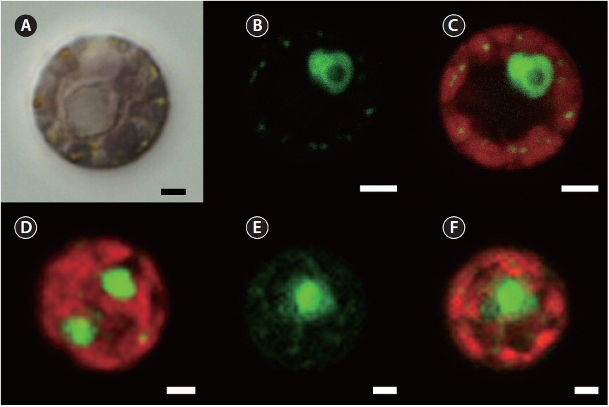
The following observations were made with the culture isolate JAW 2347 (CCMP 3129). SYBR Green stain gave a green fluorescence to nuclear DNA as well as chloro-plast and mitochondria genophores, whereas the whole chloroplast showed a red autofluorescence (Fig. 1). The nucleus of the interphase stage was always eccentric and associated with a pyrenoid (Fig. 1A-C). The single chlo-roplast had its pyrenoid centrally located and had many lobes positioned at the cell periphery. Plastid genophores were spherical to subspherical (about 0.25 μm diameter) and present in the peripheral lobes. Two nuclei were found in some dividing cells (Fig. 1D). All observed char-acters were also found in R. maculata CCMP 736 (Fig. 1E & F), without any features distinguishing it from JAW 2347 (CCMP 3129).
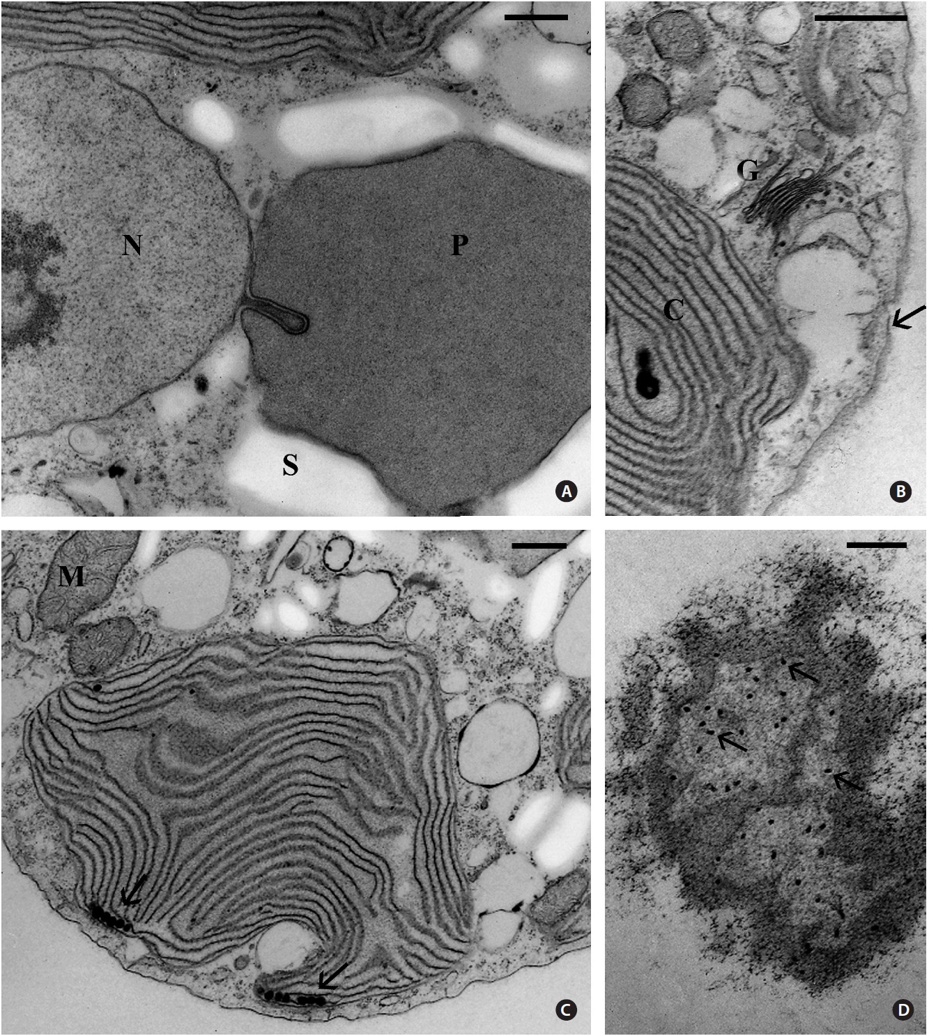
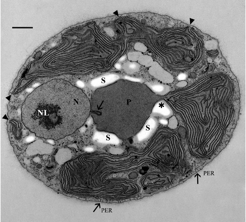
The cells were 8-12 μm in diameter and were coated by thin fibrillar material (Fig. 2). A discontinuous layer of ER was present beneath the cell membrane and appeared interconnected with the cell membrane by short tubules (Fig. 2 & 3D), as seen by Patrone et al. (1991) in Rhodella violacea, R. maculata and many other unicellular red al-gae. The spherical nucleus was approximately 3 μm in di-ameter, occupying a peripheral location and was always closely associated with the more central pyrenoid (Fig. 2). The nucleus contained one mostly central nucleolus (Fig. 2). In favorable sections usually one but occasionally two nuclear protrusions could be seen penetrating the pyre-noid as deep as 0.5-1 μm (Fig. 2 & 3A). The protrusions always contained an electron dense, fibrillar material, not well seen in Fig. 2 & 3A. In the examination of several hundred cells, nuclear protrusions were detected in over 20.
Usually one pyrenoid was found in each cell but a few cells contained two pyrenoids. The pyrenoids were com-monly larger than nuclei and were often bordered by starch that was also seen elsewhere in the cell, frequently near nuclei (Fig. 2). The moderately electron dense py-renoid matrix was devoid of thylakoids (Fig. 2 & 3A). As observed in two-dimensional sections, 1-4 chloroplast lobes extended from the pyrenoid throughout the cy-toplasm. Thylakoids were abundant and maintained consistent spacing from each other (Fig. 2, 3B & C), but phycobilisomes were not usually clearly seen. Peripheral thylakoids were absent (Fig. 2 & 3C). Electron dense clus-ters of spherical globules (plastoglobuli) were commonly seen at the outermost periphery of chloroplast lobes (Fig. 3C). Profiles of mitochondria were found throughout the cell (Fig. 2, 3B & C) as well as small electron transpar-ent vacuoles (Fig. 2 & 3B). Golgi bodies were somewhat sparse and usually located at peripheral cell regions. All observed Golgi bodies had fused or closely appressed cis-ternae (Fig. 3B).
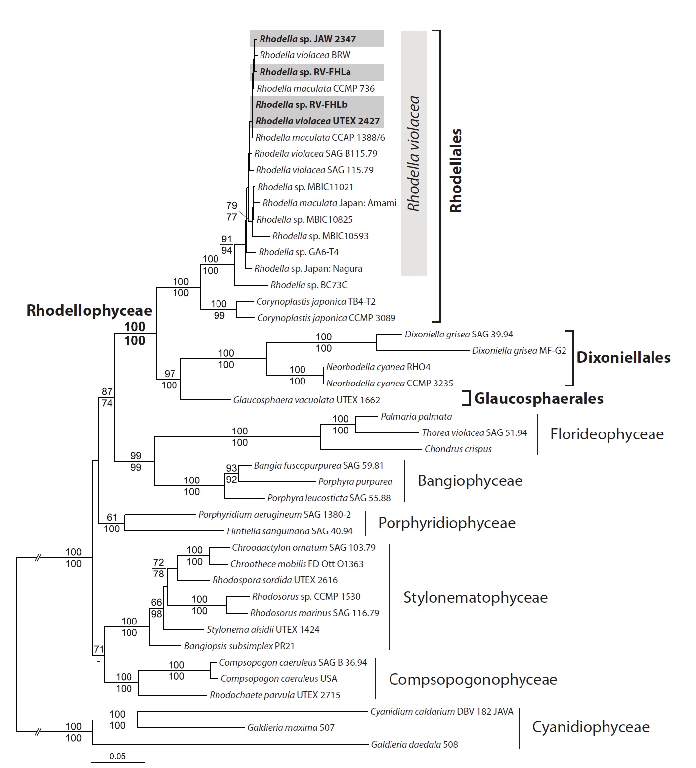
Newly determined rhodellophycean nuclear SSU rRNA and three plastid protein-coding psaA, psbA, and rbcL genes were aligned and used for the phylogenetic anal-ysis. ML trees based on DNA + protein mixed data and
DNA data were consistent with each other, except for the position of Porphyridiophyceae (Fig. 4). The Porphyrid-iophyceae grouped with a Bangiophyceae / Florideophy-ceae / Rhodellophyceae clade in the DNA + protein mixed data, whereas it grouped together with the Compsopogo-nophyceae / Stylonematophyceae clade in the DNA-only tree. However, this incongruence was not supported sta-tistically (less than 50% bootstrap value in each phylog-eny).
Multigene trees supported the monophyly of Rhodel-lophyceae strongly (MLB and mixed and DNA 100%). Within the rhodellophycean monophyly, two subclades were identified: i) the Rhodellales clade (MLB mixed and DNA 100%) including Rhodella and Corynoplastis, ii) the Glaucosphaerales and Dixoniellales clade (MLB mixed 97% and DNA 100%), including Glaucosphaera, Dixoni-ella, and Neorhodella. Within Rhodellales, all 16 Rhodella isolates were strongly grouped together (MLB mixed and DNA 100%). However, they did not make monophyletic groups for each species of R. violacea (BRW, UTEX 2427, SAG 115.79) and R. maculata (CCMP 736, CCAP 1388/6, Japan: Amami). Rhodella sp. BC73C was separated from the rest of the isolates (MLB mixed 91% and DNA 94%). Four isolates (MBIC11021, MBIC10825, MBIC10593 and Japan: Amani) were grouped together (MLB mixed 79% and DNA 77%).
Sequence divergences within Rhodella isolates ranged from 0 to 37 bp (2% p distance; between MBIC 10593 and GA6-T4) in SSU rRNA, up to 18 bp (1.5%; between JAW 2347 and UTEX 2427) in rbcL, up to 6 bp (0.7%; between RV-FHLa and SAG 115.79) in psbA, and up to 2 bp (0.1%; between CCMP 736 and SAG 115.79) in psaA. Sequence divergences between Rhodella sp. BC73C and the rest of the isolates were clearly higher, ranging from 68 (3.6%, with R. violacea UTEX 2427) to 78 bp (4.1%, with R. macu-lata Japan: Amani isolate) in the SSU rRNA sequence.
Our ultrastructural examination of JAW 2347 revealed two cell characters found in no other unicellular red al-gae except R. maculata and R. violacea (Evans 1970, Weh-rmeyer 1971, Patrone et al. 1991): the pyrenoid matrix lacked thylakoids and was penetrated by one or more pro-trusions from the closely associated nucleus. Also present in Rhodella were Golgi bodies associated only with ER, a chloroplast lacking a peripheral encircling thylakoid, and accumulations of electron dense globules (plastoglobuli) in the outer chloroplast lobes at the cell periphery.
The only other member of the Rhodellophyceae pos-sessing Golgi bodies associated with ER is Corynoplastis. Golgi bodies in Dixoniella, Glaucosphaera, and Neorho-della are consistently situated near the outer membrane of the nuclear envelope. No other red alga has this pecu-liar Golgi association. However, the fact that the ER and the outer membrane of the nuclear envelope are both functional equivalents is what unifies Golgi differences in the Rhodellophyceae (see discussion in Scott et al. 2008). In contrast, Golgi bodies in all members of the Porphy-ridiophyceae, Bangiophyceae, and Florideophyceae are always closely associated with mitochondria. Only the relatively few genera in the Stylonematophyceae and Compsopogonophyceae have an ER-Golgi alignment as is routinely found in most other eukaryotes. An additional Golgi attribute found only in the other rhodellophycean unicells and sporangia of certain multicellular red algae (Scott et al. 2008) is fusion or apposition of Golgi cister-nae. The significance of this unusual trait is unknown.
The presence or absence of a peripheral thylakoid is variable in the Rhodellophyceae. Dixoniella, Glauco-sphaera, and Corynoplastis possess a peripheral thy-lakoid, whereas Rhodella and Neorhodella do not. The four genera of Porphyridiophyceae, Erythrolobus, Flinti-ella, Porphyridium, and Timspurckia all lack this fea-ture. Within the Stylonematophyceae, Compsopogono-phyceae, Bangiophyceae, and Florideophyceae only the sporophyte (conchocelis) stages of Bangia and Porphyra (Pueschel 1990) and vegetative cells of Rhodachlya (Rho-dachlyaleales, Florideophyceae) (West et al. 2008) lack a peripheral thylakoid. The phylogenetic and functional significance of the presence or absence of this thylakoid is unknown. Electron dense globules or plastoglobuli are fairly commonly seen in red algal chloroplasts (Pueschel 1990). Plastoglobuli clusters located in the peripheral lobes of chloroplasts are present in all members of Rho-dellophyceae but are absent in other unicellular red algae (Scott et al. 2008, Yokoyama et al. 2009). See Deason et al. (1983) for a detailed discussion of plastoglobuli (stigmata = eyespots) in chloroplasts of unicellular red algae.
It is obvious that the presence or absence of starch grains closely bordering the naked pyrenoids of Rhodella is a variable. Some cells of JAW 2347 isolate (CCMP 3129) had pyrenoids free of starch, whereas other cells showed a close association. Pyrenoids in R. violacea cells observed by Wehrmeyer (1971) were surrounded by starch, where-as cells of the same culture strain studied by Patrone et al. (1991) lacked starch in the region adjacent to the nucleus where the nuclear protrusions are located. However, as Wehrmeyer (1971) did not find any nuclear protrusions
in his study, it is possible that starch could likewise be ab-sent in the protrusion zone of the isolate of R. violacea that he examined. All regions of pyrenoids in R. maculata are bordered by starch grains, even between the pyrenoids and nuclei in the protrusion zone (Evans 1970, Patrone et al. 1991). However, in R. maculata cells studied by Waller and McFadden (1995), several micrographs showed py-renoids heavily bordered by starch, whereas one micro-graph showed a starch-free pyrenoid. Most likely these observed variations in starch content and localization are more likely dependent upon differences in culture condi-tions used or cell cycle stage studied rather then reliable indicators to discriminate taxa.
The nuclear protrusions in strain JAW 2347 (CCMP 3129), R. violacea (Patrone et al. 1991), and those in R. maculata observed by Evans (1970), Patrone et al. (1991), and Waller and McFadden (1995) were variable length but all contained an electron dense material. Waller and McFadden (1995) determined that the material was non-ribosomal RNA and could possibly serve a structural role. Two protrusions were occasionally seen in a single cell of R. violacea (Patrone et al. 1991) and, two protrusions were commonly found by Waller and McFadden (1995) in R. maculata cells by serial sectioning.
The low molecular weight carbohydrate (LMWC) man-nitol is present in all Rhodellophycae including JAW 2347 (CCMP 3129) (Karsten et al. 1999, 2003, Scott et al. 2008, Nitschke et al. 2010).
JAW 2347 (CCMP 3129) is readily recognized as a species of Rhodella based on electron microscopic and molecular evidence. As seen in thin sections: (1) Golgi bodies were consistently associated with ER and no other organelles (e.g., mitochondria or the nucleus), (2) the chloroplast is highly dissected with thylakoid-filled lobes containing peripheral plastoglobuli and no peripheral thylakoid, (3) the matrix of the central to eccentric pyrenoid is devoid of thylakoids but penetrated by one or two unique digitate protrusions arising from the adjacent nucleus. No other unicellular red algal genus has this combination of ultra-structural characters (Scott et al. 2008).
The concatenated phylogeny (nuclear SSU rRNA + plastid psaA, psbA, and rbcL) is congruent with the sev-en-class system of Rhodophyta and the monophyly of Rhodellophyceae (Yoon et al. 2006, 2010). In the present study, as rhodellophycean ingroup taxa increased, there was greater ML bootstrap support for the sister relation-ship of Rhodellophyceae to the Bangio-Florideophyceae clade than that of previous studies, i.e., >50% from SSU rRNA tree in Yokoyama et al. (2009); 53% from a nine-gene tree in Yoon et al. (2006); 58% from a two-gene tree in Yang et al. (2010). These results support the idea that broad taxa sampling increased robustness of the tree (Ro-kas and Carroll 2005, Parfrey et al. 2010). Our phylogeny suggest that the Rhodellophyceae is the sister class of the Bangiophyceae-Florideophyceae clade. Within the Rho-dellophyceae, the Glaucosphaerales and Dixoniellales are sister group taxa separated from the Rhodellales.
In the Rhodella phylogeny, all Rhodella isolates (ex-cept isolate BC73C) were grouped together, and neither R. violacea nor R. maculata were a monophyletic group in the genus. Most isolates were strongly grouped together in the tree. The results showed low sequence divergences in SSU rRNA (2%) and three plastid genes (< 2%), which were significantly lower than those of other unicellular red algae (e.g., 11-15% divergence in psaA of Erythrolobus spp.) (Yang et al. 2010). However, isolate BC73C is geneti-cally distinct within Rhodella and may be a distinct spe-cies, but further investigation is required.
Wynne and Schneider (2010) indicated that the family Dixoniellaceae established by Yokoyama et al. (2009) was invalid because the family Glaucosphaeraceae, which originally included only Glaucosphaera, was established by Skuja (1954) and had precedence. However, based on the structural characters, cell activity (i.e., continuous Golgi vesicle formation and extrusion), and molecular evidence, we have placed the genus Glaucosphaera in the separate new order Glaucosphaerales Yang, Scott, Yoon and West in which the family Glaucosphaeraceae Skuja is now placed. The Glaucosphaerales is phylogenetically closely related to the Dixoniellales.
Glaucosphaerales ord. nov., Yang, Scott, Yoon and West. The order, family and genus are defined as a unicel-lular freshwater red alga, containing a blue green chloro-plast with a peripheral thylakoid, plastoglobuli clusters, no pyrenoid, active perinuclear Golgi bodies continuous-ly producing and rapidly ejecting small vesicles from the cell, cells larger than most unicellular reds (to 25 μm di-ameter) (Broadwater et al. 1995, Pickett-Heaps et al. 2001, Wilson et al. 2006), LMWCs unknown.
Glaucosphaeraceae Skuja (1954). Same characters as the order. The only taxon at present is Glaucosphaera vacuolata Korshikov (1930), known as the original type specimen from a large pond in a meadow at the outskirts of the city Charkova (Kharkov), Ukraine, in August 1929 and a single collection and culture obtained by Richard Starr in May 1968 in soil from a horse pond near Elletts-ville, IN, USA.
Dixoniellales and Dixoniellaceae Yokoyama et al. (2009). Emended ordinal and family description: single chloroplast with a single or multiple pyrenoids, plasto-globuli at chloroplast periphery, Golgi bodies perinuclear, mannitol as the only LMWC. Dixoniella and Neorhodella are now the only genera in this order and family.
Rhodellales Yoon et al. (2006). As originally defined by Yoon et al. (2006) this order includes Rhodella, Dixoniella and Glaucosphaera. Yokoyama et al. (2009) placed Dix-oniella and Neorhodella in the Dixoniellales Yokoyama et al. (2009).
Emended ordinal description: unicellular red algae, a single highly lobed plastid with single or multiple pyre-noids, plastoglobuli at plastid periphery, scattered Golgi associated with ER, contain mannitol as a LMWC.
Rhodellaceae Yoon et al. (2006) now contains only two genera Rhodella and Corynoplastis (Yokoyama et al. 2009). The genus description of Rhodella is emended to specify that digitate nuclear intrusions occur in the pyre-noid and the pyrenoid matrix is devoid of thylakoids. The two currently recognized species [R. maculata Evans and R. violacea (Kornmann) Wehrmeyer] are merged as one, R. violaceum (Kornmann) Wehrmeyer. Rhodella violacea is used because it is the basionym Porphyridium viola-ceum Kornmann (1965), and the name with priority.