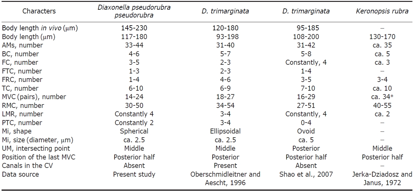
Two urostyloid ciliates, collected from brackish water in Korea, were identified as Diaxonella pseudorubra pseudorubra (Kaltenbach, 1960) Berger, 2006 and Pseudokeronopsis flava (Cohn, 1866) Wirnsberger, Larsen and Uhlig, 1987. The description was based on living, protargol impregnated specimens. These species are described as follows: Diaxonella pseudorubra pseudorubra: body size in vivo 145-230 × 40-60 μm, elongated ellipsoidal in shape. Cytoplasm reddish and flexible. Adoral zone of membranelles occupied 30-40% of the body; composed of 33-44 membranelles; 1-3 frontoterminal cirri, 1-4 frontal row cirri, 4-6 buccal cirri, 6-10 transverse cirri. Midventral rows composed of 14-24 cirri, four left marginal rows, one right marginal row. Two kinds of cortical granules; the larger one is yellowish and the smaller one is reddish. Pseudokeronopsis flava: body size in vivo 150-210 × 30-45 μm, elongated ellipsoidal shape. Cytoplasm yellowish and flexible. Adoral zone of membranelles occupied 25-30% of body; composed of 44-58 membranelles in number. Frontal cirri forming bicorona composed of 5-7 cirral pairs, 2-3 frontoterminal cirri, one buccal cirrus, and 2-3 transverse cirri. Midventral rows composed of 18-33 cirri, 34-53 left marginal cirri, and 40-58 right marginal cirri. Two kinds of cortical granules; the larger one is colorless and “blood-cell-shaped,” and the smaller one is yellowish. Diaxonella pseudorubra pseudorubra is different from the most similar subspecies, D. pseudorubra pulchra, in cytoplasmic color and number of midventral cirri. Pseudokeronopsis flava is different from its most similar congeners in pigment granular color, number of bicorona, number of midventral cirri, and position of the con-tractile vacuole.
The urostyloid ciliates are composed of a large group of hy-potrichs,
>
Sample collection and enrichment
The morphology of living specimens was observed under low (50-400×) and high (1,000×; immersion oil) magnifi-cations using a light microscope with a DIC device (Axio Imager A1; Carl Zeiss, Oberkochen, Germany) and their images were captured using a CCD camera (Axio Cam MRc; Carl Zeiss). The infraciliatures were observed after impre-gnation using the protagol method (Wilbert, 1975; Foissner, 1992). Terminology and taxonomic classification are accord-
[Table 1.] Morphometric data of Diaxonella pseudorubra pseudorubra

Morphometric data of Diaxonella pseudorubra pseudorubra
ing to Berger (2006) and Lynn (2008).
Order Urostylida Jankowski, 1979
Superfamily Urostyloidea Butschli, 1889
Family Holostichidae Faure-Fremiet, 1961
Genus 1*Diaxonella Jankowski, 1979
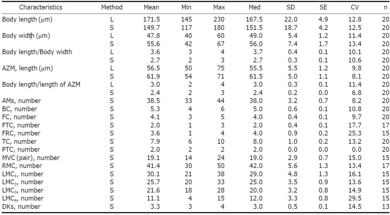
2* Diaxonella pseudorubra pseudorubra (Kaltenbach, 1960) Tables 1, 2, Figs. 1-3)
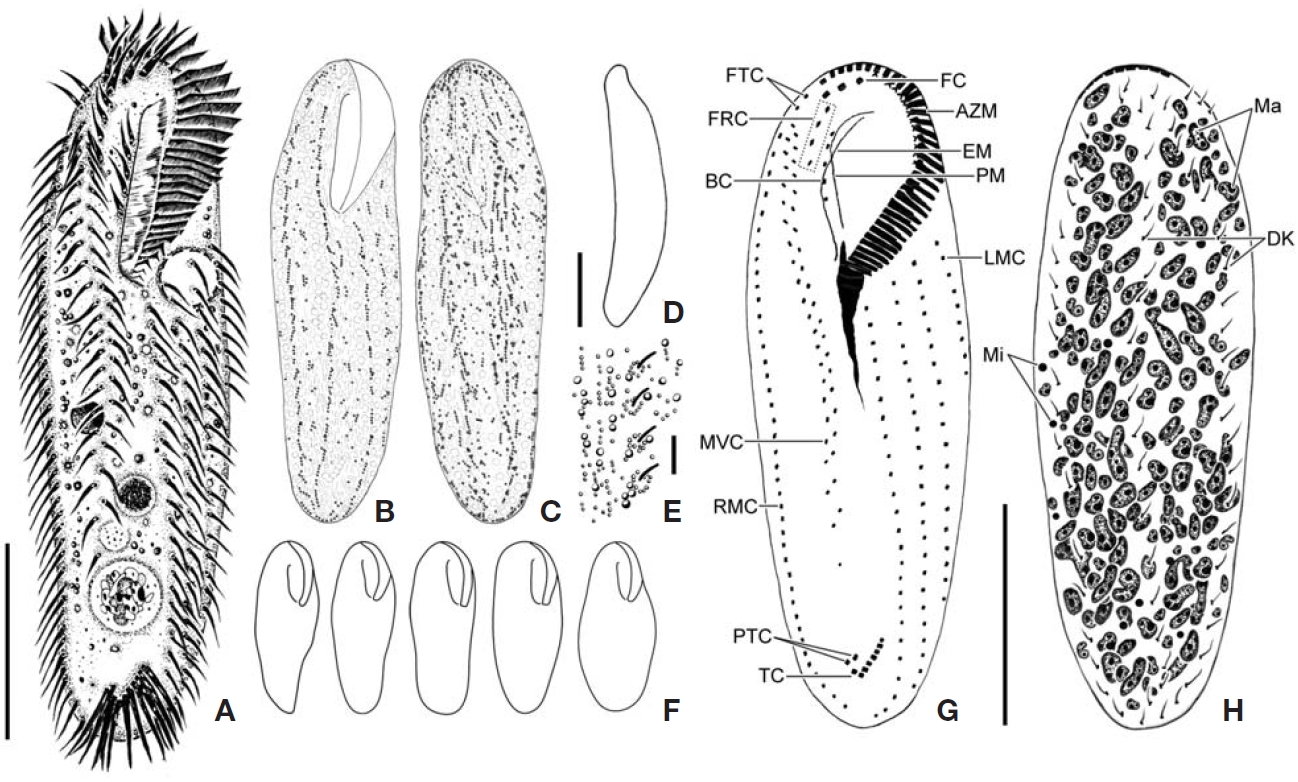
Description. General morphology and behavior : Body
[Table 2.] Comparisons of Diaxonella pseudorubra pseudorubra which has different names
Comparisons of Diaxonella pseudorubra pseudorubra which has different names
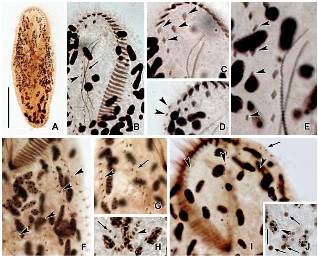
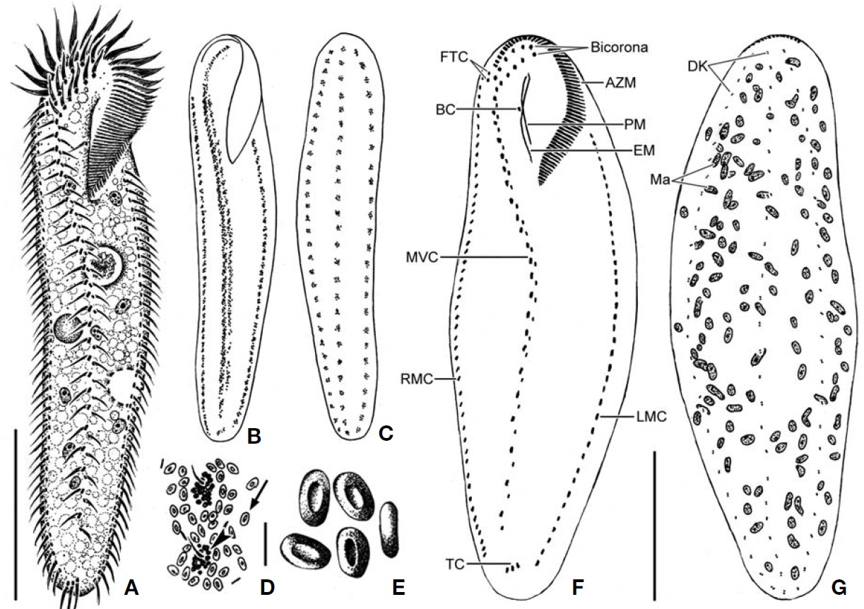
size 145-230 × 40-60 μm, usually about 170× 40 μm, length: width ratio about 4 : 1 on average in live specimens. Body shape elongated and ellipsoid with rounding at both ends(Figs.1 A, 2A, 3A), dorsoventrally flattened about 2 : 1, ven-tral side slightly concave, dorsal side convex (Figs.1 D, 2B). Cytoplasm reddish to wine color and flexible (Fig.2 D). Sin-gle contractile vacuole spherical and above the mid-body near the left margin (Figs.1 A, 2C). Locomotion usually crawl-ing on substrate. Omnivorous feeding (Fig.2 J-L).
Buccal field and oral infraciliature: Adoral zone of mem-branelles occupies 30-40% of body length (Figs.1 G, 3A), distal to proximal continuously semicircular, consists of 33-44 adoral membranelles (Fig.3 B), widest membranelle about 10 μm in length. Buccal area narrow and rather deep. Undu-lating membranes intersecting to half of the membranes, endoral membrane slightly curved and longer, paroral mem-brane anteriorly curved and shorter, distal end of paroral beyond endoral, about 45 μm in total length (Figs.1 G, 3B).
Somatic infraciliature: Usually, four frontal cirri slightly enlarged, lying on front of the anterior region but one frontal cirrus near the distal end of the adoral zone of membranelles(Figs.1 G, 3C), about 17μm long. One to three frontoterminal cirri located on the right side of frontal cirri, about 12μm long (Fig.3 D). Four to six buccal cirri arranged along paroral membrane, about 8 μm long (Figs.2F, 3B). Inconspicuous, usually four frontal row cirri starting at the same level at the distal end of the paroral membrane and terminating at the same level at intersecting point of the undulating membranes(Figs.1 G, 3E). Midventral complex composed of 14-24 pairs of midventral cirri, continued frontoterminal cirri, arranged in a zigzag pattern, terminating at the posterior half but most cirri separated from others (Figs.1 G, 3G). Six to ten trans-verse cirri arranged in a J-shape, about 18 μm long (Figs.2 I, 3H). Two pretransverse cirri located near right transverse cirrus (Fig.3 H). One right marginal row consisting of 30-50 cirri (Figs.1 G, 3G). Four left marginal rows that gradually
shorten from right to left composed of 21-38, 20-33, 18-28,and 4-15 cirri, respectively (Figs.1 G, 3F). Three dorsal kine-ties complete, but some cases of extra dorsal bristles present,bristle about 3-4 μm in length (Figs.1 H, 2H, 3I).
Cortical granules: Two kinds of cortical granules present on both sides and pigmented: larger one greenish yellow about 1 μm in diameter, arranged linearly in short groups beside the cirral rows on the ventral side and densely arranged on the dorsal side but loosely around each kineties (Fig.1 B, C). Smaller one red wine colored, about 0.3μm in diameter, scat-tered around the whole body and arranged linearly in short groups between larger granules or around each cirrus (Figs.1E, 2G).
Nuclear apparatus: Various shaped which elongated ellip-soidal to ovoid macronuclear nodules throughout the whole body, about 120 in number with several nucleoli. Spherical shaped micronuclei scattered body and 2.5 μm in diameter(Figs.1 H, 3A, J).
Distribution. Europe (Austria, Germany, and Poland), Africa (Burundi) and Asia (China, Korea [present study]).
Remarks. The taxonomy of the genus
Consequently, this Korean population of
[Table 3.] Morphometric data of Pseudokeronopsis flava
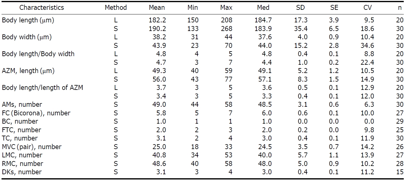
Morphometric data of Pseudokeronopsis flava
littoral zone (Berger, 2006).
Family Pseudokeronopsidae Borror & Wicklow, 1983
Genus Pseudokeronopsis Borror & Wicklow, 1983
1*Pseudokeronopsis flava (Cohn, 1866) (Tables 3, 4, Figs. 4-6)
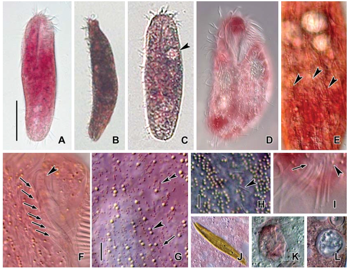
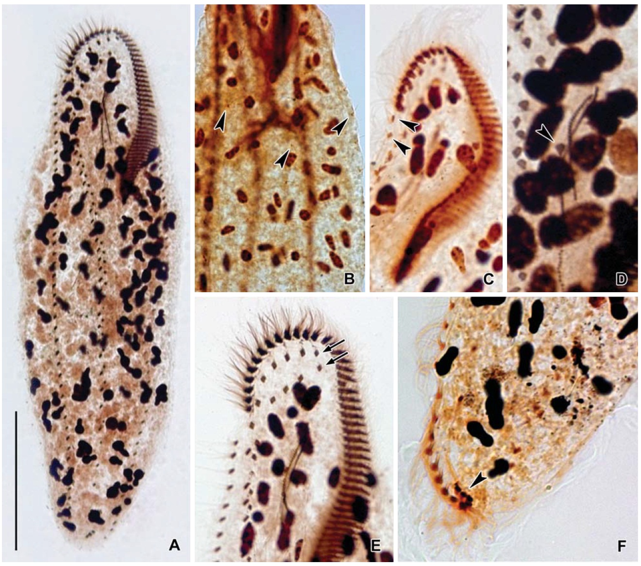
Description. General morphology and behavior: Body size 150-210× 30-45 μm, usually about 180× 40 μm, length : width ratio about 5 : 1 in live specimens. Body shape elon-gated elliptical, both ends narrowly rounded, anterior portion usually concave leftwards (Figs.4 A, 5A), dorsoventrally flat-tened about 2 : 1 (Fig.5 B). One contractile vacuole located below the mid-body near the left cell margin about 13 μm in diameter (Figs.4 A, 5E). Cytoplasm very flexible but not con-tractile(Fig.5 C), almost yellowish at low magnification. Locomotion usually crawling on substrate, wrapping to change direction (Fig.5 C). Omnivorous feeding (Fig.5 J, K).
Buccal field and oral infraciliature: Adoral zone of mem-branelles occupies 25-30% of body length, composed of 44-
[Table 4.] Comparisons of different populations of Pseudokeronopsis flava
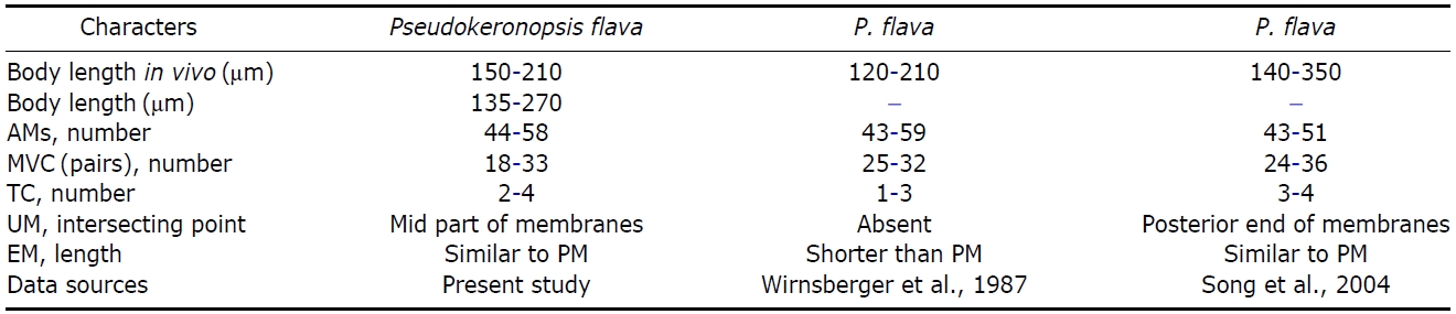
Comparisons of different populations of Pseudokeronopsis flava
58 adoral membranelles (Figs.4F, 6A). Buccal area narrow and rather deep. Paroral and endoral membranes slightly curved, intersecting to half of the membranes, endoral longer than paroral, about 35 μm in length (Figs.4F, 6A, D).
Somatic infraciliature:Frontal cirri arranged in two arcs forming bicorona comprising 5-7 cirral pairs, bicorona con-nect to midventral complex (Fig.6 E). Midventral complex extended to subposterior and consisting of 18-33 cirri; distance between posterior most cirrus of midventral cirri and upper-most transverse cirrus about 20 μm (Figs.4F, 6A). Two or
three frontoterminal cirri located at the right side of the right-most bicorona pair (Fig.6 C, E). Buccal cirrus located near the intersecting point of the undulating membranes (Fig.6 D).Two to four transverse cirri located posteriorly and distinctly separated by marginal rows (Figs.5 I, 6F). Right marginal row commenced at level of the down most bicorona cirrus,terminated at the posterior part and comprising 40-58 cirri (Figs.4F, 6A, E), left marginal row with 34-53 cirri; posterior
ends of marginal rows distinctly separated (Figs.4F, 6A). Dorsal bristles length 5-10 μm (Fig.5 D), three arranged kine-ties, dorsal kineties extending to entire dorsal surface (Figs.4G, 6B).
Cortical granules:Two types of cortical granules present on both sides (Figs.4 D, 5H); yellowish cortical granules form-ing four rows with cirral rows on ventral side (Figs.4 B, 5F), small groups with dorsal bristles on dorsal side and 0.8-1 μm in diameter (Figs.4 C, 5G, H), colorless cortical granules shaped ellipsoidal and “blood-cell-shaped” under both cortex and about 2×1.5 μm in size (Fig.5 G, H).
Nuclear apparatus: 70-100 ovoid to ellipsoidal macronuclear nodules, size 5-10 μm long in protargol-impregnated speci-mens. Micronucleus inconspicuous, about 3 μm in diameter(Figs.4 G, 6A).
Distribution. Europe (England, France, Denmark, Germany, Italy, and Poland), North America (USA), and Asia (China and Korea [present study]).
Remarks. The Korean population of
Closely-related species of
Korean name: 1*쌍열충속, 2*붉은쌍열충 Korean name: 1*노랑위각모충