검색 전체 메뉴
PDF
맨 위로
OA 학술지
Crasiella clauseni, a New Gastrotrich Species (Macrodasyida, Planodasyidae) from Jeju Island, South Korea
  • 비영리 CC BY-NC
  • 비영리 CC BY-NC
ABSTRACT
Crasiella clauseni, a New Gastrotrich Species (Macrodasyida, Planodasyidae) from Jeju Island, South Korea
KEYWORD
taxonomy , Gastrotricha , description , key , East Asia
  • INTRODUCTION

    Marine gastrotrich fauna of East Asia is still poor, in spite of serial taxonomic studies accomplished during the past 10 years and earlier. Twenty eight species of 11 genera have been recorded in five families: Cephalodasyidae, Lepidoda-syidae, Thaumastodermatidae, Chaetonotidae, and Xenotri-chulide. In Japan, 10 species of seven genera (Saito, 1937; Sudzuki, 1976, 1979; Chang et al., 2002; Lee and Chang, 2011); in Korea, 17 species of six genera (Chang et al., 1998a, 1998b; Chang and Lee, 2001; Lee and Chang, 2002, 2003, 2004, 2006, 2007; Lee et al., 2009); and in the Russian Far East, only one species, Aspidiophorus oculatusTodaro, Zotto, Maiorova and Adrianov, 2009 (Todaro et al., 2009) are currently recorded. However, planodasyid gastrotrichs have not been reported from East Asia as yet.

    Herein, we describe a new gastrotrich species of the genus Crasiella belonging to the family Planodasyidae from a sub-littoral sandy bottom at Jeju Island, South Korea. We discuss the taxonomic affinities based on the morphological charac-ters. Moreover, we provide a key to the hitherto known spec-ies of genus Crasiella.

    MATERIALS AND METHODS

    Gastrotrichs were collected from the shell-sandy bottom at about 6-7m in depth around Saeseom islet, which is located 150 m off Seogwipo Harbor at the southern coast of Jeju Is-land, South Korea.

    General sampling methods and extraction process are same with those of Lee et al. (2009), because the specimens were obtained from the same locality with the same collection data as for Ptychostomella jejuensis Lee, Hwang and Chang, 2009.

    Type specimens have been deposited in the National Insti-tute of Biological Resources (NIBR), Incheon, Korea and also in the authors' collection at the specimen room of the Department of Biology, Daegu University (DB).

    Terminology mostly follows Ruppert (1991) and Clausen (2000). Abbreviations used include: fpo, female gonopore; Lt, total length, from anterior tip of head to posterior tip of pedicles including adhesive tubes; mpo, male gonopore; Ph-JIn, junction between pharynx and intestine; TbA, anterior adhesive tubes; TbD, dorsal adhesive tubes; TbDL, dorsola-teral adhesive tubes; TbP, posterior adhesive tubes; TbV, ven-tral adhesive tubes; TbVL, ventrolateral adhesive tubes; U, percentage unit of Lt, used for the location (U-) from anterior to posterior, or for the relative length (-U).

    SYSTEMATIC ACCOUNTS

    Order Macrodayida Remane, 1925 [Rao and Clausen, 1970]

    1*Family Planodasyidae Rao and Clausen, 1970

    2*Genus Crasiella Clausen, 1968

    3*Crasiella clauseni n. sp. (Fig. 1)

    Material examined. Holotype (DBG1601), 14 paratypes (NIBRIV0000245017, 0000245018, DBG1602-13), mounted in glycerin, Korea, Jeju Island, Seogwipo, Saeseom islet, 33 ? 14'13?N, 126 ? 33' 33?E, 24 Apr 2006, leg, Lee J, Chang CY, Kim SH, in sublittoral sandy sediments containing shell crumbles, 6-7m deep.

    Etymology. The proposed specific name is in commemora-tion of the late Dr. Claus Clausen who established the genus Crasiella, and kindly guided us to study taxonomy of marine gastrotrichs.

    Diagnosis. Crasiella with somewhat oblong body, furnished with bilobed caudum; body up to 400 ㎛ long; PhJIn at about U33; dorsal surface smooth without cuticular armature such as hooks, scales and plates; lacking shallow lateral pits with ciliary tufts in cephalic region; without demarcation between TbA and TbVL, bearing about 120 ventrolateral tubes per side, extending along nearly whole body length; anterior tubes aligned in 5-7 horizontal rows, each composed of 2-6 tubes; a pair of TbV implanted mid-ventrally at one-third of pharyn-geal region; TbD (L) absent; TbP of 9-10 tubes per side, form-ing small caudal lobe, each caudal lobe comprising 2 long distal tubes, flanked by 3-4 lateral and 4-5 medial ones; in-testine somewhat curved like ‘S’ at posterior part; testes pair-ed, their tips reaching behind pharyngeal knobs; vasa defer-entia U-shaped, located at middle of intestine region; semi-nal receptacle elongate, tube-shaped with posterior end fold-ed up; copulatory organ locating just behind seminal recep-tacle, tube-shaped.

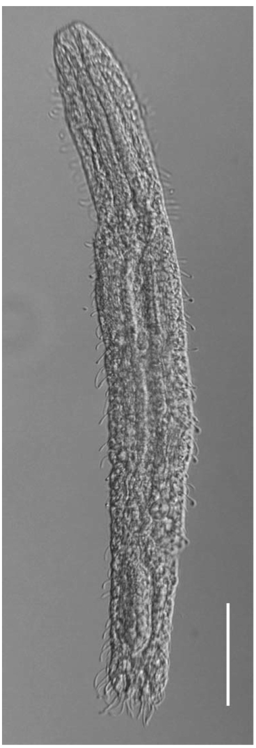
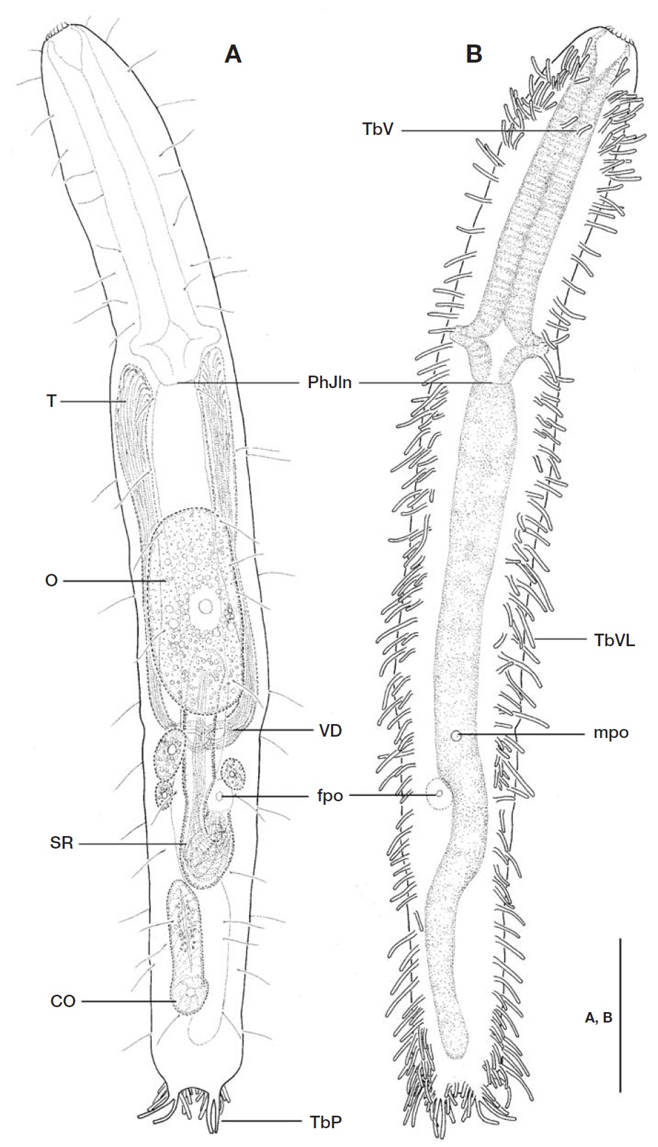
    Description. Holotype: Body (Figs. 1, 2) elongate, oblong, dorso-ventrally flattened, with small bilobed caudum, Lt 385㎛ long. Lateral sides of body nearly parallel, weakly con-stricted in the level of posterior pharyngeal region and a slightly narrowed distal head region. Head indistinct; lacking shallow lateral pits and ciliary tufts. Widths of anterior part of head/PhJIn/trunk/base of caudal lobes 28/41/42/17 ㎛ at U05/U33/U58/U96, respectively.

    Dorsal surface somewhat uneven, without cuticular arma-ture like hooks, scales and plates.

    Numerous hairs (ca. 12-18 ㎛ long) irregularly arrayed in 2-3 columns per side on subdorsal and lateral surfaces along nearly whole body length (U09-U90); several sensory hairs(ca. 5 ㎛ long) implanted sporadically on anteroventral sur-face at U01-U03.

    Locomotor ciliation covering entirely ventral surface from rear of oral opening to base of caudum.

    Adhesive tubes: TbA and TbVL not obviously separated, 119-123 tubes per side, somewhat slender, with different sizes(ranging from 5 ㎛ to 19 ㎛ long), extending over nearly whole body length (U3-U95) from rear of oral opening to base of caudal lobes. Adhesive tubes in pharyngeal region representing different arrangements of horizontal and longi-tudinal rows; foremost 2 pairs located rather ventrally at U03 and U04, respectively; 6 horizontal rows per side situating in anterior pharyngeal region from U05 to U13, each row composed of 2-6 tubes (5, 6, 5, 4, 2, 3 tubes in left side and 4, 4, 4, 3, 2, 2 in right, respectively) in different sizes (ca. 6-11 ㎛ long); followed by 14-16 tubes per side, positioned at irregular intervals, forming a longitudinal row in remaining pharyngeal region at U15-33. About 82-85 adhesive tubes per side, ca. 5-19 ㎛ long, arranged irregularly between PhJIn to front of caudal lobes, crowded near posterior end of trunk at level of anus (U94). A pair of TbV (ca. 7 ㎛ long), not foot-type, implanted mid-ventrally in anterior pharyngeal re-gion at U10. TbD (L) absent. TbP comprising 9-10 tubes per side, forming a small caudal lobe, consisting of 2 long distal tubes (15 ㎛ long), 3 lateral (ranging ca. 7-10 ㎛ long) and 4-5 medial ones (ranging ca. 5-8 ㎛ long); lateral and medial TbP gradually decreased in size towards base of caudal lobe.

    Digestive tract: Oral opening ornamented with corona of 15-18 short hook-like projections (ca. 2-3 ㎛ long) along its terminal edge; buccal cavity funnel-shaped and slightly cuti-cularized; pharynx 127 ㎛ long, with a pair of pharyngeal pores, opened laterally at U30; intestine broad and straight in its anterior region, then narrowing gradually towards anus,and curved near posterior half of seminal receptacle; anus situated ventrally at U92.

    Reproductive system: Testes paired, situated along anteri-or intestinal region, their tips reaching just behind pharyngeal knobs at U30; vasa deferentia including spermatozoa, coiled and joined at the middle of intestine region at U63-65; mpo emerging from joint of vasa deferentia at U64. Ovum well-developed,ca. 31×67 ㎛, situated dorsally in mid-intestinal region at U44-U62, followed by 3 small oocytes. Seminal receptacle elongate, 77 ㎛ long, tube-shaped with its poste-rior part a little swollen and folded; posterior end folded, opened (fpo) ventrally on two-thirds of seminal receptacle at U70; situated dorsally from posterior part of ovum to anterior

    end of copulatory organ at U57-U78. Copulatory organ (44㎛ long) tube-shaped with an internal duct, gradually nar-rowing to its anterior edge, located at U77-U89.

    Variability. Body lengths of 12 adult specimens ranged 366 to 440 ㎛ (mean 396 ㎛, standard deviation 24), and maxi-mum width 41-58 ㎛, measured in glycerin mount.

    The number of adhesive tube showed somewhat asymmet-rical array and individual variability. Especially, TbVL in the intestinal region ranged from 52 to 83 tubes. In contrast with ventrolateral tubes, the paired TbV was rather consistent, located typically in the anterior pharyngeal region, except for one specimen lacking the pair, and for four specimens with only one tube in one side.

    Epidermal glands were not clear in holotype, however, in five paratypes numerous epidermal glands were irregularly scattered at dorsal, dorsolateral, and ventrolateral surfaces, along nearly the whole body length.

    Three sub-adults were included among 15 specimens exam-ined, with the body sizes of 281 ㎛, 302 ㎛, 325 ㎛ long and 33 ㎛, 42 ㎛, 48 ㎛ wide, respectively. They showed fewer ventrolateral and posterior adhesive tubes than in adults:3-4 horizontal rows (vs. 5-7 in adults) in the anterior-most pharyngeal region; 21-39 ventrolateral tubes in the intestinal region (vs. 52-83 in adults); and 5-6 TbP per side (vs. 8-11 in adults).

    Taxonomic affinities. To date, six species have been record-ed in the genus Crasiella, since C. diplura was first describ-ed from Bergen area, Norway by Clausen (1968): C. diplura Clausen, 1968, C. oceanica d’Hondt, 1974, C. pacifica Sch-midt, 1974, C. indica Rao, 1981, C. azorensis Hummon, 2008 and C. skaia Hummon, 2010.

    Among them, C. oceanica from Roscoff, France (d’Hondt, 1971, 1974) and C. azorensis from Azore Islands (Hummon, 2008) are devoid of sensory pits with ciliary tuft in the cephal-ic region as in C. clauseni n. sp. However, C. clauseni is dis-tinguished from both the species by a pair of TbV and ven-trolateral adhesive tubes, which are not separated into TbA and TbVL, and arranged as 5-7 horizontal rows in the pha-ryngeal region. According to the d’Hondt’s original descrip-tion (1974), C. oceanica has Tb (V)L each bearing a sensory hair on its tip, however, the sensory hairs were not found in subadults from Trezen ar Skoden, France by Hummon (2008).

    Other three congeneric species, C. diplura, C. pacifica from Galapagos Island and Hawaii (Schmidt, 1974; Hummon, 2009), and C. indica from India (Rao, 1981, 1993), possess sensory pits with ciliary tufts in the cephalic region. Crasiella clauseni n. sp. shares the unipartite ventrolateral tubes with C. pacifica. However, C. clauseni differs from C. pacifica by a large number of ventrolateral adhesive tubes (about 120 tubes, vs. 50 in C. pacifica), paired TbV (against absence of TbV in C. pacifica), and joint position of vasa deferentia (at middle of intestinal region, vs. anterior third in C. pacifica).

    Crasiella diplura and C. indica are different from C. clau-seni n. sp., besides the sensory pits with ciliary tuft in the cephalic region, by absence of TbV, the arrangement of adhe-sive tubes in the anterior part of pharyngeal region (a longitu-dinal row in C. diplura and a diagonal row in C. indica, vs.5-7 horizontal rows in C. clauseni), and the shape of copula-tory organ (oblong in C. diplura and pear-shape in C. indica, vs. tube-shape in C. clauseni).

    Crasiella skaia from Florida and Bahamas (Hummon, 2010) is characteristic in having a pair of pestle organs which have not been reported in other congeneric species. This species is discernable from C. clauseni by an irregular longitudinal row of TbA. Moreover C. skaia apparently differs from C.clauseni in having fewer ventrolateral tubes, with deficiency of TbVL between anus and caudal base.

    A key to the species of genus Crasiella Clausen

    1. With pestle organ in the cephalic region ······································································Crasiella skaia Hummon With sensory pits in the cephalic region ······················2 Without sensory pits in the cephalic region ·················4

    2. TbA and TbVL separated ··········································3 TbA and TbVL not separated ··················································································Crasiella pacifica Schmidt

    3. TbA forming 2 longitudinal rows ···············································································Crasiella dipulra Clausen TbA forming 1 diagonal row ·········Crasiella indica Rao

    4. With 5-7 horizontal rows of adhesive tubes and a pair of TbV in the pharyngeal region ··············································································Crasiella clauseni n. sp.Without horizontal rows of adhesive tubes and TbV in the pharyngeal region ···········································5

    5. TbA forming 1 diagonal row ··················································································Crasiella oceanica d’Hondt TbA forming 1 longitudinal row ··········································································Crasiella azorensis Hummon

    Korean name: 1*납작털배벌레과, 2*어리납작털배벌레속, 3*어리납작털배벌레

참고문헌
  • 1. Chang CY, Kubota S, Shirayama Y 2002 New marine gastro-trichs of the family Thaumastodermatidae (Gastrotricha :Macrodasyida) from Shirahama Japan. [Proceedings of the Biological Society of Washington] Vol.115 P.769-781 google
  • 2. Chang CY, Lee JM 2001 Two new Tetranchyroderma gastro-trichs (Macrodasyidai: Thaumastodermatidae) from South Korea. [Korean Journal of Biological Science] Vol.5 P.187-194 google
  • 3. Chang CY, Lee JM, Clausen C 1998a Description of two new species thaumastodermatids (Gastrotricha Macrodasyida) from Korea. [Korean Journal of Biological Sciences] Vol.2 P.315-321 google cross ref
  • 4. Chang CY, Lee JM, Clausen C 1998b Two new species of Thaumastoderma (Gastrotricha Macrodasyida) from Korea. [Sarsia] Vol.83 P.329-336 google
  • 5. Clausen C 1968 Crasiella diplura gen. et sp. n. (Gastrotricha Macrodasyoidea). [Sarsia] Vol.33 P.59-64 google
  • 6. Clausen C 2000 Gastrotricha Macrodasyida from the Tromsøregion northern Norway. [Sarsia] Vol.85 P.357-384 google
  • 7. d?Hondt JL 1971 Gastrotricha. [Oceanography and Marine Bio-logy: an Annual Review] Vol.9 P.141-192 google
  • 8. d?Hondt JL 1974 Note sur Psammodasys cambriensis (Boaden 1963) et Crasiella oceanica n. sp. (Gastrotriches Macroda-syoides). [Bulletin de la Societe Zoologique de France] Vol.99 P.675-680 google
  • 9. Hummon WD 2008 Gastrotricha of the North Atlantic Ocean: 1. Twenty four new and two redescribed species of Macro-dasyida. [Meiofauna Marina] Vol.16 P.117-174 google
  • 10. Hummon WD 2009 (revision of 2001 2004 2007). Global data-base for marine Gastrotricha (taxonomic geographic bib-liographic and video) [Internet]. google
  • 11. Hummon WD 2010 Marine gastrotricha of the Caribbean Sea: a review and new descriptions. [Bulletin of Marine Science] Vol.86 P.661-708 google
  • 12. Lee JM, Chang CY 2002 Pseudostomella gastrotrichs (Macro-dasyida Thaumastodermatidae) from South Korea with a brief review of the genus. [Korean Journal of BiologicalSciences] Vol.6 P.207-213 google
  • 13. Lee JM, Chang CY 2003 Two new marine gastrotrichs of the genus Ptychostomella (Macrodasyida Thaumastodermati-dae) from South Korea. [Zoological Science] Vol.20 P.481-489 google cross ref
  • 14. Lee JM, Chang CY 2004 Marine chaetonotid gastrotrichs of genus Halichaetonotus (Chaetonotida: Chaetonotidae) from Korea. [Korean Journal of Systematic Zoology] Vol.20 P.55-62 google
  • 15. Lee JM, Chang CY 2006 Marine gastrotrichs of the genus Di-plodasys (Macrodasyida: Thaumastodermatidae) from Korea. [Korean Journal of Systematic Zoology] Vol.22 P.109-115 google
  • 16. Lee JM, Chang CY 2007 Two new marine gastrotrichs of the genus Tetranchyroderma (Macrodasyida: Thauma-stodermatidae) from South Korea. [Zoological Studies] Vol.46 P.474-482 google
  • 17. Lee J, Chang CY 2011 Two new species of the genus Lepido-dasys (Gastrotricha Lepidodasyidae) from Tsushima Island Japan. [Journal of Natural History] Vol.45 P.855-867 google cross ref
  • 18. Lee JM, Hwang UW, Chang CY 2009 A new gastrotrichs spec-ies of the genus Ptychostomella (Macrodasyida: Thaumas-todermatidae) from South Korea. [Animal Cells and Systems] Vol.13 P.25-30 google cross ref
  • 19. Rao GC 1981 Crasiella indica n. sp. (Gastrotricha Macrodas-yida) from Orissa coast India. [Bulletin of the ZoologicalSurvey of India] Vol.4 P.3-6 google
  • 20. Rao GC 1993 Littoral meiofauna of little Andaman. [Records of the Zoological Survey of India] Vol.155 P.1-120 google
  • 21. Ruppert EE 1991 Gastrotricha. In: Microscopic Anatomy of Invertebrates. IV. Aschelminthes (Eds. Harrison FW Rup-pert EE). P.41-109 google
  • 22. Saito I 1937 Neue und bekannte Gastrotrichen der Umgebung von Hiroshima (Japan). [Journal of Science of the HiroshimaUniversity] Vol.5 P.245-265 google
  • 23. Schmidt P 1974 Interstitielle Fauna von Galapagos. IV. Gastro-tricha. [Mikrofauna des Meeresbodens] Vol.26 P.1-76 google
  • 24. Sudzuki M 1976 Microscopical marine animals scarcely known from Japan. I. Micro- and meio-faunae around Kasado Island in the Seto Inland Sea of Japan. [Proceedings of the Japanese Society of Systematic Zoology] Vol.12 P.5-12 google
  • 25. Sudzuki M 1979 Some aspects of the haline interstitial biota from Ryuku Shoto subtropical chain islands southwest Japan. [Sesoko Marine Science Laboratory Technical Report] Vol.6 P.37-50 google
  • 26. Todaro MA, Zotto MD, Maiorova AS, Adrianov AV 2009 A new species of Aspidiophorus (Gastrotricha Chaetonotida) from the Russian Far East with a key to marine species of the genus. [Marine Biology Research] Vol.5 P.297-303 google cross ref
OAK XML 통계
이미지 / 테이블
  • [ Fig. 1. ]  Crasiella clauseni new species. A Habitus dorsal; B Habitus ventral. CO copulatory organ; fpo female gonopore; mpomale gonopore; O ovum; PhJIn junction between pharynx and intestine; SR seminal receptacle; T testis; TbP posterior adhesivetubes; TbV ventral adhesive tubes; TbVL ventrolateral adhesive tubes; VD vasa deferentia. Scale bar: A B=50 ㎛.
    Crasiella clauseni new species. A Habitus dorsal; B Habitus ventral. CO copulatory organ; fpo female gonopore; mpomale gonopore; O ovum; PhJIn junction between pharynx and intestine; SR seminal receptacle; T testis; TbP posterior adhesivetubes; TbV ventral adhesive tubes; TbVL ventrolateral adhesive tubes; VD vasa deferentia. Scale bar: A B=50 ㎛.
  • [ Fig. 2. ]  Crasiella clauseni new species habitus. Scale bar=50㎛.
    Crasiella clauseni new species habitus. Scale bar=50㎛.
(우)06579 서울시 서초구 반포대로 201(반포동)
Tel. 02-537-6389 | Fax. 02-590-0571 | 문의 : oak2014@korea.kr
Copyright(c) National Library of Korea. All rights reserved.