검색 전체 메뉴
PDF
맨 위로
OA 학술지
Modulatory Effects of Chrysanyhemi Flos Pharm-acopuncture on Nitric-oxide (NO) Production in Murin Macrophagy Cells
  • 비영리 CC BY-NC
  • 비영리 CC BY-NC
ABSTRACT
Modulatory Effects of Chrysanyhemi Flos Pharm-acopuncture on Nitric-oxide (NO) Production in Murin Macrophagy Cells
KEYWORD
CF , herb , immunomodulatory , macrophage , nitric-oxide (NO) , RAW 264.7
  • 1. Introduction

    Chrysanthemi Flos (CF), the flowers of Chrysanthemum indicum or C. morifolium, is rich in volatile oils, flavonoids and so on [1]. It is used to treat warm pathogen disease with fever and headache. It also clears eyes and dizziness [1,2].

    Macrophages are main players in the immune function as antigen-presenting cells in initiating adaptive immune responses and in the innate immune response [3]. Many studies have demonstrated that Chrysanthemum cinerari-aefolium, same genus as Chrysanthemum, increases innate immunity [4]. These immune responses are reported to be stimulated not only by intact bacterial cells but also by their components, including peptidoglycan, lipoteichoic acids, and intra-extracellular polysaccharide products, and cell wall (CW) and cell free extract (CFE) fractions [5-8].

    Much evidence exists that herbs have effective immunom-odulatory activities in both preclinical and clinical resea-rches [9-13], and studies of the modulatory effects on nitric-oxide (NO) production have been conducted [14-16]. Also, medicinal herbs, Compositae, have been studied for immune regulation [17], but few studies have addressed Chrysanyh-emi Flos regulating the production of nitric-oxide (NO). The-refore, the aim of this study was to use the lipopolysacch-aride (LPS)-treated RAW 264.7 murine macrophage model to further explore the potential immunomodulation of CF, especially for the regulation of NO production.

    2. Material and methods

       2.1. Sample preparation

    Chrysanthemi Flos was purchased from Omniherb (Korea). CF was prepared as follows: Chrysanthemi Flos, 100 g, was added to 2,000 ml of distilled water and boiled in a heating extractor for 3 hours. The extract was filtered and concentrated by using a rotary evaporator and was lyophilized by using a freeze dryer (17.9 g). The lyophilized extract was dissolved in water and filtered three times through microfilter paper (What-man no. 2, 0.45-0.2 μm). Then, it was placed in a disinfected vial and sealed for further study.

       2.2. Cell culture

    Mouse monocyte/macrophage cell line RAW 264.7 (ATCC TIB-71) was grown in Dulbecco's Modified Eagle's Medium (DMEM), supplemented with 10% fetal bovine serum (FBS), streptomycin (100 mg/ml) and penicillin (100 U/ml), at 37℃ with 5% CO2. Cells were transferred to 96-well culture plates at a density of 1 x 10⁵ cells/well and were allowed to adhere for 24 hours prior to sample treatment. The culture supernatant was collected and stored at -80℃ until it was analyzed for nitric-oxide (NO), interleukin-6 (IL-6) and tumor necrosis factor-alpha (TNF-α). Samples were incubated with cells for 24 hours, and cells cultured with LPS from Escherichia coli serotype O127:B8 (Sigma-Aldrich, Missouri, USA) at a concentration of 1 μg/ml [18] were used as a control.

       2.3. Cell viability

    The general viability of cultured cells was determined by reduction of 3-(4, 5-dimethylthiazol-2-yl)-2, 5-diphenyl-tetrazolium bromide (MTT) to formazan. MTT in living cells is reduced to formazan crystals, and the amount of formazan dissolved in dimethyl sulfoxide (DMSO), as measured by using the by spectroscopic method, shows the growth of cells. The cell were seeded in 96-well plates at a density of 1 x 10⁵ cells/well and were cultured at 37℃ in 5% CO2. Cells were pretreated with the sample at concentration, of 0, 50, 100 and 200 μg/ml with or without 1 μg/ml of LPS for 24 hours. Then, cells were treated with 0.05 mg/ml (final concentration) of MTT and incubated at 37℃ for an additional 4 hours. The medium containing MTT was discarded, and the MTT formazan that had been produced was dissolved in 200 μl of DMSO. The absorba-nce was read at 595 nm with a reference wavelength of 690 nm. The cell viability was calculated as follows:

    Cell viability (%) = [(optical Density 595 of sample)/(optical density 595 of control)]×100.

       2.4. Phagocytosis analysis

    RAW 264.7 cells were seeded in triplicates at a density of 5 x 10⁴ cells/well in 96-well plates with complete DMEM and were allowed to adhere for 24 hours. Cells were cultured with LPS (1μg/ml) as control. Various concentrations of CF were incubated with cells for 24 hours. After that, the cells were washed three times with phosphate buffered saline, (pH 7.2), and the phagocytic ability of the macrophages was measured by using a phagocytosis assay kit (Cayman Chemical, Michigan, USA) according to the procedure described by the manufacturer.

       2.5. NO determination

    The NO concentration was determined by measuring the amount of released nitrite with Griess reagent (Sigma-Aldrich, Missouri, USA) according to the Griess reaction [19]. Griess rea-gent produced a chemical reaction with nitrite and formed a purple azo salt that was consistent with the NO concentration. Briefly, 50 μl of cell culture supernatant was added to new 96-well plates and mixed with 50 μl of modified Griess reagent. After incubation at room temperature for 15 min, the absorban-ce was measured in a plate reader at 540 nm. Nitrite concent-rations were calculated on the basis of a NaNO2 standard curve.

       2.6. Cytokine measurement

    The concentrations of IL-6 and TNF-α in the culture supernat-ant were measured by using a IL-6 kit (Thermo scientific, Illinois, USA) and a TNF-α kit (R&D systems, Minnesota, USA), respectively, according to the procedure described by the manufacturer.

       2.7. Statistical analysis

    The results were expressed as means ± standard deviations (SD). Significant changes were evaluated by using the one-way ANOVA with Dunnett’s post-hoc test. Values of p < 0.05 were considered significant.

    3. Results

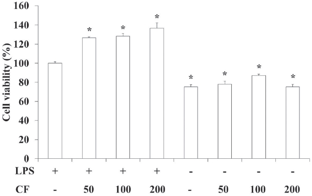
       3.1. Cytotoxicity on RAW 264.7 cells

    In order to evaluate the cytotoxicity of CF, samples were prepared at various concentrations and used to treat RAW 264.7 cells. The results of this evaluation are shown in Fig. 1 at conce-ntrations of 50, 100 and 200 μg/ml. The cell viability was recalc-ulated into 100 % of LPS-treated control group. The cell's viabi-lities treated with LPS, both LPS and CF 50 μg/ml, both LPS and CF 100 μg/ml, both LPS and CF 200 μg/ml, vehicle, CF 50, CF 100 and CF 200 μg/ml were respectively 100.0 ± 1.6, 126.7 ±1.0, 128.5 ± 2.7, 136.7 ± 5.5, 75.3 ± 2.2, 78.0 ± 3.2, 87.3 ± 1.3 and 75.2 ± 2.6 %. LPS with or without CF treatments significan-tly increased the cell's viabilities compared to vehicle with or without CF treatments. Moreover all groups treated with both LPS and CF (LPS(+) CF(50, 100 and 200)) showed significantly increased cell viabilities compared to control group treated with LPS.

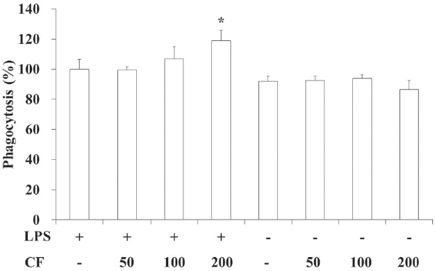
       3.2. Phagocytic activity of RAW 264.7 cells

    In order to evaluate the phagocytic activity of CF, we prepared samples at various concentrations and used them to treat RAW 264.7 cells. The results of this evaluation are shown in Fig. 2 at concentrations of 50, 100 and 200 μg/ml. The phagocytic activity was normalized to 100% for the LPS-treated control group. The phagocytic activities treated with LPS, both LPS and CF (50 μg/ml), both LPS and CF (100 μg/ml), both LPS and CF (200 μg/ml), vehicle, CF (50 μg/ml), CF (100 μg/ml) and CF (200 μg/ml) were, respectively, 100.0 ± 6.4, 99.4 ± 2.1, 106.7 ± 8.2, 119.0 ± 6.7, 92.0 ± 3.4, 92.3 ± 3.0, 93.8 ± 2.5 and 86.5 ± 5.9 %.

    Both LPS and CF treatments increased the phagocytic activities compared to LPS treatments in a dose-dependent manner. Especially both LPS and CF (200 μg/ml) treatments showed statistical difference compared to the LPS-treated control group (p < 0.05, Fig. 2). However, groups treated without LPS showed no changes at all.

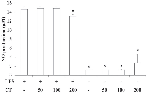
       3.3. NO production of RAW 264.7 cells

    In order to evaluate the NO production of CF, we prepared samples at various concentrations and used them to treat RAW 264.7 cells. The results of this evaluation are shown in Fig. 3 at concentrations of 50, 100 and 200 μg/ml. The NO productions treated with LPS, both LPS and CF (50 μg/ml), both LPS and CF (100 μg/ml), both LPS and CF (200 μg/ml), vehicle, CF (50 μg/ml), CF (100 μg/ml) and CF (200 μg/ml) were, respectively, 14.6 ±0.4, 14.8 ± 0.2, 14.8 ± 0.2, 13.0 ± 0.4, 1.2 ± 0.0, 1.3 ± 0.0, 1.2± 0.1 and 2.8 ± 1.9 μM.

    LPS treatment significantly increased the NO production compared to vehicle treatment (p < 0.05), but both LPS and CF (200 μg/ml) treatment could significantly reduce the NO production induced by LPS treatment (p < 0.05, Fig. 3).

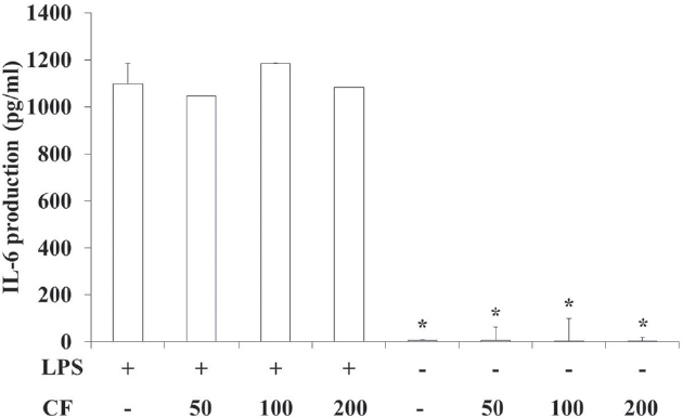
       3.4. IL-6 production of RAW 264.7 cells

    In order to evaluate the IL-6 production of CF, we prepared samples at various concentrations and used them to treat RAW 264.7 cells. The results of this evaluation are shown in Fig. 4 at concentrations of 50, 100 and 200 μg/ml. The IL-6 production treated with LPS, both LPS and CF (50 μg/ml), both LPS and CF (100 μg/ml), both LPS and CF (200 μg/ml), vehicle, CF (50 μg/ml), CF (100 μg/ml) and CF (200 μg/ml) were, respectively, 1097.1 ±88.0, 1045.7 ± 0.3, 1185.7 ± 1.4, 1081.4 ± 0.3, 5.1 ± 3.3, 6.2 ±55.0, 3.9 ± 95.0 and 3.2 ± 14.3 pg/ml.

    LPS treatment significantly increased the IL-6 production compared to vehicle treatment (p < 0.05). However, CF treatme-nt did not change the IL-6 production induced by LPS treatment (Fig. 4).

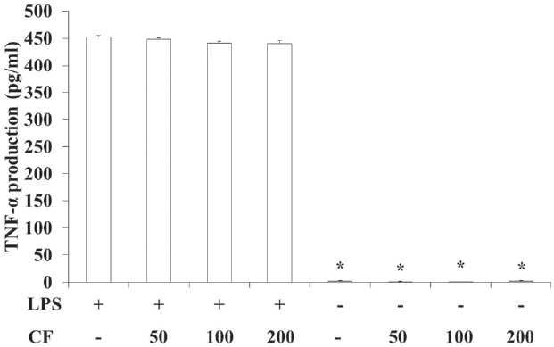
       3.5. TNF-α production of RAW 264.7 cells

    In order to evaluate the TNF-α production of CF, we prepared samples at various concentrations and used to treat RAW 264.7 cells. The results of this evaluation are shown in Fig. 5 at concentrations of 50, 100 and 200 μg/ml. The TNF-α production treated with LPS, both LPS and CF (50 μg/ml), both LPS and CF (100 μg/ml), both LPS and CF (200 μg/ml), vehicle, CF (50 μg/ml), CF (100 μg/ml) and CF (200 μg/ml) were, respectively, 452.7 ±3.0, 448.3 ± 2.5, 441.3 ± 3.4, 440.7 ± 5.4, 1.9 ± 1.1, 1.5 ± 0.7, 1.0 ± 0.6 and 2.4 ± 0.6 pg/ml.

    LPS treatment significantly increased the TNF-α production compared to vehicle treatment (p < 0.05). However, CF treatme-nt did not change the TNF-α production induced by LPS treatment (Fig. 5).

    4. Discussion and conclusion

    Chrysanthemi Flos, the flowers of Chrysanthemum indicum or C. morifolium, is rich in borneol, camphor, chrysanthenone, cosmosiin, apigenin, quercitrin, luteolin, thymol, heneicosane, tricosane and hexacosane [1]. It is traditionally used to disperse wind and clear heat. Also, it is used clinically for elimination of wind-heat patterns associated with a common cold or warm pa-thogen disease with fever and headache. It is also used to calm the liver and extinguish wind, whose symptoms are dizziness, headache and deafness due to ascendant liver yang. In addition, it is effective in clearing red, swollen, dry and painful eyes from wind-heat in the liver channel or ascendant liver yang. It can be used for spots in front of the eyes, blurry vision, or dizziness due to liver and kidney yin deficiency [1,2,20].

    LPS is present in the cell's outer membrane of gram negative bacteria and augments pro-inflammatory cytokines, such as TNF-α, IL-6 and IL-1β, at macrophages or monocytes [21]. One important role of the macrophage is the phagocytosis of pathogens and necrotic cellular debris. When a macrophage ingests a pathogen, the pathogen becomes trapped in a phagosome, which then fuses with a lysosome. Within the phagolysosome, enzymes and toxic peroxides digest the path-ogen. Macrophages can digest more than 100 bacteria before they finally die due to their own digestive compounds [22].

    LPS as endotoxin [23] causes an immune response, but excessive production of inflammatory mediators can destroy normal tissues and cause shock [24]. Thus, herbs that regulate the immune response are important. Many studies concerning the immunomodulatory effects of herbs, such as immunomodu-latory effects on healthy volunteers, anti-tumor immune response, splenocyte proliferation, macrophage function, NK anti-tumor activities and so on [9-13], have been performed.

    To investigate the in this study, CF on LPS-stimulated RAW 264.7 macrophages were examined immunomodulatory effects by measuring the cytotoxicity, the phagocytic activity and the amount of NO, IL-6 and TNF-α. No cytotoxicity was observed for all CF-treated concentrations. However, LPS treatment incre-ased the cell viability, and both LPS and CF treatment augme-nted the cell viability which was increased by LPS treatment. Although a high-concentration LPS treatment (e.g., 100 μg/ml) showed cytotoxicity in RAW 264.7 cells, the low concentration used in this study could stimulate the proliferation of macroph-age cells [25]. CF treatment could significantly augment the stimulation of proliferation only in the presence of LPS. For the phagocytic activity of CF, although CF treatment without LPS did not change the phagocytic activity compared to the vehicle-treated group, CF treatment with LPS could significantly augment the phagocytic activity compared to the LPS-treated control group in a dose-dependent manner.

    NO is generated by macrophages as part of the human immu-ne response. The immune system may regulate the armamen-tarium of the phagocytes that play a role in the inflammation and the immune responses with NO. NO secreted as an immune response acts as a free radical and is toxic to bacteria. The mechanisms for this include DNA damage [26-28] and degrada-tion of iron-sulfur centers into iron ions and iron-nitrosyl compounds [29]. NO shows an anti-cancer or an anti-microbial effect, but excessive secretion can kill normal cells or promote inflammation [30]. In this study, LPS treatment significantly increased the NO production compared to vehicle treatment (p < 0.05). However, both LPS and CF (200 μg/ml) treatment signifi-cantly reduced the NO production induced by LPS treatment (p < 0.05).

    IL-6 is an interleukin that acts as both a pro-inflammatory and an anti-inflammatory cytokine. It is secreted by macrophages and T cells to stimulate immune response, e.g., during infection, but excessive secretion causes rheumatoid arthritis and inflammatory diseases, such as Crohn's disease [31]. TNF-α is a cytokine involved in systemic inflammation and is a member of cytokine groups that stimulate an acute phase reaction. It is produced chiefly by activated macrophages. The effect of TNF-α is to stimulate phagocytosis of macrophages, but excessive secretion causes decreased myocardial contractility, low blood pressure and metabolic process damage [32].

    LPS treatment significantly increased the IL-6 and the TNF-α productions compared to vehicle treatment (p < 0.05). However, CF treatment did not change the IL-6 and the TNF-α productions induced by LPS treatment. The results for IL-6 and TNF-α may reveal the characteristics of that CF immunomodulatory effect; that is, it does not affect IL-6 and TNF-α-induced immune responses.

    In conclusion, CF regulates the production of NO and can prevent the pathological phenomena caused by excessive production of NO, such as expansion of blood vessels, mutation and damage to organization and nerve by inflammation. CF may play an important role in intensity modulated NO-induced immune responses and may have potential for use as an immunoenhancing pharmacopuncture. Further studies will be needed to unravel the characteristics of the effects of CF and the mechanisms of its functions.

참고문헌
  • 1. Kim IR, Kim HC, Kuk YB, Park SJ, Park YK, Park JH 2007 Boncho-Hak. P.182-3 google
  • 2. Chen JL, Chen TT. 2004 Chinese Medical Herbology and Pharm-acology. P.58-61 google
  • 3. Laskin DL. 2009 Macrophages and inflammatory mediators in chemical toxicity: a battle of forces. [Chem Res Toxicol.] Vol.22 P.1376-85 google cross ref
  • 4. Harikrishnan R, Heo J, Balasundaram C, Kim MC, Kim JS, Han YJ 2010 Effect of traditional Korean medicinal TKM triherbal extract on the innate immune system and disease resistance in Paralichthys olivaceus against Uronema marinum. [Vet Parasitol.] Vol.170 P.1-7 google cross ref
  • 5. Hatcher GE, Lambrecht RS. 1993 Augmentation of macrophage phagocytic activity by cell-free extracts of selected lactic acid-producing bacteria. [J Dairy Sci.] Vol.76 P.2485-92 google cross ref
  • 6. Matsuguchi T, Takagi A, Matsuzaki T, Nagaoka M, Ishikawa K, Yokokura T 2003 Lipoteichoic acids from Lactobacillus strains elicit strong tumor necrosis factor alpha-inducing activities in macrophages through Toll-like receptor 2. [Clin Diagn Lab Immunol.] Vol.10 P.259-66 google
  • 7. Amrouche T, Boutin Y, Moroni O, Kheadr E, Fliss I. 2006 Product-ion and characterization of anti-bifidobacteria monoclonal antibodies and their application in the development of an immuno-culture detection method. [J Microbiol Methods.] Vol.65 P.159-70 google cross ref
  • 8. Shida K, Kiyoshima-Shibata J, Kaji R, Nagaoka M, Nanno M. 2009 Peptidoglycan from lactobacilli inhibits interleukin-12 prod-uction by macrophages induced by Lactobacillus casei through Toll-like receptor 2-dependent and independent mechanisms. [Immunology.] Vol.128 P.858-69 google cross ref
  • 9. Zhuang SR, Chiu HF, Chen SL, Tsai JH, Lee MY, Lee HS 2012 Effects of a Chinese medical herbs complex on cellular immunity and toxicity-related conditions of breast cancer patients. [Br J Nutr.] Vol.107 P.712-8 google cross ref
  • 10. Mondal S, Varma S, Bamola VD, Naik SN, Mirdha BR, Padhi MM 2011 Double-blinded randomized controlled trial for immunomodulatory effects of Tulsi (Ocimum sanctum Linn.) leaf extract on healthy volunteers. [J Ethnopharmacol.] Vol.136 P.452-6 google cross ref
  • 11. Amirghofran Z, Javidnia K, Bahmani M, Azadmehr A, Esm-aeilbeig M. 2011 The effect of the methanol extract of Galium mite on the cellular immunity and antibody synthesis. [J Immuno-assay Immunochem.] Vol.32 P.157-69 google cross ref
  • 12. Cai Y, Xiong S, Zheng Y, Luo F, Jiang P, Chu Y. 2011 Trichosanthin enhances anti-tumor immune response in a murine Lewis lung cancer model by boosting the interaction between TSLC1 and CRTAM. [Cell Mol Immunol.] Vol.8 P.359-67 google cross ref
  • 13. Majdalawieh AF, Hmaidan R, Carr RI. 2010 Nigella sativa modulates splenocyte proliferation, Th1/Th2 cytokine profile, macrophage function and NK anti-tumor activity. [J Ethnopharmacol.] Vol.131 P.268-75 google cross ref
  • 14. Kim SH, Kang MY, Nam SH. 2005 Modulatory Effects of 21 Kinds of Medicinal Herbs Including Herba Pogostemi (Agastache rugosa) on nitric-oxide Production in Macrophage Cell Line RAW 264.7 Cells. [J Korean Soc Appl Biol Chem.] Vol.48 P.411-7 google
  • 15. Shin SW, Lee YS, Park JH, Kwon TK, Suh SI, Kwon YK. 2004 Com-parison of Immunomodualtory. Effects of Waterextracted Ginseng Radix, Pilose Asia-bell, Astragali Radix, Astracty-lodes Rhizoma alba and Dioscoreae Rhizoma. [Korean J Oriental Physiology & Pathology.] Vol.18 P.1140-6 google
  • 16. Ryu HS, Kim HS. 2006 Effect of Zingiber officinale and Hizikia fusiforme Water Extracts on NO Production in Macrophage of Mice. [Korean J food & Nutr.] Vol.19 P.327-31 google
  • 17. Lee MK, Moon HC, Lee JH, Kim JD, Yu CY, Lee HY. 2002 Screen-ing of immune enhancing activities in medicinal herbs, Compositae. [Korean J Medicinal Crop Sci.] Vol.10 P.51-7 google
  • 18. Okada Y, Tsuzuki Y, Hokari R, Komoto S, Kurihara C, Kawag-uchi A 2009 Anti-inflammatory effects of the genus Bifido-bacterium on macrophages by modification of phospho-I kappaB and SOCS gene expression. [Int J Exp Pathol.] Vol.90 P.131-40 google cross ref
  • 19. Green LC, Wagner DA, Glogowski J, Skipper PL, Wishnok JS, Tannenbaum SR. 1982 Analysis of nitrate, nitrite, and [15N] nitrate in biological fluids. [Anal Biochem.] Vol.126 P.131-8 google cross ref
  • 20. 2004 Herbalogy. P.184-5 google
  • 21. Willeaume V, Mijatovic T, Huez G. 1995-1996 Tumor necrosis factor alp-ha production induced by viruses and by lipopolysaccharides in macrophages: similarities and differences. [J Inflamm.] Vol.46 P.1-12 google
  • 22. 2011 Wikipedia. google
  • 23. Shin HT. 2002 Internal Medicine Understanding with Pathophys-iology (Epidemiology). P.9 google
  • 24. Akira S, Takeda K, Kaisho T. 2001 Toll-like receptors: critical proteins linking innate and acquired immunity. [Nature Imm-unology.] Vol.2 P.675-80 google cross ref
  • 25. Kim ID, Ha BJ. 2009 Paeoniflorin protects RAW 264.7 macroph-ages from LPS-induced cytotoxicity and genotoxicity. [Toxico-logy in Vitro.] Vol.23 P.1014-9 google cross ref
  • 26. Wink DA, Kasprzak KS, Maragos CM, Elespuru RK, Misra M, Dunams TM 1991 DNA deaminating ability and genotoxicity of nitric oxide and its progenitors. [Science.] Vol.254 P.1001-3 google cross ref
  • 27. Nguyen T, Brunson D, Crespi CL, Penman BW, Wishnok JS, Tannenbaum SR. 1992 DNA damage and mutation in human cells exposed to nitric oxide in vitro. [Proc Natl Acad Sci USA.] Vol.89 P.3030-4 google cross ref
  • 28. Li CQ, Pang B, Kiziltepe T, Trudel LJ, Engelward BP, Dedon PC 2006 Threshold effects of nitric-oxide-induced toxicity and cellular responses in wild-type and p53-null human lymphoblastoid cells. [Chem Res Toxicol.] Vol.19 P.399-406 google cross ref
  • 29. Hibbs JB Jr, Taintor RR, Vavrin Z, Rachlin EM. 1988 Nitric oxide: a cytotoxic activated macrophage effector molecule. [Biochem Biophys Res Commun.] Vol.157 P.87-94 google cross ref
  • 30. Lowenstein CJ, Dinerman JL, Snyder SH. 1994 Nitric oxide; a physiologic messenger. [Ann Intern Med.] Vol.120 P.227-37 google
  • 31. Nishimoto N, Kishimoto T, Yoshizaki K. 2000 Anti-interleukin 6 receptor antibody treatment in rheumatic disease. [Ann Rheum Dis.] Vol.59 P.i21-7 google cross ref
  • 32. Eigler A, Sinha B, Hartmann G, Endres S. 1997 Taming TNF: strate-gies to restrain this proinflammatory cytokine. [Immunology Today.] Vol.18 P.487-92 google cross ref
OAK XML 통계
이미지 / 테이블
  • [ Figure 1 ]  Cell viability of CF on RAW 264.7 cells.
    Cell viability of CF on RAW 264.7 cells.
  • [ Figure 2 ]  Phagocytic activity of CF on RAW 264.7 cells.
    Phagocytic activity of CF on RAW 264.7 cells.
  • [ Figure 3 ]  NO production of CF on RAW 264.7 cells.
    NO production of CF on RAW 264.7 cells.
  • [ Figure 4 ]  IL-6 production of CF on RAW 264.7 cells.
    IL-6 production of CF on RAW 264.7 cells.
  • [ Figure 5 ]  TNF-α production of CF on RAW 264.7 cells.
    TNF-α production of CF on RAW 264.7 cells.
(우)06579 서울시 서초구 반포대로 201(반포동)
Tel. 02-537-6389 | Fax. 02-590-0571 | 문의 : oak2014@korea.kr
Copyright(c) National Library of Korea. All rights reserved.